
Pindamoraria, Reid & Rocha, 2003
|
publication ID |
https://doi.org/ 10.1046/j.1096-3642.2003.00068.x |
|
DOI |
https://doi.org/10.5281/zenodo.5490968 |
|
persistent identifier |
https://treatment.plazi.org/id/03AE87C8-FF9F-CF51-FEF1-F9A8132889B2 |
|
treatment provided by |
Carolina |
|
scientific name |
Pindamoraria |
| status |
gen. nov. |
PINDAMORARIA GEN. NOV.
Diagnosis: Canthocamptidae . Body small, slender. Posterior margins of urosomites ventrally with spines. Anal operculum produced posteriorly, free margin hyaline and crenate. Caudal ramus long-ovate, with longitudinal dorsal keel; sexually dimorphic with additional medial spine row in female. Antennule 8-segmented in female. Antennal exopodite 1- segmented, with 3 terminal setae. Mandibular palp 1-segmented, with 3 terminal setae. Maxilliped dimorphic, claw greatly enlarged in male. Legs 1–4 all with 3-segmented exopodites and 2-segmented endopodites; except leg 3 endopodite of male 3- segmented. Leg 1 exopodite segment 2 without, legs 2–4 exopodite segment 2 each with medial seta. Legs 1–4 exopodite segment 3 each with 4 lateral and terminal setae. Leg 1 endopodite not prehensile. Legs 2–4 endopodites sexually dimorphic. Leg 5, baseoendopodite and exopodite distinct. Leg 5 of male, baseoendopodite medial expansion with 1, exopodite with 5 setae; leg 5 of female, baseoendopodites partly fused medially, medial expansion of each with 4 setae and expanded crenate distomedial margin covering insertions of 2 medial setae; exopodite with 4 setae.
Type species: Pindamoraria boraceiae sp. nov. (by monotypy).
Etymology: The name derives from the Guarani Indian word ‘pindá’, hook or claw, describing the modified claw of the maxilliped in the male; joined to the genus Moraria . The gender is feminine.
PINDAMORARIA BORACEIAE SP. NOV.
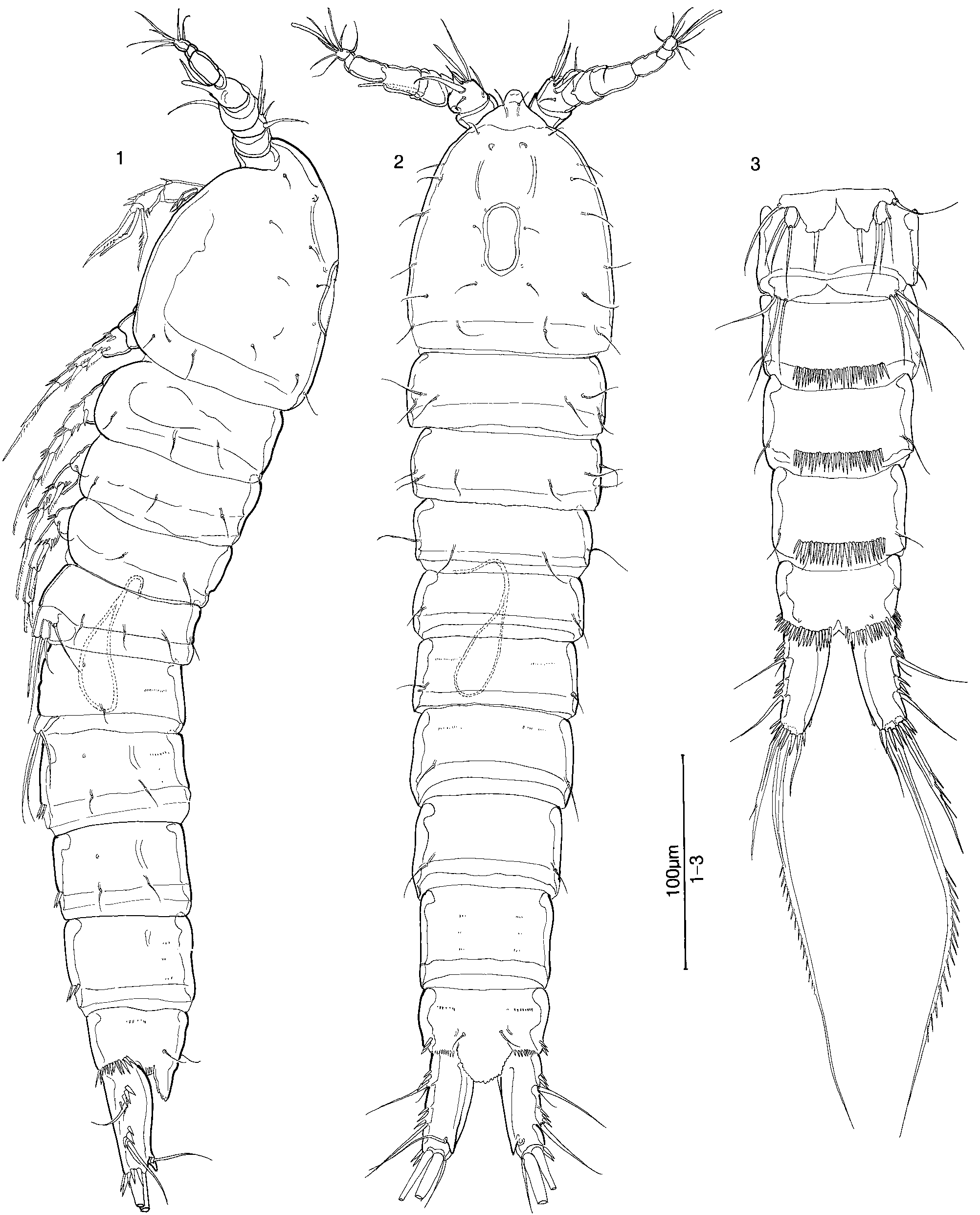
( FIGS 1–35 View Figures 1–3 View Figures 4–8 View Figures 9–15 View Figures 16–24 View Figures 25–31 View Figures 32–35 )
Material examined: Male holotype ( MZUSP 15370 View Materials ) and female allotype ( MZUSP 15371 View Materials ), each dissected on slide, and 13 males, 13 females, and four copepodids, paratypes ( MZUSP 15379 View Materials ), all from sample SP99-5-6, along the Trilha da Clareira (‘ Clearing Trail’ ), beside last of three unnamed streamlets before clearing at end of trail (23∞40¢08.5¢¢S, 045∞53¢54.2¢¢W; UTM 23K0408370/7382086), damp moss growing on tree trunks at heights of 5 cm to 1 m above the ground, 24.viii.1999 . Additional paratypes: one female and one male ( MZUSP 15372 View Materials ), sample SP99-1-5, combined water from tanks of two arboreal bromeliads along shaded trail leading to Poço Verde waterfall on Rio Claro (23∞38¢56.3¢¢S, 045∞52¢49.5¢¢W; UTM 23K0410050/7384157), 23.viii.1999 . One female and one male ( MZUSP 15373 View Materials ), sample SP99-1-11, tank of large bromeliad at base of tree trunk , same locality and date as SP99-1-5. One male ( MZUSP 15374 View Materials ), sample 99-1-17, in Sphagnum moss in small puddle near base of waterfall , same locality and date as SP99-1-5. One male ( MZUSP 15375 View Materials ), sample SP99-3-1, saturated moss on small rocks by riverside, bank of stream Ribeirão do Campo , just downstream from waterfall (23∞38¢10.3¢¢S, 045∞49¢56.6¢¢W; UTM 23K 0415023/ 7385808), 23.viii.1999 . One female ( MZUSP 15376 View Materials ), sample SP99-3-2, shallow shaded pool containing decomposing leaves and sand, on large rock beside stream , same locality and date as SP99-3-1. Four females, six males, and one copepodid (NHM reg. no. 2002.1095–2005), sample SP99-3-3, among roots of small plant on rock at base of waterfall , same locality and date as SP99-3-1. Two females ( MZUSP 15377 View Materials ), sample SP99-5-1, sand from bed of first tiny unnamed creek after clearing , same locality and date as SP99-5- 6. One female and One male ( MZUSP 15378 View Materials ), sample SP99-5-4, wet moss and plant roots on bank of tiny creek , same locality and date as SP99-5-6. 6 females and one copepodid ( MZUSP 15380 View Materials ), sample SP99-5-8, in organic detritus at base of large arboreal bromeliad , same locality and date as SP99-5-6. Three females, five males, and one copepodid ( VMNH Crustacea Catalogue no. 440), sample SP99-6-2, in decaying leaves in small debris dam in creek , same locality and date as SP99-5-6. One female ( MZUSP 15381 View Materials ), sample SP99-9-1, Trilha da Clareira, head of trail about 10 m from SABESP ( Companhia de Saneamento Básico do Estado de São Paulo) pipe (23∞40¢07.8¢¢S, 045∞53¢57.6¢¢W; UTM23K 0408315/7382105), saturated sandy-muddy soil with odour of sulphide, 24.viii.1999. Collectors C. E. F. Rocha and J. W. Reid.
Description of male
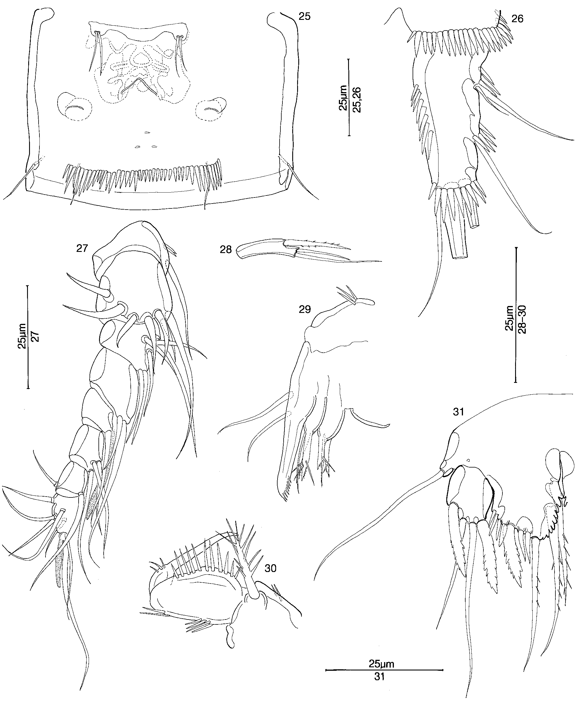
Length of holotype 490 Mm; lengths of four paratypes from Sample SP99-5-6, 455–483, mean 473 Mm. Body ( Figs 1–3 View Figures 1–3 ) slender. Cephalosome (Figs 1,2) with dorsomedial integumental window, ovoid and slightly constricted at midlength; no lateral window present. Hyaline frills of all somites smooth. Posterior margins of urosomites 2–4 each with ventral row of spines. Urosomite 5 ( Figs 1–6 View Figures 1–3 View Figures 4–8 ) with ventrolateral row of spines, and dorsal row of tiny hyaline spines along posterior margin. Anal operculum ( Fig. 5 View Figures 4–8 ) produced posteriorly, free margin hyaline, rounded and crenate.
Caudal ramus ( Figs 1–6 View Figures 1–3 View Figures 4–8 ) long-ovate, with longitudinal dorsal keel ending in acute prominence near distal end of ramus. Rows of large spines present at insertions of lateral setae and along posteroventral margin of ramus; medial surface bare. Inner terminal seta slightly longer than half length of ramus, slender and hyaline. Middle and outer terminal setae stout, without breaking planes, set with strong spinules along approximately middle third.
Rostrum ( Fig. 2 View Figures 1–3 ) subtriangular, reaching distal end of antennule segment 1, bearing two subapical sensilla.
Antennule (Figs 1,2,7,8) about 2/3 length of cephalosome, 9-segmented with well-developed basal prominence, geniculate, with long aesthetasc on segment 4 and short aesthetasc on segment 9, and bifurcate spiniform seta on segment 4.
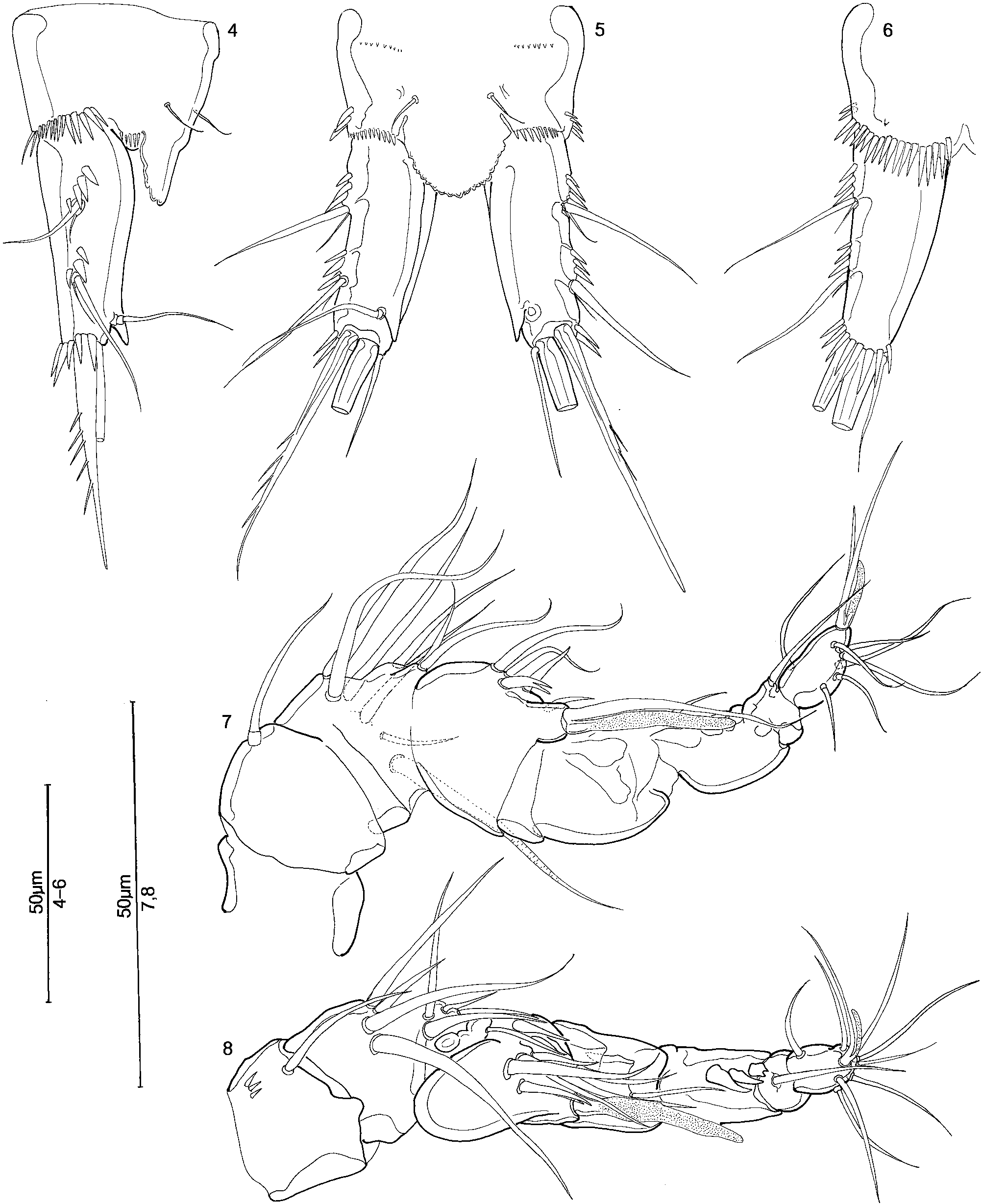
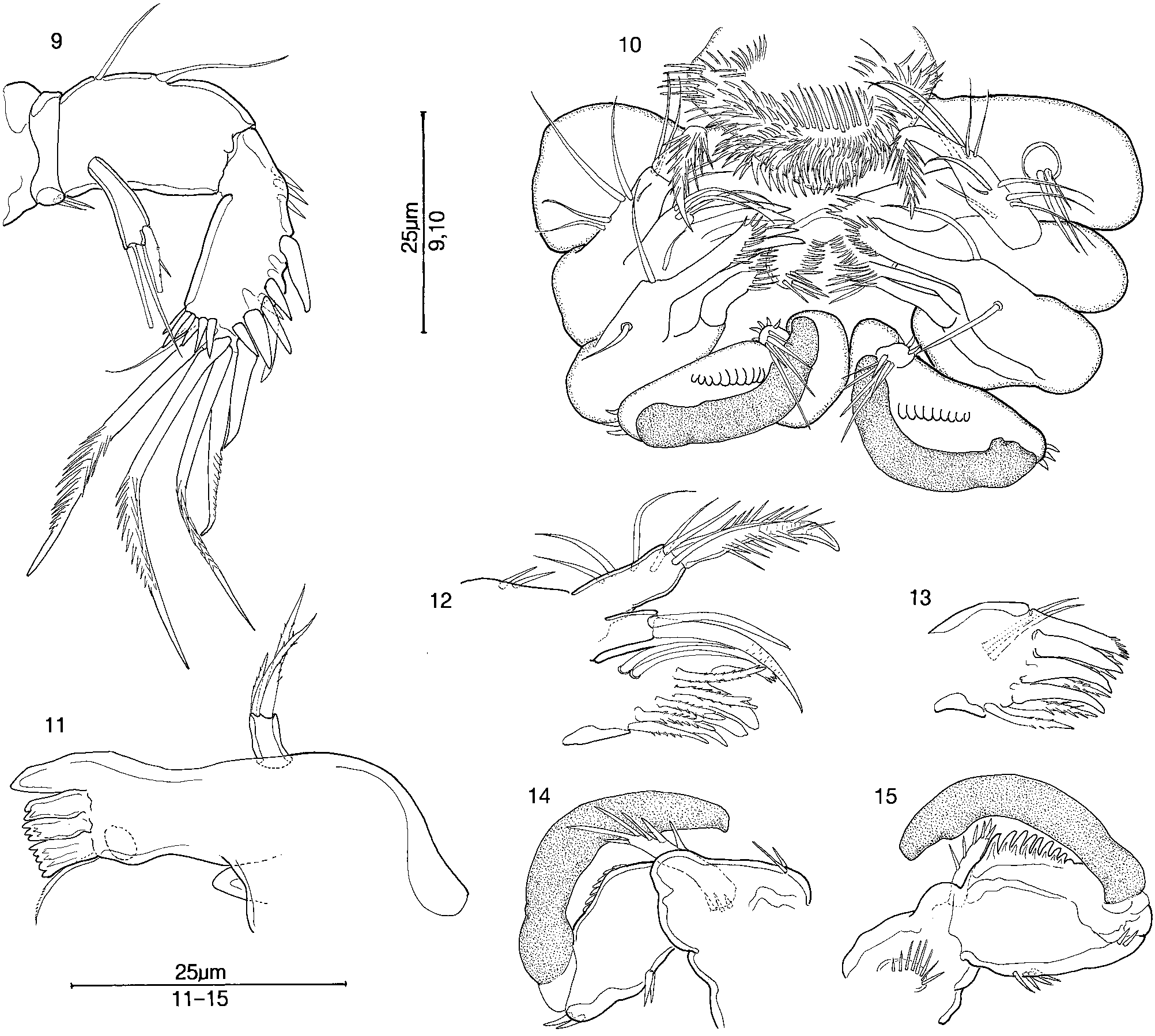
Antenna ( Fig. 9 View Figures 9–15 ) with allobasis; exopodite 1- segmented, with three terminal setae.
Labrum ( Fig. 10 View Figures 9–15 ) diamond-shaped in ventral view, thickly set with spines.
Mandible ( Fig. 11 View Figures 9–15 ), gnathobase with one large conical tooth, three bifid to multicuspidate spines, and one finely serrate seta; palp 1-segmented, with three terminal setae.
Maxillule (Figs 12,13), precoxal arthrite with four stout spines fused to arthrite and five spinulose setae along inner margin, and pair of smooth setae on distal margin. Coxal endite with two terminal setae. Palp bearing stout spinulose spine and two setae at apex; outer margin with four setae. Epipodite represented by two short setae.
Maxilla as in female ( Fig. 29 View Figures 25–31 ).
Maxilliped (Figs 10,14,15), syncoxa with crescentic row of spines, and stout spinulose distal seta with several spines at its base; basis with two groups of spines on outer margin, and inner margin with wide, deeply crenate hyaline flange; endopodite lacking seta, with claw enormously enlarged and sclerotized, recurved, with rounded inner subterminal expansion and blunt recurved tip.
Legs 1–4 ( Figs 16–23 View Figures 16–24 ) with 3-segmented exopodites and 2-segmented endopodites, except leg 3 endopodite 3-segmented. Couplers (intercoxal sclerites) bare. Leg 1 basis with spiniform seta on mediodistal corner. Leg 1 exopodite segment 2 without, legs 2–4 exopodite segment 2 each with medial seta. Legs 1–4 exopodite 3 each with four lateral and terminal setae, lateral setae inserted near apex of exopodite. Leg 1 endopodite not prehensile, segment 1 broad. Leg 2 endopodite ( Fig. 18 View Figures 16–24 ), segment 1 short and broad with large, stout, medially curved spine on laterodistal corner, flanked medially by two slightly smaller stout spines, and with two tiny spines on medial surface; segment 2 narrower and shorter, with two long terminal spinulose setae and tiny spine on medial surface. Leg 3 basis ( Fig. 19 View Figures 16–24 ) with two spines on medial surface. Leg 3 endopodite ( Fig. 20 View Figures 16–24 ) 3-segmented, segment 1 bare, segment 2 with stout medial apophysis having slender, simple tip, and segment 3 with stout apical seta flanked by longer, medial subapical plumed seta and two more proximal, short smooth hyaline setae on medial margin. Leg 4 endopodite (Figs 22,23) 2-segmented; segment 1 with stout seta on mediodistal corner; segment 2 with two slender apical setae and one short stout apical spine, and at about midlength, one slender smooth seta, two stout denticulate setae, and two stout spines.
Leg 5 ( Fig. 24 View Figures 16–24 ), baseoendopodites partly fused medially; baseoendopodite and exopodite distinct, with bare surfaces. Medial expansion of baseoendopodite broadly triangular, with one simple subapical seta and tiny papilla lateral to insertion of seta. Exopodite subquadrate, shorter than baseoendopodite, with five setae of which middle seta is longest. Medialmost seta curved toward median line.
Leg 6 ( Fig. 3 View Figures 1–3 ) a simple broad plate bearing three long setae.
Observed variation: Leg 2 endopodite segment 1 may have 1–3 large spines along the outer distal corner; two is the most usual number. The usual number of setae on the leg 5 exopodite is 5; the holotype has three setae on the right exopodite and five on the left; of the males in Sample SP99-5-6, 2 specimens had three or four setae on one exopodite.
Description of female
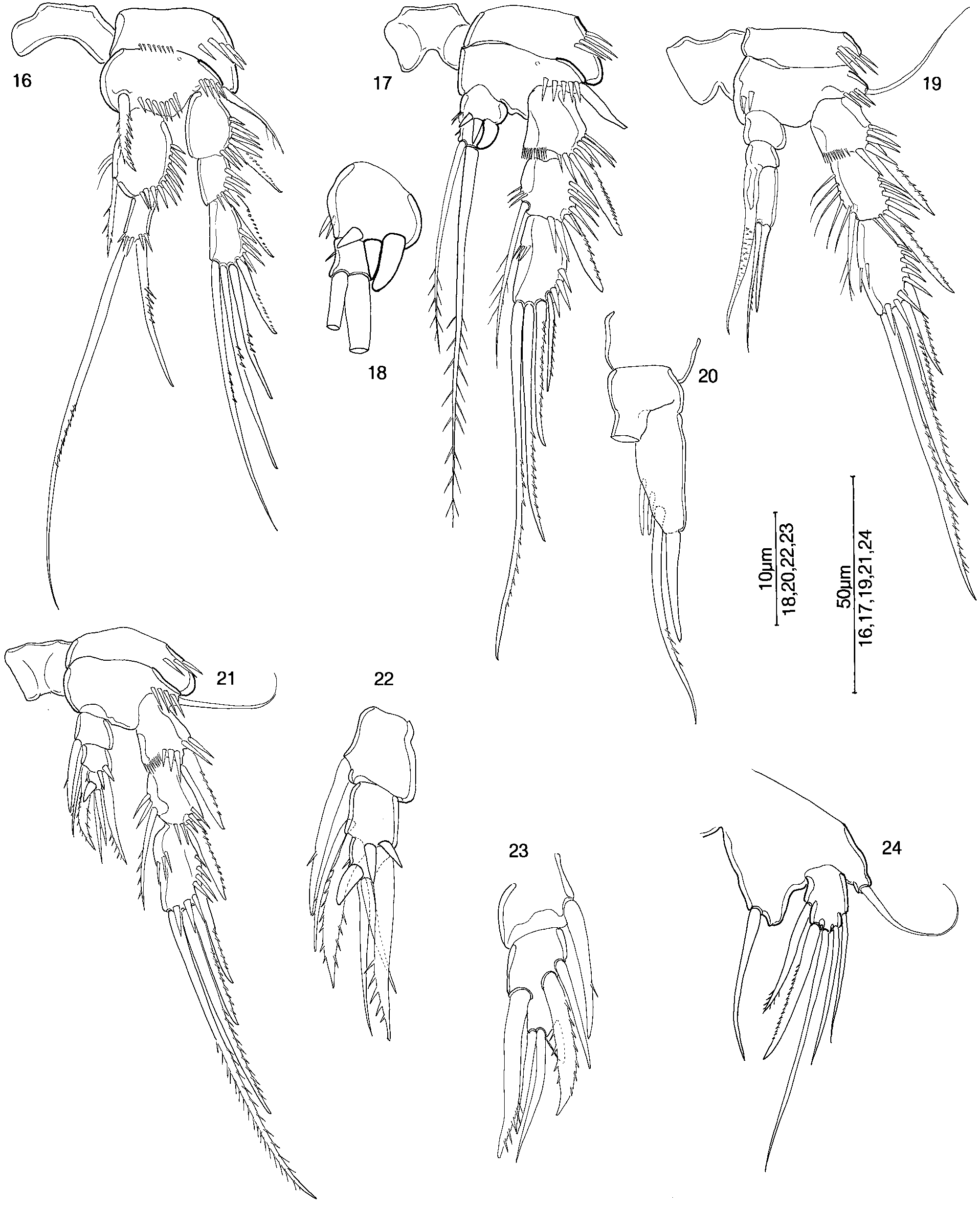
Length of allotype 510 Mm; lengths of nine paratypes in Sample SP99-5-6, 460–508, mean 489 Mm. Habitus and ornamentation of urosome as in male, except for fused genital double-somite ( Fig. 25 View Figures 25–31 ). Genital field ( Fig. 25 View Figures 25–31 ) occupying anterior half of genital doublesomite, with short wide funnel leading to genital pore, and large paired dorsal receptacula seminis.
Caudal ramus ( Fig. 26 View Figures 25–31 ) as in male, except with additional row of spines along middle third of medial surface. Caudal setae as in male.
Antennule ( Fig. 27 View Figures 25–31 ) 8-segmented, with long slender aesthetasc on segment 4, and short slender aesthetasc on segment 8.
Antennal exopodite ( Fig. 28 View Figures 25–31 ) as in male.
Maxilla ( Fig. 29 View Figures 25–31 ), syncoxa with two endites, each with three apical setae; basis with two setae and stout claw ending in broad, deeply serrated, spatulate tip.
Maxilliped ( Fig. 30 View Figures 25–31 ), syncoxa with group of spines, and stout spinulose distal seta; basis with two groups of spines on outer margin, and inner margin produced, bearing row of strong spines; endopodite with tiny seta and few spines, claw slender, tapering, acute, slightly recurved near tip.
Leg 5 ( Fig. 31 View Figures 25–31 ), baseoendopodites partly fused medially. Medial expansion of baseoendopodite with four setae, lateralmost seta spatulate and serrate, three medial setae slender, with stiff setules; mediodistal margin deeply crenate and expanded, covering insertions of two medial setae. Exopodite quadrate, shorter than baseoendopodite, with four setae of which two outer setae are spatulate and serrate, and two middle setae are slender and smooth.
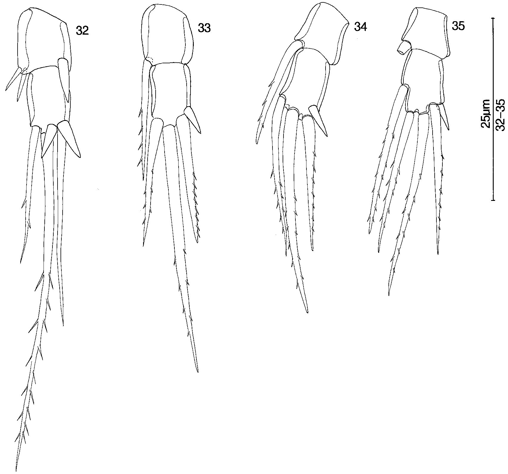
Legs 1–4 basipodites, exopodites, leg 1 endopodite, spiniform seta on distomedial corner of leg 1 basipodite, and couplers all as in male. Leg 2 endopodite ( Fig. 32 View Figures 32–35 ), segment 1 with three or four spines on distal margin, outer one or two spines very stout; segment 2 with three terminal and subterminal setae, and two subterminal spines. Leg 3 endopodite ( Fig. 33 View Figures 32–35 ), segment 1 with seta on mediodistal corner; segment 2 with three apical and subapical setae of which apical seta is longest, and subapical spine. Leg 4 endopodite (Figs 34,35), segment 1 with seta on distomedial corner; segment 2 with three apical and subapical setae, apical seta longest, and subapical spine; in allotype, right segment 2 with, left segment 2 without medial seta.
Leg 6 ( Fig. 25 View Figures 25–31 ) consisting of tiny plate bearing two setae.
Observed variation: Of the female paratype specimens in Sample SP99-5-6, all had leg 4 endopodite segment 2 bearing three setae.
Etymology: The species name is given for the collection locality, with profound appreciation for the long efforts of staff members of the Museum of Zoology to maintain the Boracéia preserve.
| VMNH |
Virginia Museum of Natural History |
No known copyright restrictions apply. See Agosti, D., Egloff, W., 2009. Taxonomic information exchange and copyright: the Plazi approach. BMC Research Notes 2009, 2:53 for further explanation.
|
Kingdom |
|
|
Phylum |
|
|
Class |
|
|
Order |
|
|
Family |