
Protimesius junina, Villarreal-Manzanilla, Osvaldo & Pinto-Da-Rocha, Ricardo, 2006
|
publication ID |
https://doi.org/10.5281/zenodo.174062 |
|
DOI |
https://doi.org/10.5281/zenodo.6255234 |
|
persistent identifier |
https://treatment.plazi.org/id/03AE87D7-FFE8-FF88-FEE3-F888FB80A730 |
|
treatment provided by |
Plazi |
|
scientific name |
Protimesius junina |
| status |
sp. nov. |
Protimesius junina View in CoL n. sp.
Figs 8–14 View FIGURE 8 View FIGURES 9 – 14 , 38–39 View FIGURES 36 – 45
Typematerial: Male holotype (MZSP), Brazil, Bahia, Pau Brasil (Gruta Califórnia, Atlantic Rain Forest, 15°27´S, 39°39´W), 28.IX.1997, B.S. Santos leg. Paratypes: Female, same data as for holotype; male (MZSP19345), Itororó (Serra do Oricama), III.2000, G. Machado leg.
Diagnosis: It differs from all other species of Protimesius by the presence of one retrolateral row and one prolateral row of tubercles on basal part of male femur IV; rest of femur, patella and tibia IV smooth.
Etymology: The “festa junina ” is a Brazilian festival, which is held, as the name implies, in June. It is in honor of the Saints Anthony, Peter and John. The specific epithet is a noun in apposition.
Description of male ( holotype):
Measurements: Dorsal scute length 5.0; prosoma length 2.4; dorsal scute width 4.0; prosoma width 3.9; interocular distance 2.6; chelicera: II 6.1; III 2.5; pedipalpus 17.5; leg I 20.5; II 37.0; III 33.5; IV 32.5.
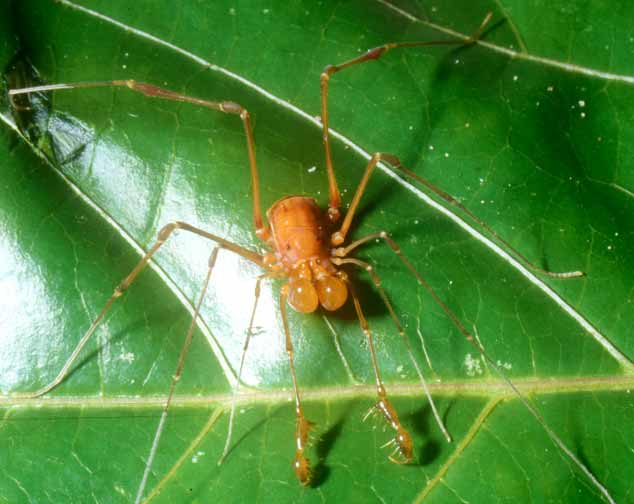
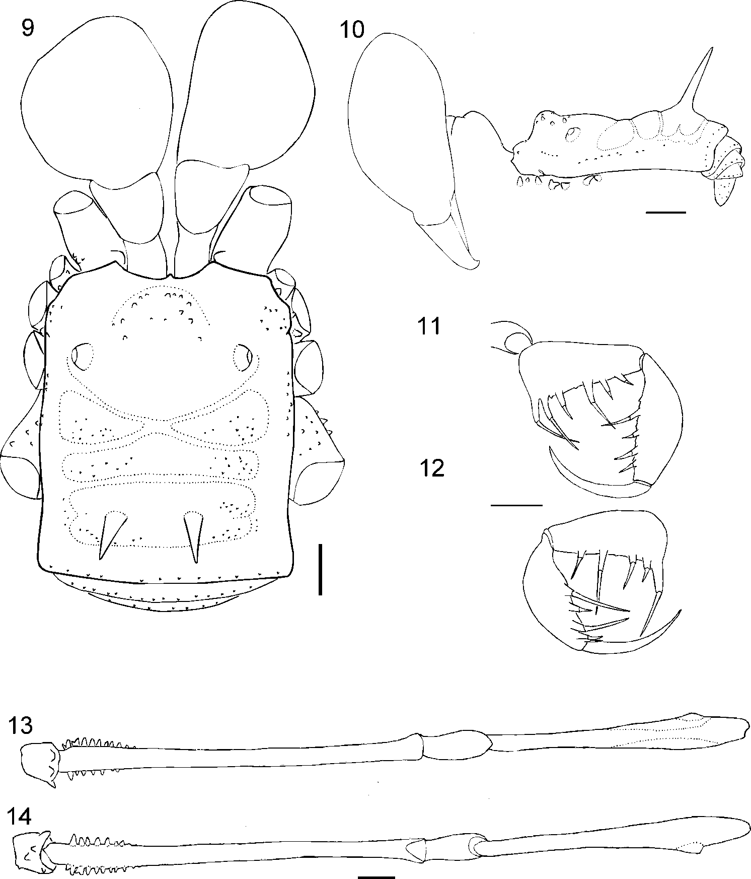
Dorsum ( Figs 8–10 View FIGURE 8 View FIGURES 9 – 14 ): Prosoma with 1 high and tuberculated anterior eminence. Ocularium smooth. Lateral margin with 1 row of tubercles between grooves I and II. Area III partially divided by a transversal groove, with 2 high spines. Areas, posterior margin and free tergites minutely granular.
Venter: Coxa I with 1 median row of 6–7 tubercles and 2 apical tubercles; II with median row of 9 tubercles and 3 apical tubercles; III with median row of 8 tubercles and 3 apical tubercles; IV irregularly tuberculated.
Chelicera: Swollen. Segment I smooth; II with finger carrying 1 large tooth followed by 1 small tooth; III with 2 large teeth followed by 2 small teeth.
Pedipalpus ( Figs 11–12 View FIGURES 9 – 14 ): Base of coxa with mesal apophysis, ectal side with 3 small tubercles; ventral side with 1 median row of 3–5 tubercles and 1 apical tubercle. Trochanter with 2 ventral tubercles. Femur with 4 ventrobasal tubercles (basal one longest). Tibia with 8 ventral tubercles; mesal IiiIi (1>4>3=5>2), ectal IIiIii (1=2>4>3>5=6). Tarsus with 2 ventral rows of setiferous tubercles; mesal Iiii, ectal Iiii.
Legs ( Figs 13–14 View FIGURES 9 – 14 ): Coxa I with 2 anterior and 1 posterior tubercles; II with 1 large anterior tubercle, 1 posterior tubercle bifid, fused with 1 tubercle of III; III with 1 tubercle fused with 1 tubercle of coxa IV; IV irregularly tuberculate. Trochantera I and II with 3 ventral tubercles; III with 4 ventral and 1 retrolateral tubercles; IV with 2 large ventral, 1 large retrolateral and 2 wide apical tubercles. Femora III and IV with retrolateral and prolateral rows of tubercles on basal fourth. Tibia IV swollen subapically, 1 large ventroectal tubercle present. Tarsal segmentation: 7, 15, 7,?[missing; 8 in paratype].
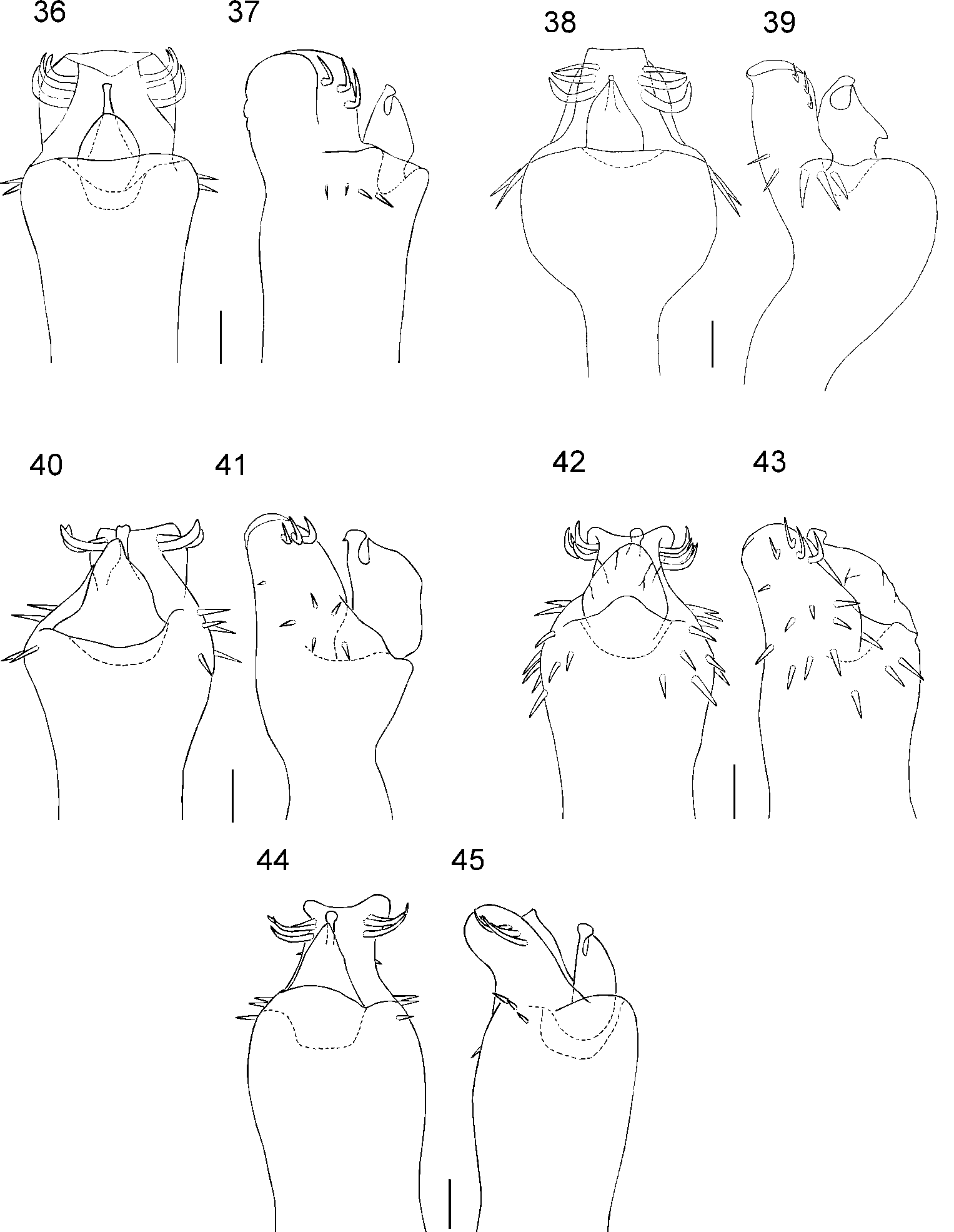
Penis ( Figs 38–39 View FIGURES 36 – 45 ): Truncus strongly swollen distally. Ventral plate with lateral margin converging, apical and distal margin straight, 2 curved distal setae and 1 pair of straight setae, without intermediary pair, 5 basal pairs of straight setae. Dorsal process present. Stylus swollen apically.
Color ( Fig. 8 View FIGURE 8 ): Mostly brown in alcohol and alive. Sulci III and IV, surroundings of each ocularium, legs I–IV, anterior and lateral margin with fine small pigmentation. Chelicera with black reticulation. Base of femora III and IV, apical third of tibia IV and cheliceral fingers reddish brown.
Description of female ( paratype):
Measurements: Dorsal scute length 4.5; prosoma length 1.9; dorsal scute width 3.7; prosoma width 3.4; interocular distance 1.9; chelicera: II 2.7; III 1.5; pedipalpus 15.5; leg I 17.5; II 35.0; III 24.0; IV 32.5.
Somatic morphology: Similar to male, except for: Chelicera not swollen; prosoma without large eminence, carrying 6 tubercles; body sligthly darker than in male; legs and chelicerae of same color as body. Pedipalpal tibia mesal IiiIi, ectal IiIii/Iiii; tarsus mesal IiIii, ectal Iiii. Legs finely granular; tibia IV cylindrical.
Natural history: Only two specimens were collected at the entrance of the California Cave. They do not show any sign of troglomorphism (morphological modification usually found in cave animals) and the species was not collected in other caves of the region ( Trajano 2000). It is not clear if this is a cavernicolous species or not.
No known copyright restrictions apply. See Agosti, D., Egloff, W., 2009. Taxonomic information exchange and copyright: the Plazi approach. BMC Research Notes 2009, 2:53 for further explanation.
|
Kingdom |
|
|
Phylum |
|
|
Class |
|
|
Order |
|
|
Family |
|
|
Genus |