
Penia smetanai Schimmel, 1996
|
publication ID |
https://doi.org/ 10.11646/zootaxa.5375.3.1 |
|
publication LSID |
lsid:zoobank.org:pub:27D02F09-01B7-457A-8A99-D8644B7B6ADE |
|
DOI |
https://doi.org/10.5281/zenodo.10201490 |
|
persistent identifier |
https://treatment.plazi.org/id/03AE8A2D-FFB2-CF7C-FF47-ED05FD37FDD0 |
|
treatment provided by |
Plazi |
|
scientific name |
Penia smetanai Schimmel, 1996 |
| status |
|
( Figures 14 View FIGURE 14 , 15 View FIGURE 15 )
Penia smetanai Schimmel, 1996: 187 (original description; type locality: Taiwan, Taichung City, Heping District, Mt. Anma Shan); Schimmel, 2001: 221, 227 (comparison with the other species); Kundrata et al., 2018: 55 View Cited Treatment (catalogue).
Type material. Paratype. Male, Taiwan, Taichung City, Heping District, Mt. Anma Shan , 2225 m, 11–15 V 1992, A. Smetana leg. [ BMNH; PSS01 View Materials ] .
Male. Diagnosis. This species is characterized by the following features: eyes 0.5 x longer than interocular distance in dorsal view; antennae extending beyond pronotum posterior lateral apices by antennomere VI, surpassing elytral half by antennomere XI; antennomeres III distinctly longer than II; IV 1.5 x longer than III, almost as long as II – III combined; apical maxillary palpomere 2.8 x longer than wide, shorter than maximum length of eye; pronotum roundly narrowed and distinctly constricted ahead of hind angles; posterior edge of pronotum with a sublateral incision near each hind angle; hind angles of pronotum acute, strongly protruding posterolaterad; hypomeron with slight mesial projection; anterior angle of hypomeron rounded; hind angle of hypomeron claw-like shaped; scutellar shield almost as long as wide; mesosternal process between mesocoxae lower than mesocoxae, not visible in lateral view; posterior edge of mesosternal process less than 0.1 x wider than total width of mesosternum; elytron 4.7 x longer than wide, 4.4 x longer than pronotum length; abdominal ventrite V semicircular but slightly incurved laterally ahead of apex, rounded apically; phallobase almost as long as wide; apex of parameres beyond preapical expansions bullet-like shaped; apex length 2.4 x width of parameres at expansions in ventral side.
In Taiwan, three species with elongate bodies are known: P. alishanensis , P. elongata , and P. smetanai . Their elytra are more than 4× longer than their widths. They are separated from the other species, whose elytra are less than 4× longer than their widths.
Penia smetanai is more similar to P. alishanensis in the relative length of the basal antennomeres and the development of the mesosternal process between mesocoxae, but is distinguished from P. alishanensis by the following contrasting characters ( P. alishanensis in parentheses): apical maxillary palpomere 2.8 x longer than wide (apical maxillary palpomere 2.3 x longer than wide); hypomeron with slight mesial projection (hypomeron with distinct mesial projection); anterior angle of hypomeron rounded (anterior angle of hypomeron biangular); scutellar shield almost as long as wide (scutellar shield 1.2 x longer than wide); abdominal ventrite V semicircular but slightly incurved laterally ahead of apex (abdominal ventrite V curved triangular); apex of parameres beyond preapical expansions bullet-like shaped (apex of parameres beyond preapical expansions elongated fan-shaped); apex length 2.4 x width of parameres at expansions in ventral side (apex length 2.7 x width of parameres at expansions in ventral side).
Penia smetanai is distinguished from P. elongata by many characters ( P. elongata in parentheses): antennomeres III distinctly longer than II (antennomeres II and III similar, short); antennomeres IV 1.5 x longer than III, almost as long as II – III combined (antennomeres IV 3.3 x longer than III, 1.8 x longer than II – III combined); apical maxillary palpomere 2.8 x longer than wide, shorter than maximum length of eye (apical maxillary palpomere 3.7 x longer than wide, longer than maximum eye length); posterior edge of pronotum with sublateral incisions (posterior edge of pronotum without sublateral incisions); hypomeron with slight mesial projection (hypomeron without mesial projection); anterior angle of hypomeron rounded (anterior angle of hypomeron biangular); hind angle of hypomeron claw-like shaped (hind angle of hypomeron narrowly triangular); abdominal ventrite V semicircular (abdominal ventrite V trapezoidal); apex of parameres beyond preapical expansions bullet-like shaped (apex of parameres beyond preapical expansions blade-like shaped); apex length 2.4 x width of parameres at expansions in ventral side (apex length 1.8–1.9 x width of parameres at expansions in ventral side).
Measurements. BL: 10.5, BW: 3.42, MAE: 1.46, MBE: 0.72, OI: 203, PL: 1.86, PML: 1.53, PW: 2.45, PAW: 1.51, PLI: 75.8, PWI: 162, EL: 8.12, EW: 1.73, EI: 471, BI: 438.
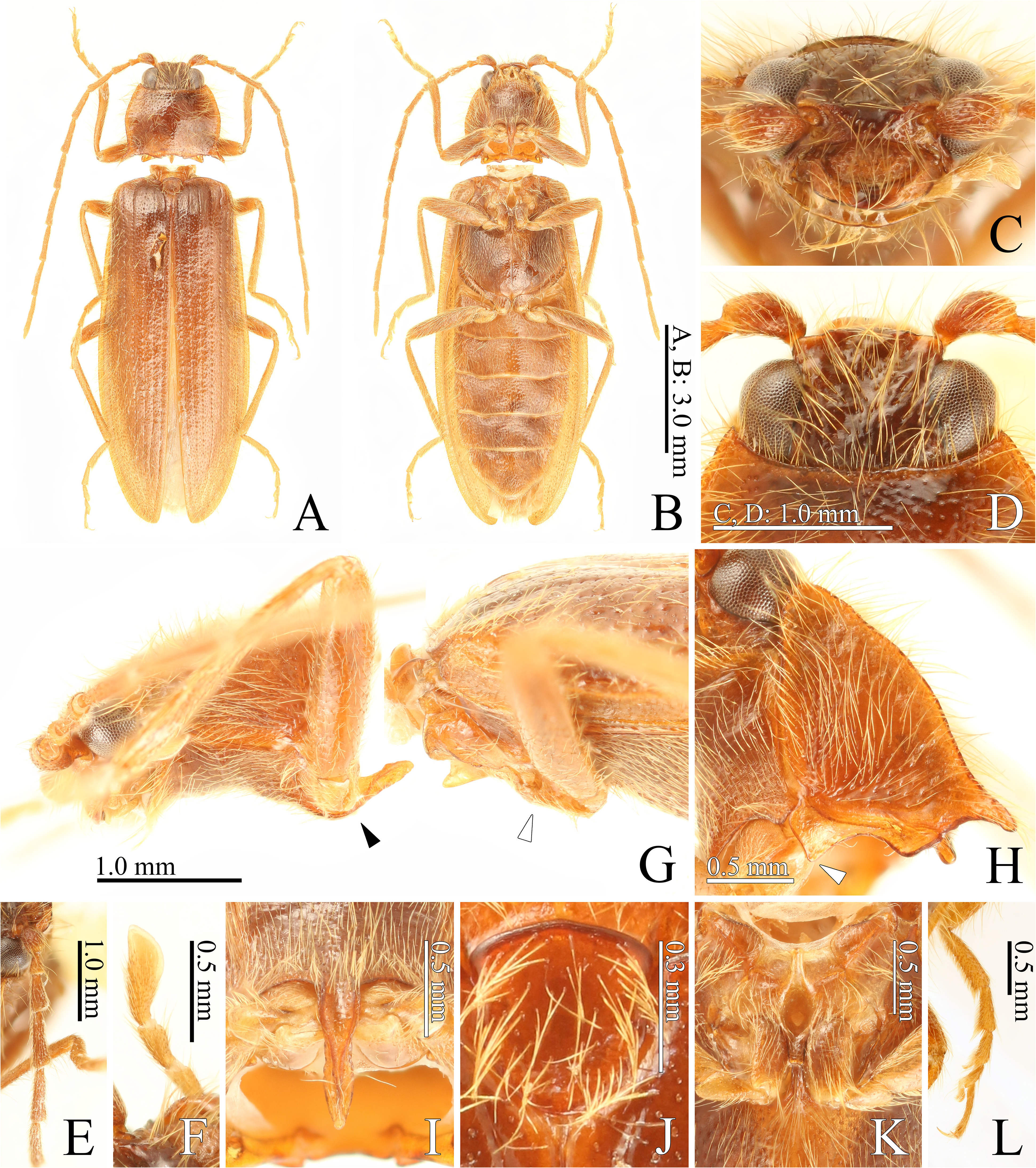
Redescription. Body elongated, widest behind elytral midlength ( Fig. 14A View FIGURE 14 ); surface surface generally smooth but prothorax and abdomen with microstructures; interspaces between punctures distinctly larger than fine puncture diameter ( Fig. 14A, B View FIGURE 14 ). Color. Body, antennae and legs brown. Lateral margins of pronotum and elytra, external margins of hypomeron and prosternum, and posterior margin of abdominal ventrite V yellowish. External edges of mandible and pronotum, posterior edge of prosternum, outer and posterior edges of hypomera, and anterior edges of scutellum and elytra blackish. Body covered with long yellow setae.
Head. Frons flatted medially ( Fig. 14C, D View FIGURE 14 ); frontal carina not complete ( Fig. 14C View FIGURE 14 ); frontal margin rectangular, nearly straight apically but weakly protruding medially in dorsal view ( Fig. 14D View FIGURE 14 ); frontoclypeal region protruding beyond base of labrum. Eyes protuberant, 0.5 x longer than interocular distance in dorsal view ( Fig. 14D View FIGURE 14 ). Antennae extending beyond pronotum posterior lateral apices by antennomere VI, surpassing elytral half by antennomere XI; antennomeres longer than wide; II obconical, shortest, 1.1 x longer than wide; III weakly serrated, 2.2 x longer than wide, 2.4 x longer than II; IV weakly serrated, 3.3 x longer than wide, 1.5 x longer than III, almost as long as II–III combined ( Fig. 14E View FIGURE 14 ); V–XI filiform; V 3.7 x longer than wide, almost as long as x longer than IV; XI 7.9 x longer than wide, 1.2 x longer than X. Mandible bidentate ( Fig. 14C View FIGURE 14 ). Apical maxillary palpomere elongated securiform ( Fig. 14F View FIGURE 14 ), 2.8 x longer than wide, shorter than maximum length of eye; anterior edge rounded.
Prothorax. Pronotum hexagonal, 0.8 x longer than wide, roundly widening anteriorly and then roundly narrowed, distinctly constricted ahead of hind angles ( Fig. 14A View FIGURE 14 ), widest across posterior lateral apices, tallest behind midlength ( Fig. 14G View FIGURE 14 ), without median longitudinal depression posteriorlyn; anterior edge weakly concave; anterior angles simple, nearly right angle; punctate lateral ridge extending from anterior angles to hind angles ( Fig. 14A View FIGURE 14 ); hind angles simple, acute, strongly protruding posterolaterad; posterior edge with a moderate sublateral incision near each hind angle, without carinae next to sublateral incisions ( Fig. 14A View FIGURE 14 ). Hypomeron with slight mesial projection ( Fig. 14H View FIGURE 14 : arrow); anterior angle rounded; mesial edge weakly sinuate; mesial and posterior margins with impunctate ridge ( Fig. 14H View FIGURE 14 ); posterior margin with triangular projection between two large emarginations; hind angle claw-like shaped. Prosternum strongly incurved ventrally in lateral view; anterior lobe distinctly protruding beyond prosternal ventral line in lateral view ( Fig. 14G View FIGURE 14 ); anterior edge rounded in ventral view ( Fig. 14B View FIGURE 14 ). Prosternal process slender, 1.9 x longer than procoxal cavity length, concave between procoxae, strongly curved dorsad from middle of procoxal cavities in lateral view ( Fig. 14G View FIGURE 14 ), without subapical tooth; dorsal lobe slightly roundly expanded anterior to apex in ventral view ( Fig. 14I View FIGURE 14 ); ventral lobe weakly roundly expanded near base and then abruptly narrowed posterad in ventral view ( Fig. 14I View FIGURE 14 ); ventral margin almost straight but slightly expanded medially in lateral view ( Fig. 14G View FIGURE 14 ); apex rounded in lateral and ventral views ( Fig. 14G, I View FIGURE 14 ). Pronotosternal sutures not grooved ( Fig. 14H View FIGURE 14 ), straight anteriorly and curved posteriorly in ventral view ( Fig. 14B View FIGURE 14 ), slightly opened anteriorly. Scutellar shield tongue-shaped ( Fig. 14J View FIGURE 14 ), almost as long as wide, widest anteriorly, weakly constricted anteriorly, almost parallel-sided and then narrowed posteriad, flat, inclined anterior-downwards, not visible in lateral view ( Fig. 14G View FIGURE 14 ); anterior margin triangular; posterior edge rounded. Mesosternum: borders of mesosternal cavity straight anteriorly and then obtusely curved in lateral view ( Fig. 14G View FIGURE 14 ); mesosternal process between mesocoxae lower than mesocoxae, not visible in lateral view ( Fig. 14G View FIGURE 14 ); posterior edge less than 0.1 x wider than total width of mesosternum, emarginate ( Fig. 14K View FIGURE 14 ). Mesepisternum reaching mesocoxal cavity ( Fig. 14K View FIGURE 14 ). Metasternum sulcate medially and anterior to metacoxal cavities ( Fig. 14B View FIGURE 14 ). Metacoxal plate narrowed toward outer side, becoming like a parallel-sided bar at its outer 7/ 10 in ventral view ( Fig. 14B View FIGURE 14 ). Elytron convex but flat in median area, with outer margin narrowly depressed, widest behind middle, 4.7 x longer than wide, 4.4 x longer than pronotum length; apex rounded; elytral striae defined by lines of elongated punctures. Hind wings fully developed. Tibiae with paired spurs; relative tarsomere lengths: IV<III II<V<I; tarsomeres III and IV with lobe ventrally ( Fig. 14L View FIGURE 14 ).
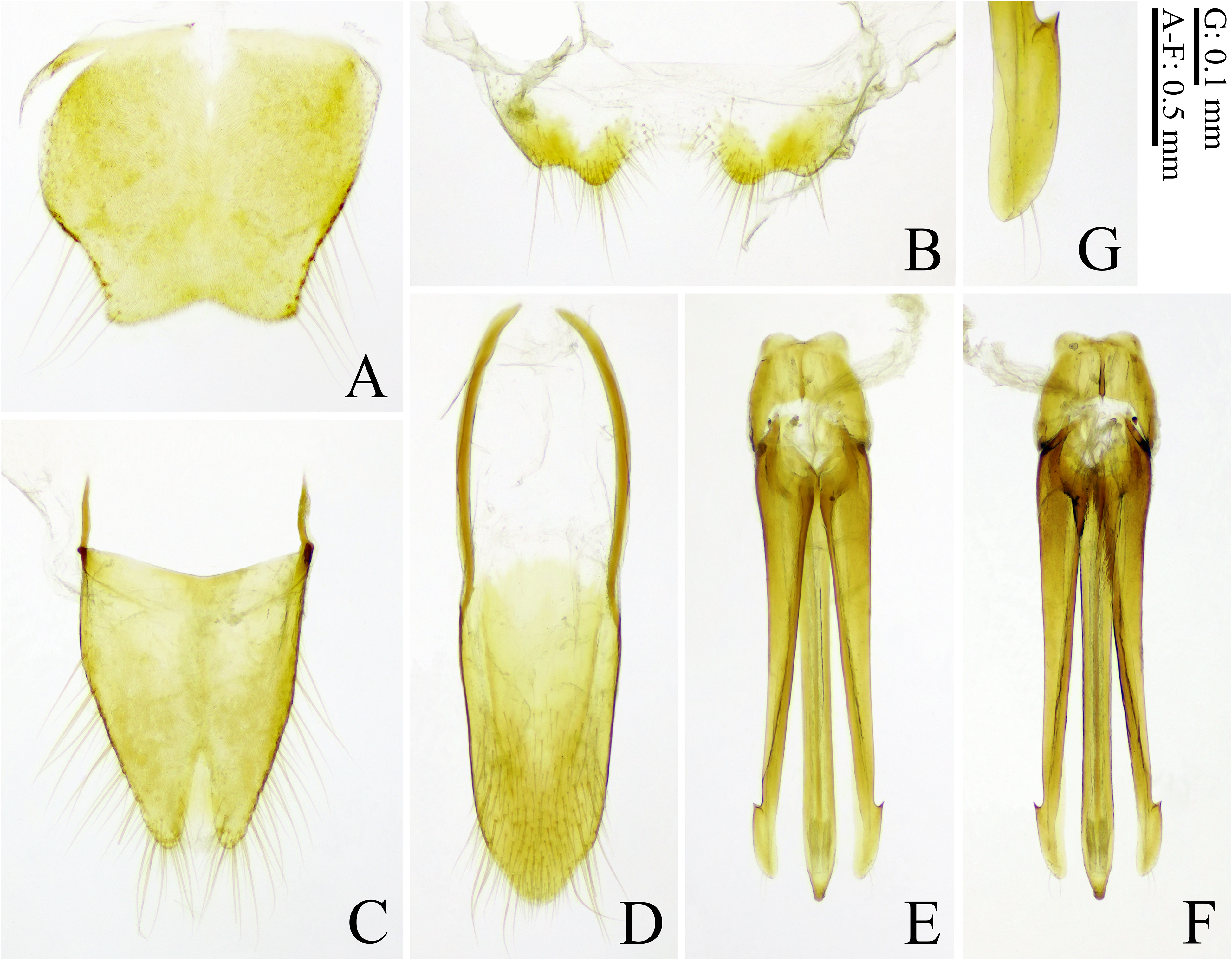
Abdomen. Ventrite V semicircular but slightly incurved laterally anterior to apex, rounded apically ( Fig. 14B View FIGURE 14 ), 0.6 x longer than wide. Tergites and sternites VIII‒X yellow. Tergite VIII trapezoidal, 0.85 x longer than wide, narrowed posteriad, weakly incurved laterally anterior to posterior lateral apices; posterior edge emarginate ( Fig. 15A View FIGURE 15 ). Sternite VIII posteriorly widely concave between two projections ( Fig. 15B View FIGURE 15 ); posterior lateral angle weakly protruding posterad. Tergite IX 1.3 x longer than wide; median notch 1/3 x total length of tergite IX ( Fig. 15C View FIGURE 15 ). Tergite X longer than wide, rounded apically ( Fig. 15C View FIGURE 15 ). Sternite IX 3.45 x longer than wide, constricted anterior to midlength ( Fig. 15D View FIGURE 15 ), rounded apically. Aedeagus yellow ( Fig. 15E, F View FIGURE 15 ). Phallobase 0.2 x total length of aedeagus, almost as long as wide. Median lobe exceeding apices of parameres by apical 1/20; basal struts 0.2 x total length of median lobe. Parameres elongtae, not fused ventrally ( Fig. 15F View FIGURE 15 ); preapical expansions projecting anterolaterad ( Fig. 15G View FIGURE 15 ); apex beyond preapical expansions bullet-like shaped ( Fig. 15G View FIGURE 15 ), rounded apically, with a seta dorsally, with two or four setae ventrally; apex length 2.4 x width of parameres at expansions in ventral side.
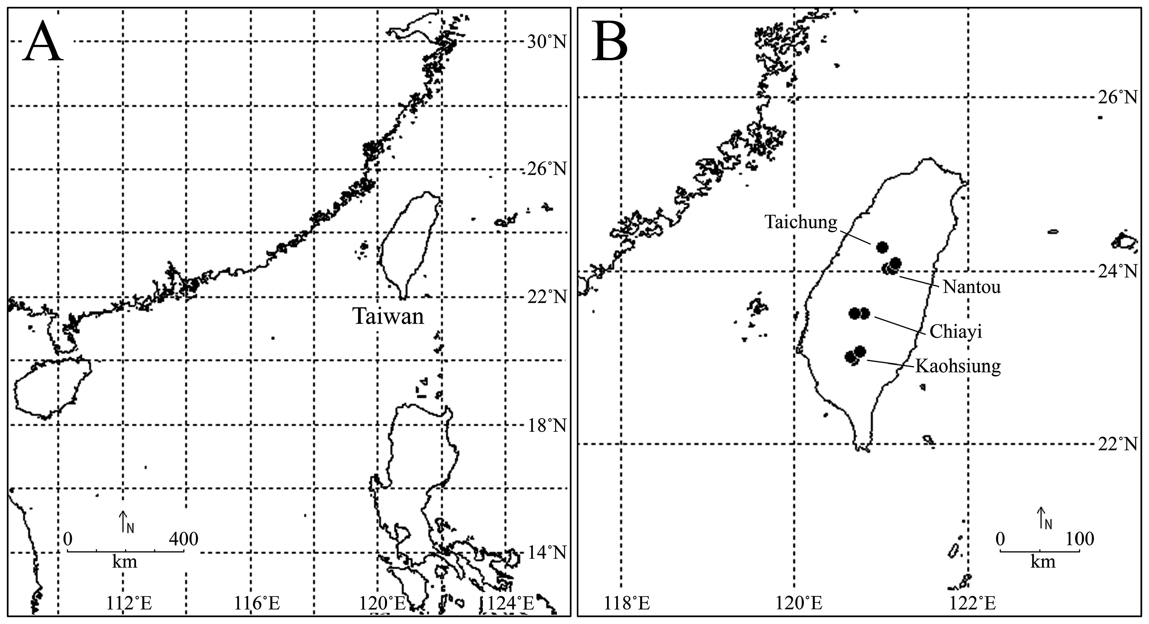
Distribution. Taiwan: Taichung City ( Fig. 1 View FIGURE 1 ).
| V |
Royal British Columbia Museum - Herbarium |
| VI |
Mykotektet, National Veterinary Institute |
No known copyright restrictions apply. See Agosti, D., Egloff, W., 2009. Taxonomic information exchange and copyright: the Plazi approach. BMC Research Notes 2009, 2:53 for further explanation.
|
Kingdom |
|
|
Phylum |
|
|
Class |
|
|
Order |
|
|
Family |
|
|
Genus |
Penia smetanai Schimmel, 1996
| Arimoto, Kôichi 2023 |
Penia smetanai
| Kundrata, R. & Musalkova, M. & Kubaczkova, M. 2018: 55 |
| Schimmel, R. 2001: 221 |
| Schimmel, R. 1996: 187 |