
Parastenhelia bulbosa, Gee, 2006
|
publication ID |
https://doi.org/10.1080/00222930601108194 |
|
persistent identifier |
https://treatment.plazi.org/id/03AF8548-BD25-310E-FE12-FE474843FE72 |
|
treatment provided by |
Felipe |
|
scientific name |
Parastenhelia bulbosa |
| status |
sp. nov. |
Parastenhelia bulbosa sp. nov.
( Figures 14–19 View Figure 14 View Figure 15 View Figure 17 View Figure 18 View Figure 19 )
Synonyms
Parastenhelia spinosa in Bozic (1955). Parastenhelia spinosa forma bulbosa in Wells (1963). Parastenhelia spinosa in Wells (1970) from Porth Hellick.
Material examined
Holotype; 1♀ (dissected onto four slides) from St. Martins Flat , Isles of Scilly, NHM Reg. No. 2006.1989 . Paratypes, from the same locality as the holotype, 1♀ (dissected onto three slides) and 2 „ (each dissected onto three or four slides) NHM Reg Nos 2006.1990–1992; 4♀, 4 „ and 3 copepodites (spirit preserved), NHM Reg. No. 2006.1993–2002 . Other material, 1 „ (spirit preserved) collected by University of London Sub-Aqua Club in gravel at low water at Porth Hellick, St Marys, Isles of Scilly, NHM Reg. No. 1967.10.31.50 .
Description of female
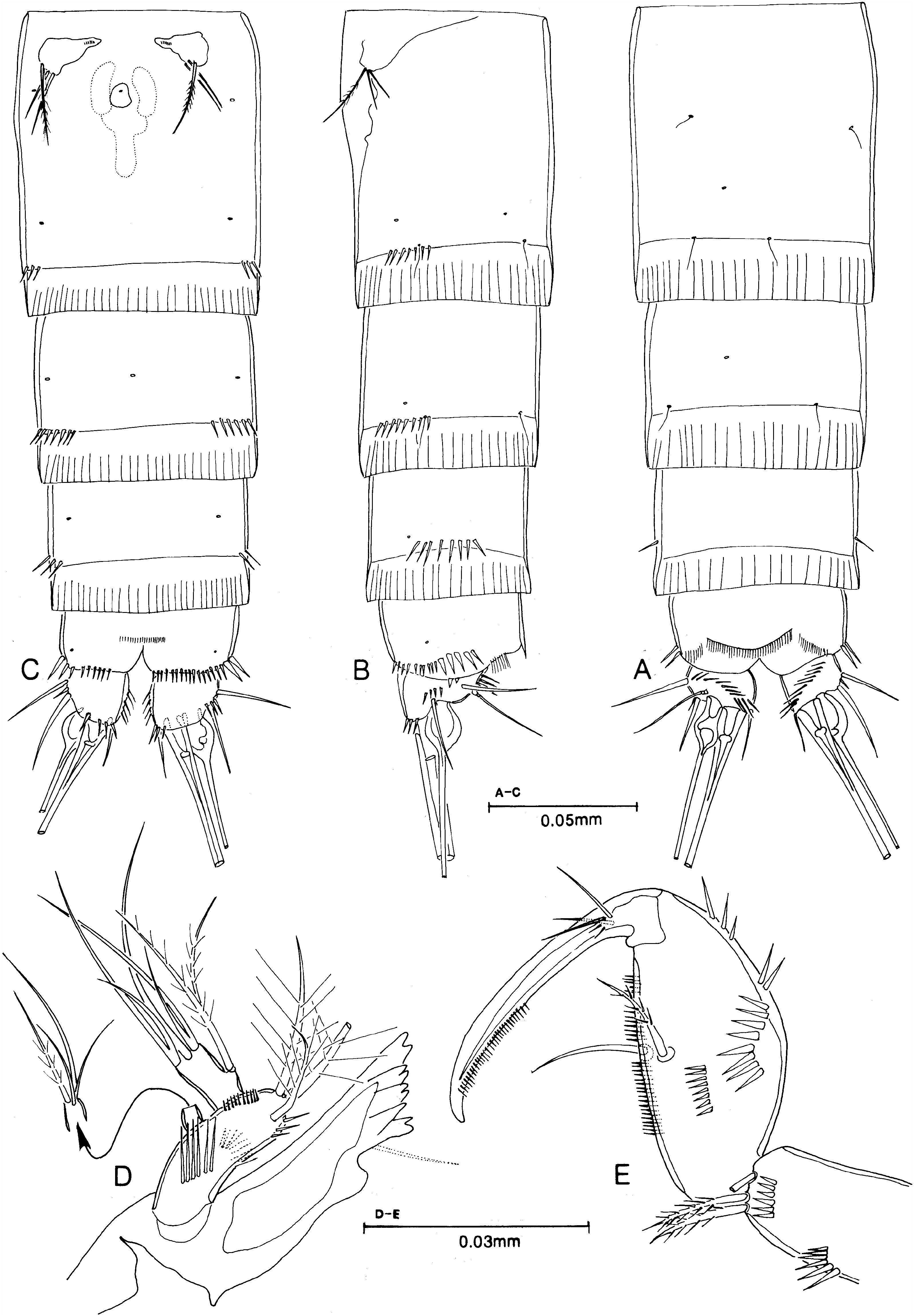
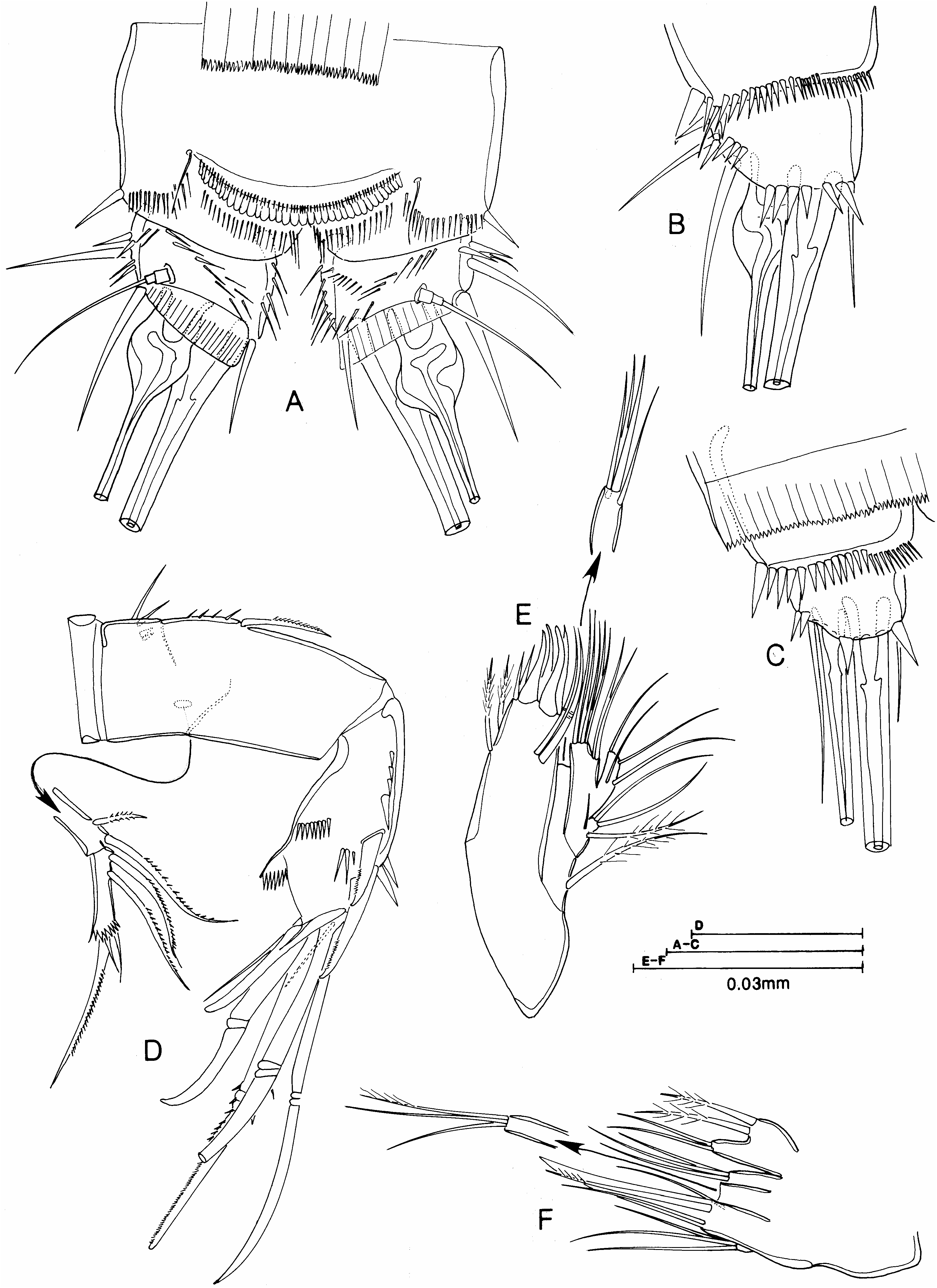
Body. Length (of contracted specimens) 0.41–0.49 mm (mean 50.46 mm, n 54), semicylindrical, widest at posterior margin of cephalothorax, tapering posteriorly without clear distinction between prosome and urosome. Urosomites ( Figure 14A–C View Figure 14 ) with wide, palisade hyaline frills finely dentate on posterior margin ( Figure 15A View Figure 15 ). Genital doublesomite ( Figure 14A–C View Figure 14 ) completely fused without trace of sub-cuticular rib, ornamented only with a short ventro-lateral row of spinules on posterior margin. Genital apparatus ( Figure 14C View Figure 14 ) with median ventral copulatory pore and separate anterior gonopores, each armed internally with a row of teeth and covered by a vestigial P6 bearing three setae. Urosomites 4 and 5 with short lateral row of spinules on posterior margin. Anal somite with median ventral row of short spinules and, on posterior margin, a ventral and lateral row of stronger spinules and setules on dorsal margin; anal operculum ( Figure 15A View Figure 15 ) semi-circular with a sub-marginal row of minute spinules and a marginal row of about 35 larger spinules. Caudal rami ( Figure 15A, B View Figure 15 ) broader than long, with rows of spinules on inner and outer margin, diagonally across dorsal surface and around ventral posterior margin; minute seta I and larger seta II implanted anteriorly on lateral margin; robust seta III implanted at distal outer corner; terminal seta IV moderately slender but with characteristic swollen bulbose base; terminal seta V robust, large without noticeably swollen base; terminal, inner seta VI small and slender and triarticulated seta VII implanted on dorsal surface.
Figure 16. Parastenhelia bulbosa sp. nov. ♀: (A) P1; (B) P2.
Rostrum ( Figure 19B View Figure 19 ). Moderately small, only reaching a short way past proximal margin of second antennular segment, defined at base, triangular with rounded tip and a pair of subapical sensilla.
Antennule ( Figure 19B View Figure 19 ). Distinctly nine-segmented, combined length of distal five segments markedly longer than segment 4; all segments without pinnate setae, aesthetascs on segments 4 and 9. Tentative setal formula as follows 1-[1], 2-[10], 3-[8], 4–[2+(1+a)], 5–[2], 6-[4?], 7-[2?], 8-[2?], 9-[5+(2+a)].
Antenna ( Figure 15D View Figure 15 ). Allobasis partially divided, abexopodal margin with a proximal transverse row of spinules and a more distal vertical row of spinules below a pinnate seta. Exopod two-segmented, proximal segment with two setae, distal segment with five setae (two proximal, lateral setae and one large and two small setae on distal margin) and a subdistal row of spinules. Endopod with row of spinules and two minutely pinnate subdistal spines on outer margin; distal margin with two rows of spinules and seven elements, a pinnate spine, four geniculate setae (inner with large pinnules at geniculation) and two plain setae, one of which is fused to base of inner geniculate seta.
Mandible ( Figure 14D View Figure 14 ). Coxal gnathobase well developed with bicuspid and unicuspid teeth and a seta at distal corner. Basis with rows of spinules on outer and inner margin and across anterior and posterior face and with three pinnate setae on distal margin; endopod elongate, one-segmented, with two lateral and five terminal setae (two pairs fused at base); exopod small, one-segmented, with four setae.
Maxillule ( Figure 15E View Figure 15 ). Praecoxal arthrite with six slender elements on distal margin, two geniculate surface setae and two pinnate setae on inner margin. Coxal endite with four setae on distal margin; coxal epipodite represented by one seta. Basis with seven setae (five on distal margin and two subdistally); rami fused to basis but clearly discerned, endopod one-segmented, with four setae; exopod one-segmented with four setae.
Maxilla ( Figure 15F View Figure 15 ). Coxa with three endites on inner margin, proximal endite broad and bicuspid with two pinnate setae on inner cusp and two naked setae on outer cusp; middle and outer endite each with three setae; allobasal endite with one fused spine, one articulating, pinnate, spine and two naked setae; endopod with four setae.
Maxilliped ( Figure 14E View Figure 14 ). Syncoxa with two rows of short surface spinules and three pinnate setae on distal margin. Basis with row of spinules on outer margin, on lateral face and on palmar margin, which also bears two setae. Endopod represented by a well-developed claw with small teeth on distal inner margin and three accessory setae proximally.
P1 (Figure 16A). Intercoxal sclerite small, unadorned. Praecoxa, small, triangular with row of spinules on distal margin. Coxa almost square with row of setules on outer margin, rows of strong spinules on inner and outer distal margin and three rows of spinules on anterior face. Basis also almost square, with row of strong spinules at base of inner and outer pinnate spine and on distal margin at base of endopod. Exopod three-segmented, proximal segment with row of spinules on outer margin and a spine at outer distal corner; middle segment markedly elongate, three times longer than proximal segment, with row of spinules on outer margin, a small spine at outer distal corner and a small seta at inner distal corner; distal segment very small, bearing a short, slender geniculate seta and one geniculate and two non-geniculate, recurved, short, strong spines. Endopod two-segmented, proximal segment longer than entire exopod with a small, pinnate seta implanted within proximal third of segment; distal segment small with a few spinules, one minute seta and two short, recurved, dentate spines.
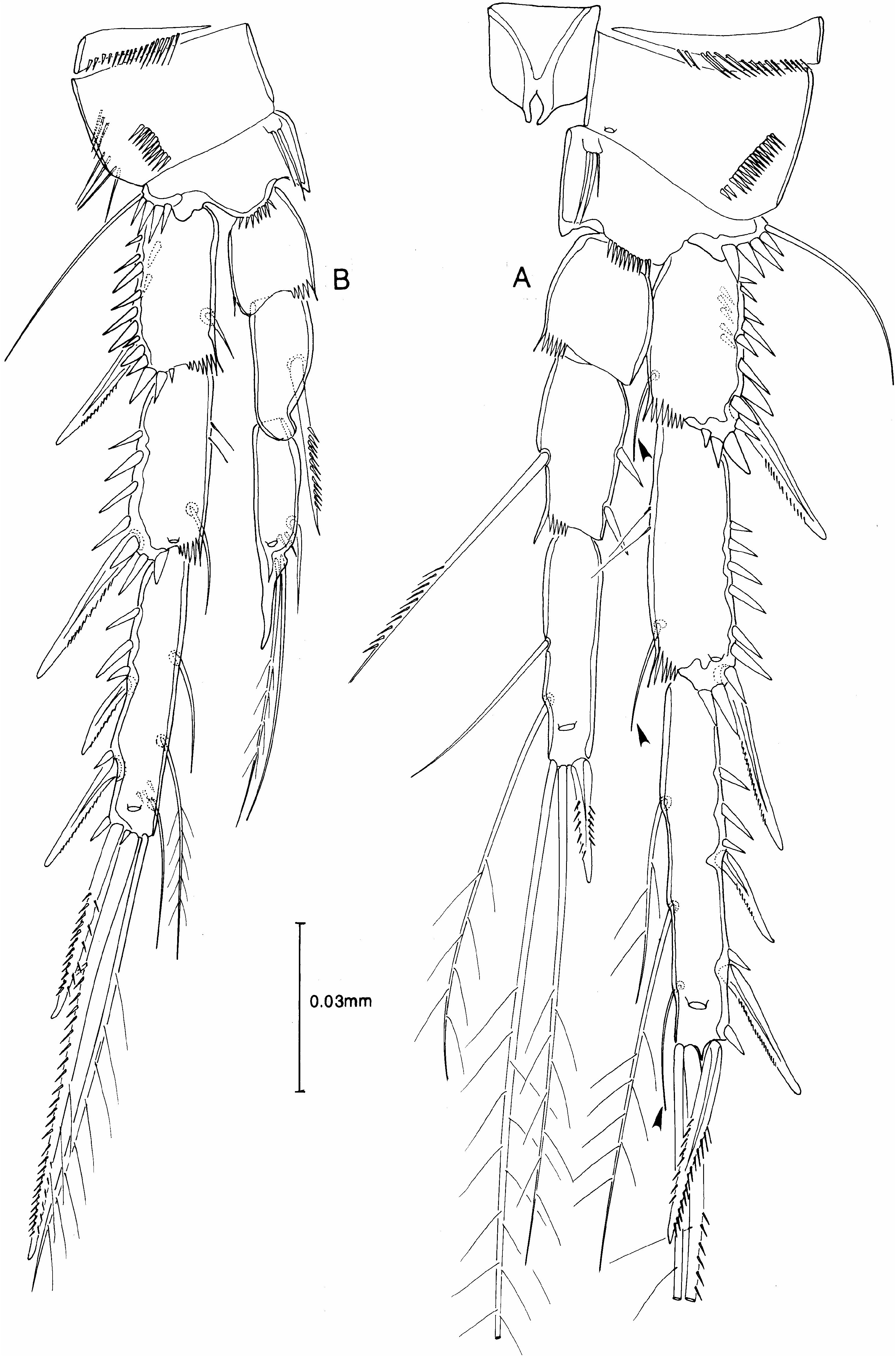
P2–P4 (Figures 16B, 17A, 18B). Intercoxal sclerites unadorned; praecoxa small triangular with spinule row on distal margin; coxa broader than long with row of setules on outer margin and row of spinules on anterior face; basis with row of setules near proximal inner margin, row of fine spinules distally at base of exopod and row of strong spinules near base of endopod, with pinnate outer spine on P2 and slender naked outer seta on P3 and P4. Rami three-segmented, segments with variable row of spinules on outer margin, exp 2 and 3 and enp 3 with pore on anterior face; exp 1 with inner seta on P2 and P3. The inner seta on exp 1 and exp 2 and the distal inner seta on exp 3 are extremely small or slender, are inserted on the posterior face and often lie along the posterior face of the succeeding segment. They are therefore, sometimes very difficult to see even with x100 oil immersion objective. The setal formula is as follows:
Exopod Endopod
P1 0.1.022 1.111
P2 1.1.123 0.1.021
P3 1.1.323 0.1.221 „ 0.1.12+apophysis
P4 0.1.323 1.1.221 „ 1.1.121
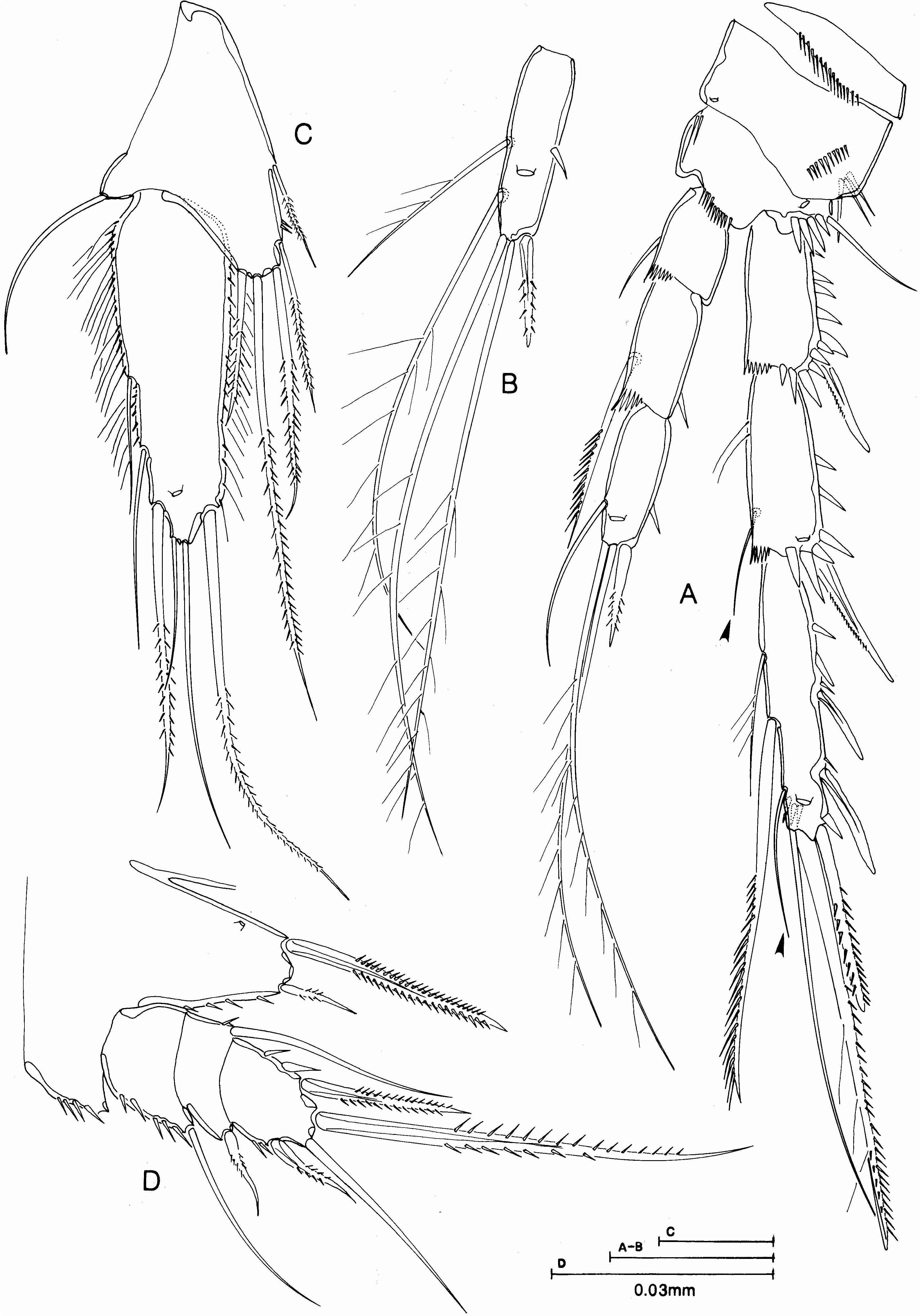
P5 ( Figure 18C View Figure 18 ). Benps of each side not fused medially and exopods also separate. Benp with well-developed inner expansion triangular in shape with row of setules on inner margin and bearing five pinnate setae, lengths as shown in Figure 18C View Figure 18 . Outer basal peduncle of benp with a naked seta. Exopod slender, three times longer than wide, with row of pinnules on outer margin and spinules on inner margin, and bearing six setae (one pinnate seta on inner margin, two naked setae on distal margin and one pinnate and two naked setae on outer margin).
Description of male
As in female except for urosome, caudal ramus, antennule, P3, P4, P5 and P6.
Body. Slightly smaller than female, contracted length 0.34–0.36 mm (mean 50.35 mm, n 55) and urosomites 2 and 3 not fused. Body ornamentation ( Figure 19A View Figure 19 ) as in female except urosomite 3 with row of small spinules near ventral anterior margin and urosomites 3 and 4 with complete row of spinules on ventral posterior margin. Caudal ramus seta IV not swollen at base ( Figure 19A View Figure 19 ).
Antennule ( Figure 19C View Figure 19 ). Eleven-segmented, haplocer with moderately swollen segments 5– 7, with major articulation between segments 7 and 8 and with four segments distal to articulation; aesthetascs on segments 5 and 11 and all setae naked; tentative setal formula as follows: 1-[1], 2-[10], 3-[8], 4-[4?], 5-[2+(1+a)], 6-[4?], 7-[2], 8[2 modified spines], 9,[1], 10-[4], 11-[5+(2+a)].
P3 ( Figure 17B View Figure 17 ). Exopod as in female. Enp 2 more slender than female with a shorter, stouter inner seta; enp 3 shorter than female, with one small seta on inner margin, two setae on distal margin and the outer spine fused to the segment to form an apophysis at outer distal corner.
P4 ( Figure 18A View Figure 18 ). As in female except that enp 3 has only one inner naked seta.
P5 ( Figure 18D View Figure 18 ). Benps of each side fused medially, inner expansions more rounded than in female, with row of spinules on outer margin and bearing two pinnate setae separated by a pore, inner twice as long as outer; outer peduncle bearing long naked seta (lost in Figure 18D View Figure 18 ). Exopod three-segmented, oval, overall not quite twice as long as wide; proximal segment with row of spinules on outer margin and a naked seta at outer distal corner; middle segment with long naked seta on inner margin and short pinnate seta at outer distal corner; distal segment with spinule row on inner and outer margin, and with three pinnate and one naked seta.
P6 ( Figure 19A View Figure 19 ). A single plate with three setae on each side.
Variability
In one dissected male the P2 enp 2 lacked the inner terminal seta on one side and the outer seta on the benp of the P5 on one side was implanted close to the inner seta and was twice as long as shown in Figure 18D View Figure 18 . In one dissected female the P3 exp 1 lacked the inner seta on both sides.

| V |
Royal British Columbia Museum - Herbarium |
| VI |
Mykotektet, National Veterinary Institute |
No known copyright restrictions apply. See Agosti, D., Egloff, W., 2009. Taxonomic information exchange and copyright: the Plazi approach. BMC Research Notes 2009, 2:53 for further explanation.
|
Kingdom |
|
|
Phylum |
|
|
Class |
|
|
Order |
|
|
Family |
|
|
Genus |
Parastenhelia bulbosa
| Gee, J. Michael 2006 |
bulbosa
| Gee 2006 |
Parastenhelia spinosa
| Lang 1944 |
Parastenhelia spinosa
| Lang 1944 |
Parastenhelia spinosa
| Lang 1944 |