
Proctophyllodes truncatilobus, Hernandes, 2023
|
publication ID |
https://doi.org/10.1080/00222933.2023.2174459 |
|
DOI |
https://doi.org/10.5281/zenodo.7741788 |
|
persistent identifier |
https://treatment.plazi.org/id/03AF9F75-0A17-006E-FE56-FDE1FF7BFA8B |
|
treatment provided by |
Plazi |
|
scientific name |
Proctophyllodes truncatilobus |
| status |
sp. nov. |
Proctophyllodes truncatilobus sp. nov.
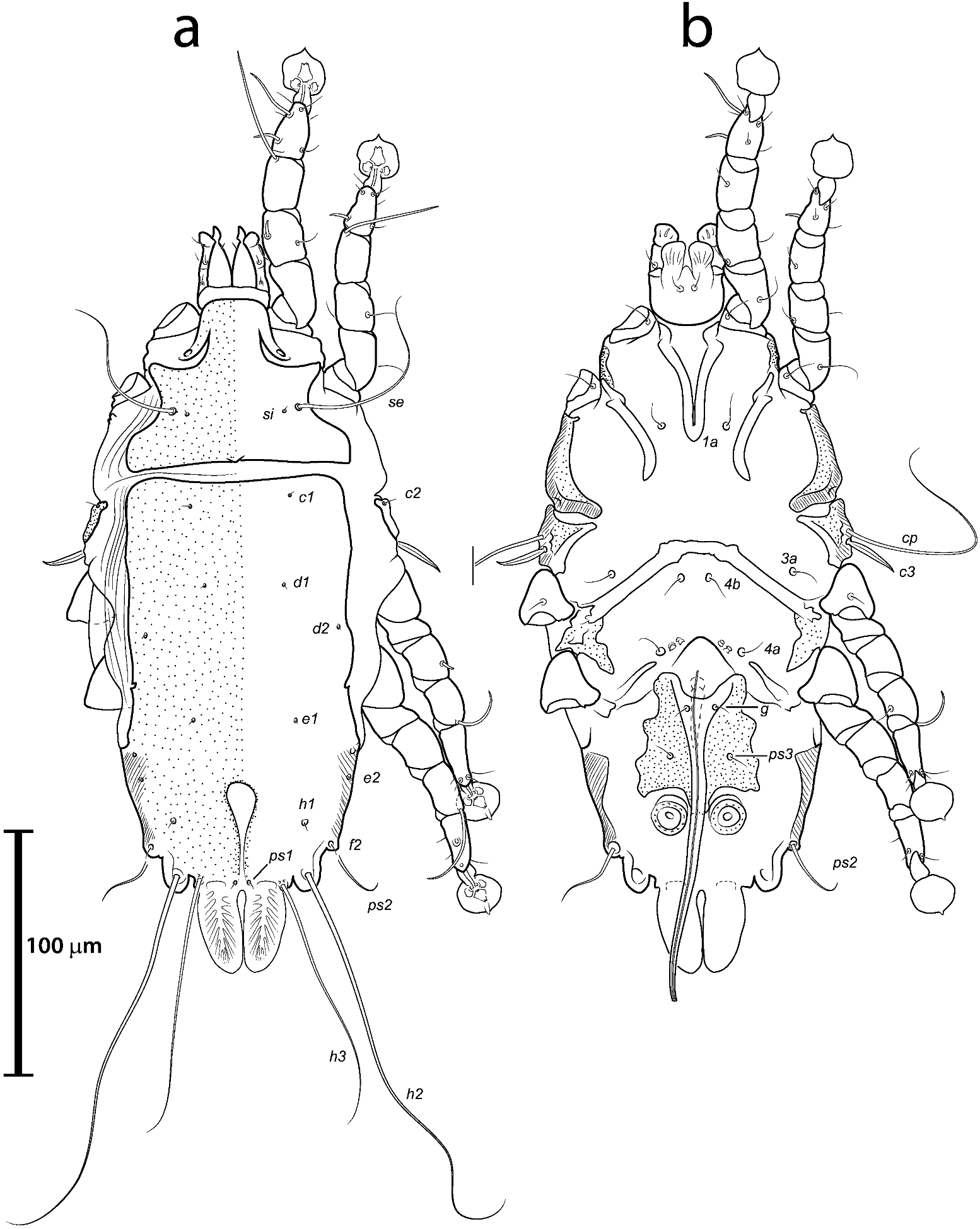
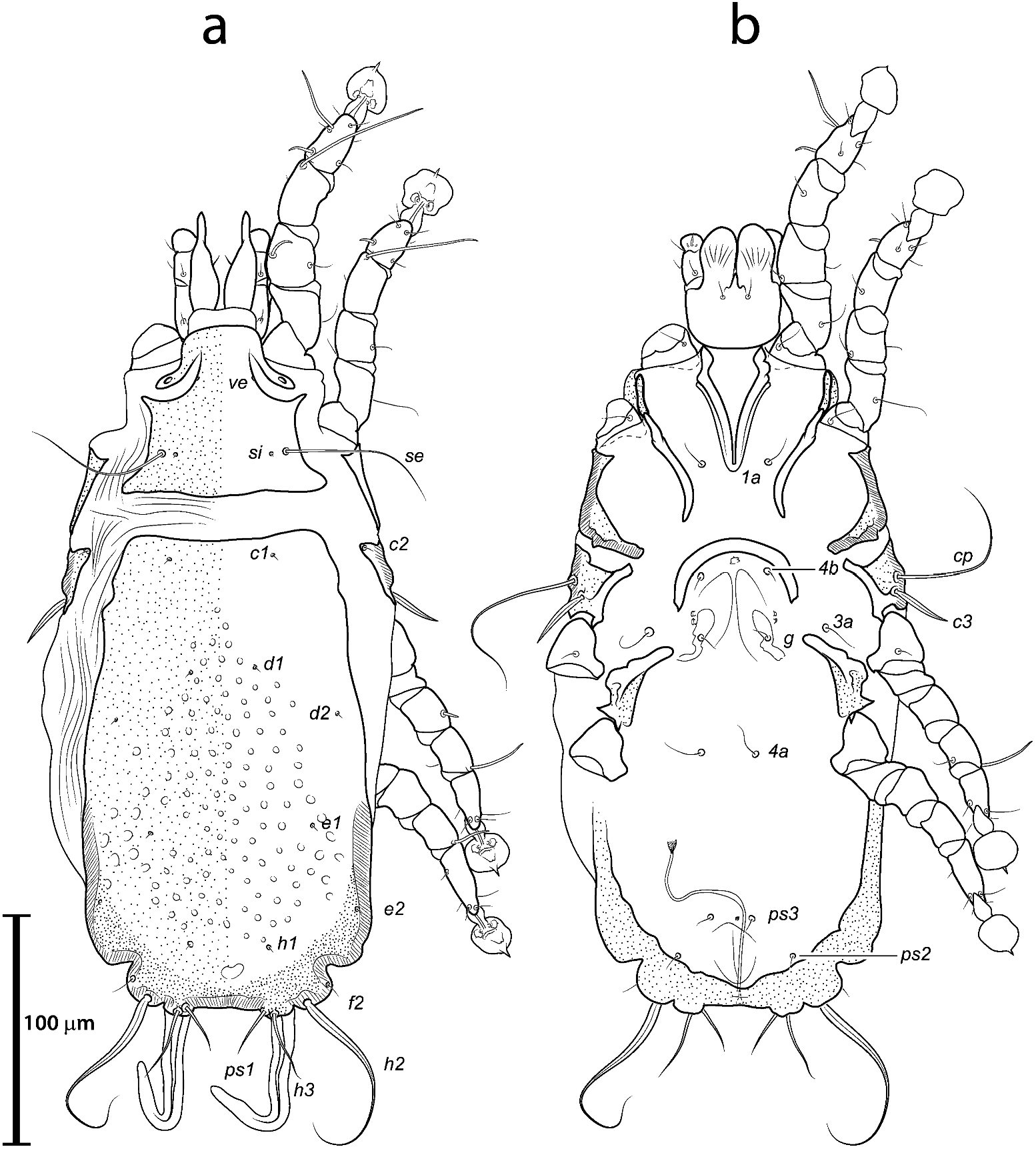
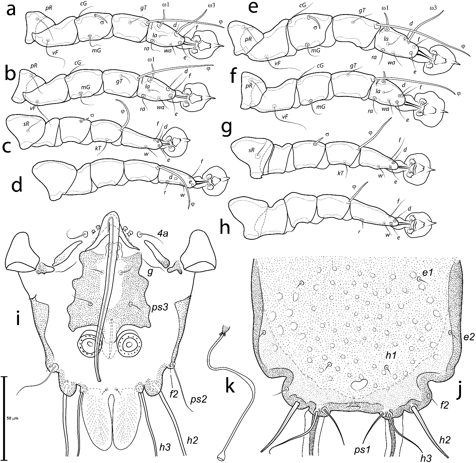
( Figures 1 View Figure 1 –3,13(a))
Type material
Holotype male (#5371), paratypes 9 males and 9 females (#5372–5380) ex Cacicus cela (Linnaeus, 1758) ( Passeriformes : Icteridae ), BRAZIL, Bahia State, Ilhéus, campus of Universidade Estadual de Santa Cruz ( UESC), 14.7597222°S, 039.2302778°W, found dead on the ground, 14 December 2020, Anibal R. Oliveira coll.; GoogleMaps paratypes 4 males and 3 females (#5381–5387), same host species, BRAZIL, Pará State, Fazenda Fartura, 9.666667°S, 50.383333°W, Santana do Araguaia , dead after collision with glass window, 11 September 2013, D. V. Boas-Filho coll. (#557). GoogleMaps
Description
Male ( Figures 1 View Figure 1 (a,b), 3 (a–3,i), 13(a)), holotype (range for 8 paratypes in parentheses). Idiosoma, length × width, 236 (235–243) × 127 (124–132); length of hysterosoma excluding lamellae 165 (166–171). Prodorsal shield: setae vi absent, anterolateral extensions acute, lateral margins entire with two semicircular concavities at the level of scapular setae, posterior margin straight, posterior corners right-angled, greatest length 65 (61–70), greatest width 90 (88–94), surface without ornamentation or lacunae. Distance between scapular setae se 51 (50–55). Scapular shields not developed dorsally. Humeral shields small, fused with epimerites III, setae c2 on anterior margins of humeral shields. Subhumeral setae c3 lanceolate, 21 (20–24) in length, 2 (2–3) in width. Hysteronotal shield: anterior margin slightly concave, anterior angles rounded, length 165 (161–167), width at anterior margin 89 (82–92), surface without ornamentation. Supranal concavity shaped like an inverted teardrop, closed terminally, its anterior end at level of setae e2, length from anterior end to bases of setae ps1 36 (37–43). Posterior margin of opisthosoma between setae h3 straight. Terminal lamellae small, tongue-shaped, with inner margins almost touching, with pennate venation; length 33 (32–37), greatest width 16 (16–20). Setae ps1 minute. Distances between hysteronotal setae: c2:d2 51 (49–54), d2:e2 61 (59– 62), e2:h3 43 (37–43), d1:d2 19 (19–22), e1: e2 24 (24–30), h1:h3 27 (23–27), h2:h2 52 (51–53), h3:h3 34 (32–36), ps2:ps2 72 (68–72).
Coxal apodemes I (epimerites) fused into a narrow V without lateral extensions, inner ends of epimerites IIIa connected transverselly to each other above genital apparatus. Setae 4b and 3a situated almost at the same transverse level. Genital arch 25 (22–26) in length from tips of its branches to bend of aedeagus, 28 (25–28) in width, with arch base at midlevel of trochanters IV and with arch apex at midlevel between trochanters III and IV. Aedeagus stylet-shaped, directed backward from the genital arch apex, extending to the level of setae h2, 102 (99–110) in length from bend to tip ( Figure 3 View Figure 3 (i)). Setae 4a and posterior pair of genital papillae situated at midlevel of genital arch. Paragenital and pregenital apodemes absent. Genital papillae not connected. Opisthogastric shield roughly trapezoidal with strongly sinuous margins and poorly sclerotised central area; greatest length of opisthogastric shields 42 (42–47), width in anterior part 45 (39–48). Setae g and ps3 filiform, arranged in a high trapezoid, setae g on inner margins of the opisthogastric shield, distances between these setae: g:g 14 (11–18), g:ps3 19 (14–20), ps3: ps3 25 (24–27). Adanal suckers cylindrical, 14 (12–15) in diameter, corolla with 13–15 small teeth.
Femora II without ventral crests. Solenidion σ 7 (6–8) long, about half the length of genu I and situated at midlevel of this segment ( Figure 3 View Figure 3 (a)). Solenidion σIII 4 (4–6) long, situated in middle part of genu III, tibial solenidion φIV 33 (31–36). Tarsus IV 27 (26–30) in length, button-like seta d about as wide as seta e and situated at midlength of this segment, seta e situated distally ( Figure 3 View Figure 3 (d)).
Female ( Figures 2 View Figure 2 (a,b), 3 (e–3,j,3), 13(a)), range for 8 paratypes. Length of idiosoma 298–312, width 141–150, length of hysterosoma 209–224. Prodorsal shield: setae vi absent, anterolateral extensions acute, not extending to bases of epimerites I, lateral margins with concavities at level of scapular setae, posterior margin straight, length 76– 81, width 88–93, surface without ornamentation ( Figure 2 View Figure 2 (a)). Distance between scapular setae se 54–57. Scapular shields not developed dorsally. Humeral shields fused with epimerites III; setae c2 on anterior margin of this shield. Subhumeral setae c3 lanceolate, 20–28 in length, 3–4 in width. Anterior hysteronotal shield roughly rectangular, length of hysteronotal and lobar shields combined 200–218, width at level of setae c1 73–84, anterior margin slightly concave, surface with numerous small circular lacunae. Lobar region of opisthosoma short and truncate, well delimited from remaining part of hysterosoma by lateral concavities, opisthosomal lobes and terminal cleft not developed. Hysteronotal shield entire, not split into anterior and lobar parts. Supranal concavity present, oval, situated between levels of setae h1 and f2. Width of lobar region at level of setae f2 87–94, area between setae ps1 straight. Setae h2 with enlargement on the anterior half and with filiform apical part; setae h3 filiform, 19–20 in length, about 1/3 the length of terminal appendages. Distance between dorsal setae: c2:d2 69–75, d1:d2 19–28, d2:e2 80–90, e1: e2 31–36, e2:h2 36–45, h1:h2 18–26, h1:h1 29–36, h2:h2 69–74.
Coxal apodemes I shaped as in males. Epigynum semicircular, tips barey reaching the level of genital papillae, lateral extensions absent, length 26–31, width 47–54. Copulatory opening situated apically between level of setae ps1. Head of spermatheca simple, triangular, primary duct with a noticeable enlargement at its midlength, secondary spermaducts short ( Figure 3 View Figure 3 (k)). Genital setae g at the same level as setae 3a. Translobar apodemes stretching along the posterior margin of opisthosoma and fused to each other posterior to the anal opening. Setae ps2 situated at midlevel of anal opening.
Femora I, II without ventral crests. Solenidion σ of genu III situated in distal half of segment. Length of genual solenidia: σI 11–13, σIII 6–8. Length of tibial solenidia φIV 10– 15. Legs IV with ambulacral discs extending to level of setae f2.
Differential diagnosis
Proctophyllodes truncatilobus sp. nov. is very similar to P. attenuatus Trouessart, 1899 , described from the Montezuma oropendola, Psarocolius montezuma (Lesson, 1830) (Icteridae) , in Mexico, in having a distinctly truncate opisthosoma – almost flat posteriorly – in females, giving the two species almost indistinct opisthosomal lobes. The new species, however, differs from the latter in having, in males, the aedeagus distinctly longer, reaching the level of bases of setae h2, epimerites IIIa connected transverselly to each other anterior to the genital apparatus, and the apices of terminal lamellae rounded. In males of P. attenuatus , the aedeagus extends only slightly beyond the level of setae ps3, epimerites IIIa have at most surface fields at their inner ends, and the terminal lamellae are acute apically. Females of the new species are considerably smaller, with the idiosoma length × width 298–312 × 141–150, while those of P. attenuatus are about 398 × 155 (Atyeo and Braasch, 1986).
Etymology
The species name is a combination of truncata (L., truncate, mutilate) + lobus (L., lobe, rounded projection), referring to the abruptly ending lobar region of the opisthosoma in females.
| UESC |
Universidade Estadual de Santa Cruz |
| R |
Departamento de Geologia, Universidad de Chile |
| V |
Royal British Columbia Museum - Herbarium |
No known copyright restrictions apply. See Agosti, D., Egloff, W., 2009. Taxonomic information exchange and copyright: the Plazi approach. BMC Research Notes 2009, 2:53 for further explanation.
|
Kingdom |
|
|
Phylum |
|
|
Class |
|
|
SuperOrder |
Acariformes |
|
Order |
|
|
SubOrder |
Astigmata |
|
Family |
|
|
SubFamily |
Proctophyllodinae |
|
Genus |