
bradyi M’Intosh, 1885
|
publication ID |
https://doi.org/ 10.11646/zootaxa.4174.1.22 |
|
publication LSID |
lsid:zoobank.org:pub:C7196500-B74B-423D-9FE1-3EB079B7F106 |
|
DOI |
https://doi.org/10.5281/zenodo.6071961 |
|
persistent identifier |
https://treatment.plazi.org/id/03AFBB1A-FFB1-FF80-0D8F-FA09FE93FABC |
|
treatment provided by |
Plazi |
|
scientific name |
bradyi M’Intosh, 1885 |
| status |
|
Trophoniphila bradyi M’Intosh, 1885
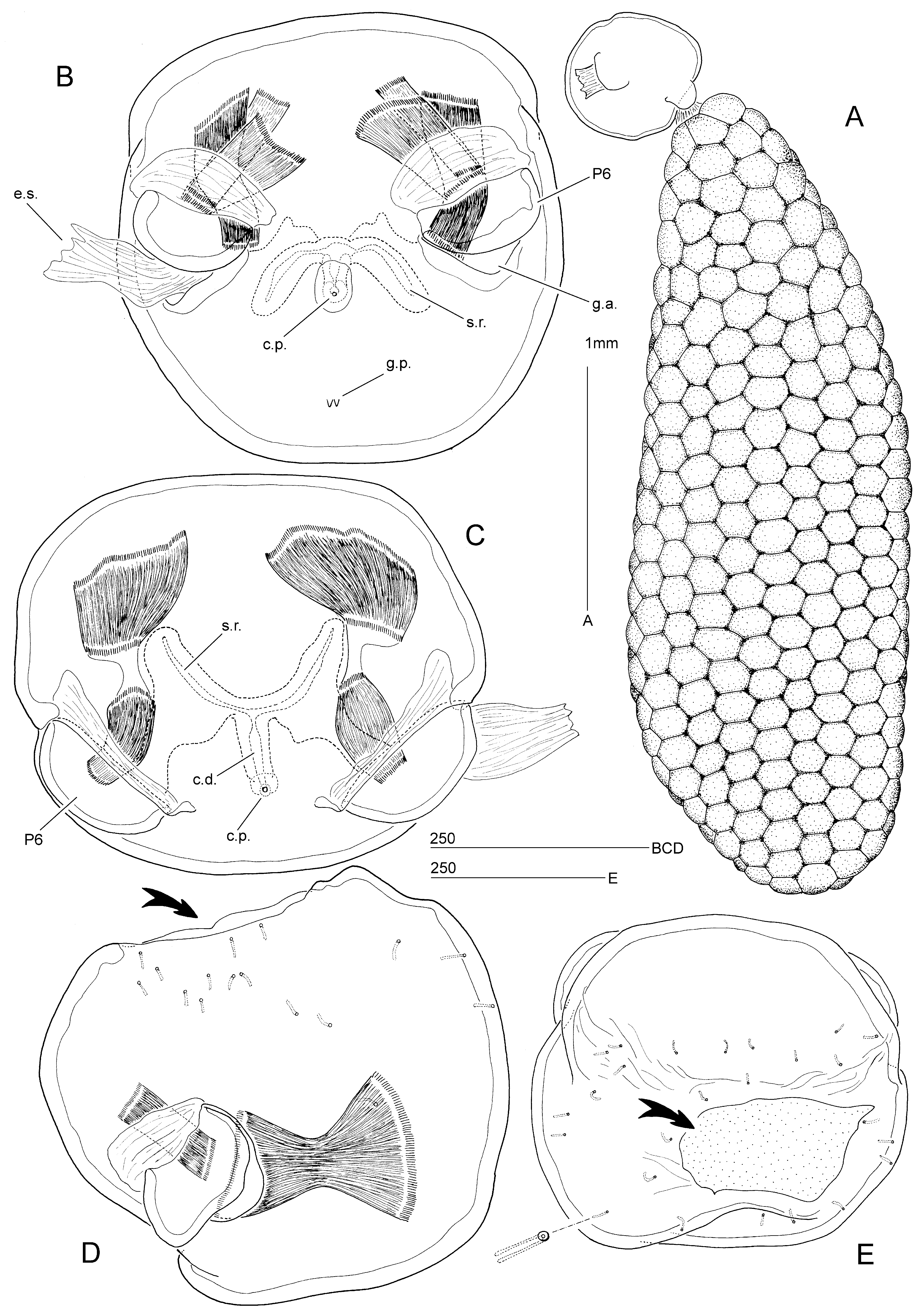
( Fig. 12 View FIGURE 12 )
Trophoniphila bradii M’Intosh, 1885 —incorrect original spelling.
Trophoniphila Bradii M’Intosh, 1885 — Hansen (1892: 21): incorrect subsequent spelling. Trophoniphila Bradyi M’Intosh, 1885 : Hansen (1923: 79) incorrect subsequent spelling. Trophonophila bradii M’Intosh, 1885 : Conradi et al. (2015: 153) incorrect subsequent spelling.
Original description. M’Intosh (1885): 368, Plate XXXVIa, fig. 4.
Host. Ilyphagus wyvillei (M’Intosh, 1885) [as Trophonia wyvillei ] (family Flabelligeridae ).
Type locality. Embedded in specimen of I. wyvillei trawled at H.M.S. Challenger station 157 (Antarctic Ocean); 53º55’ S, 108º 35’ E; depth 1,950 fathoms (3,566 m); diatom ooze. GoogleMaps
Material examined. Holotype ♀ in alcohol (NHMUK reg. no. 1939.4.24.1); attached to the bases of the gills; collected 03 March 1874. Inspection of the dissected holotype of I. wyvillei (NHMUK reg. no. 85.12.1.261) failed to reveal the second specimen mentioned by M’Intosh (1885).
Redescription of female. Body highly transformed and lacking any external trace of segmentation or appendages; consisting of two parts, endosoma and ectosoma, possibly connected by short frontal stalk; stalk and endosoma presumably torn off during dissection. Ectosoma ( Fig. 12 View FIGURE 12 B) almost spherical, about 520 µm in diameter; frontal surface with numerous minute pores around connection with endosoma ( Fig. 12 View FIGURE 12 D, E).
Genital apertures paired, located ventrolaterally almost on opposite side of stalk ( Fig. 12 View FIGURE 12 B–D) and carried on highly sclerotized genital swellings; closed off by strongly developed semicircular opercula, derived from sixth legs; opercula with large membranous insert at base; opening and closing by strong muscles inserting on opercula and posterior wall of genital antra, respectively ( Fig. 12 View FIGURE 12 B–D). Small median copulatory pore located in shelf-like depression between genital apertures ( Fig. 12 View FIGURE 12 B, C), leading via short copulatory duct to seminal receptacle(s); small spiniform papillae discernible, positioned posteriorly to copulatory pore ( Fig. 12 View FIGURE 12 B).
Egg sacs paired, very large, multiseriate, containing several hundreds of eggs; about 3.6 mm long (approx. 7.0 times diameter of ectosoma) and 1.2 mm wide; clavate.
Male. Unknown.
Remarks. M’Intosh’s (1885) original description is restricted to a single text paragraph and an illustration of the female habitus ( Fig. 1 View FIGURE 1 D). He described the egg sac as fusiform or elongate-ovoid, yellowish, and projecting into the cephalic cage of the polychaete. M’Intosh (1885) referred to Levinsen’s (1878) description of Bradophila but remarked that the size of the ectosoma and the attachment site of the egg sacs were different between both genera. He also used the number of egg sacs as a discriminating feature, erroneously assuming that only one was present in T. kroyeri , and believed the latter was closer to “… the larval form of Levinsen’s species” which actually represents the adult male.
The presence of a median copulatory pore in the adult females of both Bradophila and Trophoniphila is of considerable significance in determining the familial position of these highly modified genera. This character is not displayed by any of the other mesoparasitic families that utilize polychaete hosts; instead the presumably paired copulatory pores are contained within the paired genital apertures of the female. Although Levinsen (1878) did not use the correct descriptive terminology, his account of the female genital system in B. pygmaea was remarkably informative. Between the two lateral “protrusions” (genital apertures) he observed another smaller round protrusion with a central opening (copulatory pore), which he assumed to be the genital opening. After clearing in potassium hydroxide this opening was seen to continue upwards as a tube (copulatory duct). Neither a discrete copulatory pore nor duct were observed by Marchenkov (1999b) who overlooked both structures. Re-examination of B. pygmaea and T. kroyeri showed a striking resemblance in the female genital system, suggesting that both species are closely related and belong to the same family. Additional evidence in support of the assignment of Trophoniphila to the Bradophilidae is provided by the disproportionately large egg sacs, the shape of the ectosoma and host utilization.

No known copyright restrictions apply. See Agosti, D., Egloff, W., 2009. Taxonomic information exchange and copyright: the Plazi approach. BMC Research Notes 2009, 2:53 for further explanation.
bradyi M’Intosh, 1885
| Huys, Rony 2016 |
Bradii M’Intosh, 1885
| Conradi 2015: 153 |
| Hansen 1923: 79 |
| Hansen 1892: 21 |