
Trichoderma virens (J.H. Mill., Giddens & A.A. Foster) Arx,
|
publication ID |
https://doi.org/ 10.11646/phytotaxa.587.3.4 |
|
DOI |
https://doi.org/10.5281/zenodo.7753001 |
|
persistent identifier |
https://treatment.plazi.org/id/03B07573-A443-FFC5-B7A0-FE04E5BFFB49 |
|
treatment provided by |
Plazi |
|
scientific name |
Trichoderma virens (J.H. Mill., Giddens & A.A. Foster) Arx, |
| status |
|
Trichoderma virens (J.H. Mill., Giddens & A.A. Foster) Arx, View in CoL View at ENA Beih. Nova Hedwigia 87: 288 (1987) ( Figure 6 View FIGURE 6 )
Index Fungorum number: IF 128198
Isolated from intestinal contents of dead American bullfrog larvae. Sexual morph: Undetermined. Asexual morph on PDA: Aerial mycelium abundant on PDA, fast-growing, forming cream-yellow to green sporulation with maturity, with a distinctive odour, sometimes producing a farinose to granular mat. Conidiophores 20–30 μm high, irregularly branched in a dendriform structure. Vegetative hyphae 4–8 μm wide (x̅ = 6 μm, n = 30), branched, hyaline, smooth and thick-walled, septate, narrow and flexuous, terminal branched, often curved. Conidiogenous cells 5.5–8 × 3–5 μm (x̅ = 6.5 × 4 μm, n = 20), occurring in lateral and terminal clusters, pyramidal. Conidia 4–6 × 3–4 μm (x̅ = 5 × 3.5 μm, n = 20), catenated, obovoid to globose, hyaline to olivaceous, delicately roughened, aseptate, smooth-walled. Chlamydospores 8–11 × 7–9 μm (x̅ = 9.5 × 8.5 μm, n = 20), hyaline, thick-walled, globose to subglobose, terminal.
Culture characteristics: Colonies growing on PDA reach 70–80 mm in diameter after one week at 27 °C, forming the hyaline to olivaceous to green sporulation in PDA. Obverse: aerial, fluffy, hyaline mycelium, peripheral fertile, creamy green to dark green. Reverse olivaceous to pale brown. Without pigments produced in PDA.
Known substratum: Theobroma cacao (Hanada et al. 2010) , Betula pendula & Pinus sylvestris (Mulenko et al. 2008) , Soil samples ( Arx 1987, Kindermann et al. 1998, Chaverri et al. 2001, Zeng et al. 2016), intestinal contents of dead bullfrog larvae (This study).
Known distribution: Unites States ( Chaverri et al. 2001, Arx 1987), New Zealand (Kindermann et al. 1998), Poland (Mulenko et al. 2008), Brazil (Hanada et al. 2010), China (Zeng et al. 2016, This study).
Material examined: China, Yunnan Province, Qujing Normal University, intestinal contents of dead American bullfrog larvae, GPS: 103°44’35”E, 25°30’46”N, 1856.6 m. Wen-hua Lu, ER2 (Herb. HKAS 125765 View Materials ) GoogleMaps living culture KUNCC22-12508 .
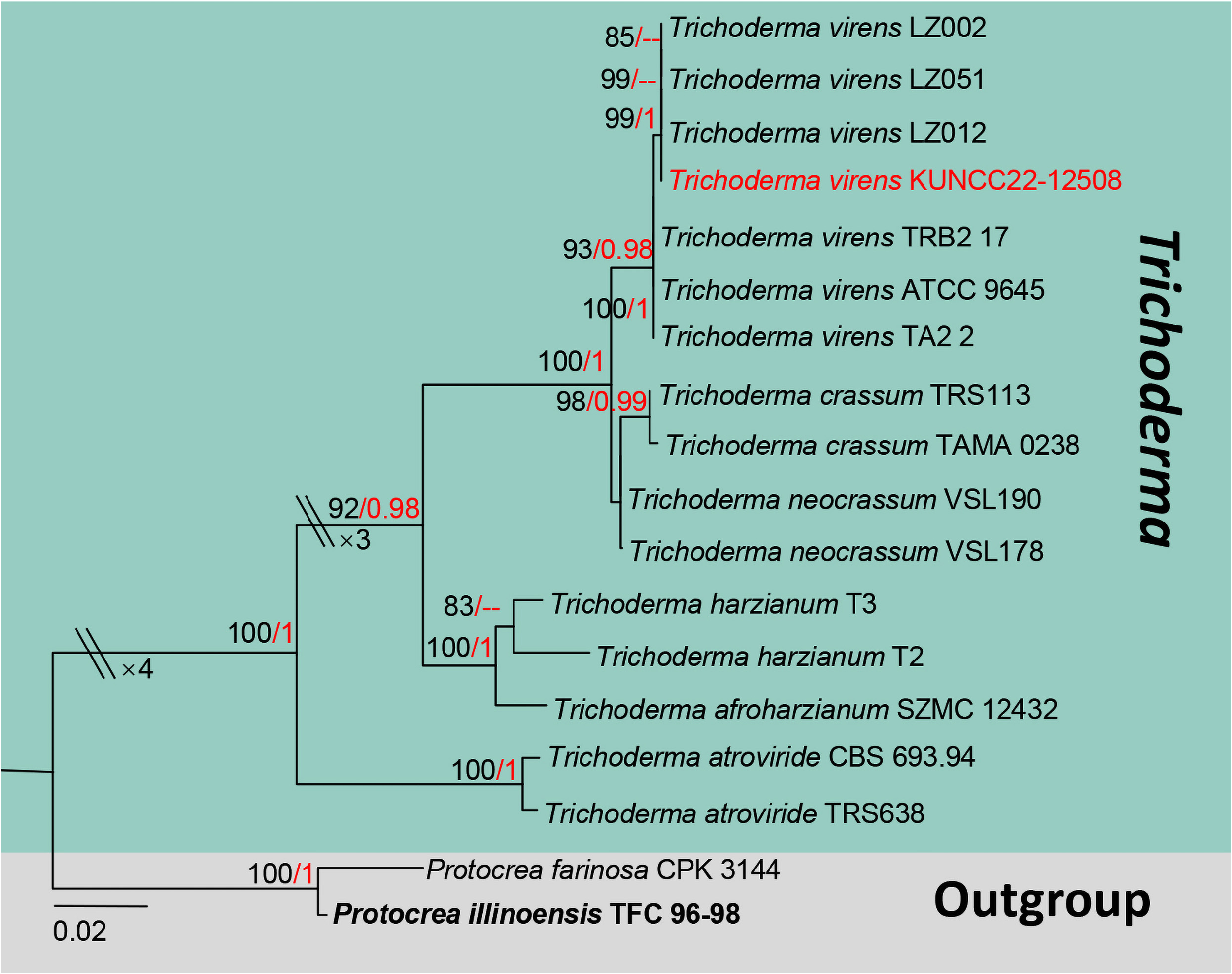
Notes: Morphologically, our isolate KUNCC22-12508 well fits the concept of Trichoderma virens by fastforming typical green sporulation in vitro, with obovoid, hyaline to olivaceous, slightly roughened conidia ( Arx 1987, Chaverri et al. 2001). In addition, its chlamydospores were easily formed between the hyphae or formed terminally in the hyphae tip, similar results were also observed by Abd-Aziz et al. (2008). Phylogenetically, KUNCC22-12508 has placed within Trichoderma virens strains, and separated well with neighbor species T. crassum and T. neocrassum with high statistical supports (100%ML/1 BYPP) ( Figure 3 View FIGURE 3 ). Moreover, the BLASTn results of ITS, LSU, tef1-α and rpb2 expect our isolate KUNCC22-12508 100% similar to Trichoderma virens strains (LZ002, LZ012, Z051). Therefore, our isolate KUNCC22-12508 is identified as Trichoderma virens , a new host record based on morphological features and phylogenetic evidence.

No known copyright restrictions apply. See Agosti, D., Egloff, W., 2009. Taxonomic information exchange and copyright: the Plazi approach. BMC Research Notes 2009, 2:53 for further explanation.
|
Kingdom |
|
|
Phylum |
|
|
Class |
|
|
Order |
|
|
Family |
|
|
Genus |