
Macrophthalmus (Macrophthalmus) manggala, Murniati & Asakura & Nugroho & Hernawan & Dharmawan, 2022
|
publication ID |
https://doi.org/ 10.26107/RBZ-2022-0026 |
|
publication LSID |
lsid:zoobank.org:pub:2E07F63A-50B5-42CE-98FA-027B0BF38756 |
|
DOI |
https://doi.org/10.5281/zenodo.7502088 |
|
persistent identifier |
https://treatment.plazi.org/id/7AA8CCB6-8BC4-4CEE-9A55-90C45BF5CB88 |
|
taxon LSID |
lsid:zoobank.org:act:7AA8CCB6-8BC4-4CEE-9A55-90C45BF5CB88 |
|
treatment provided by |
Felipe |
|
scientific name |
Macrophthalmus (Macrophthalmus) manggala |
| status |
sp. nov. |
Macrophthalmus (Macrophthalmus) manggala View in CoL , new species
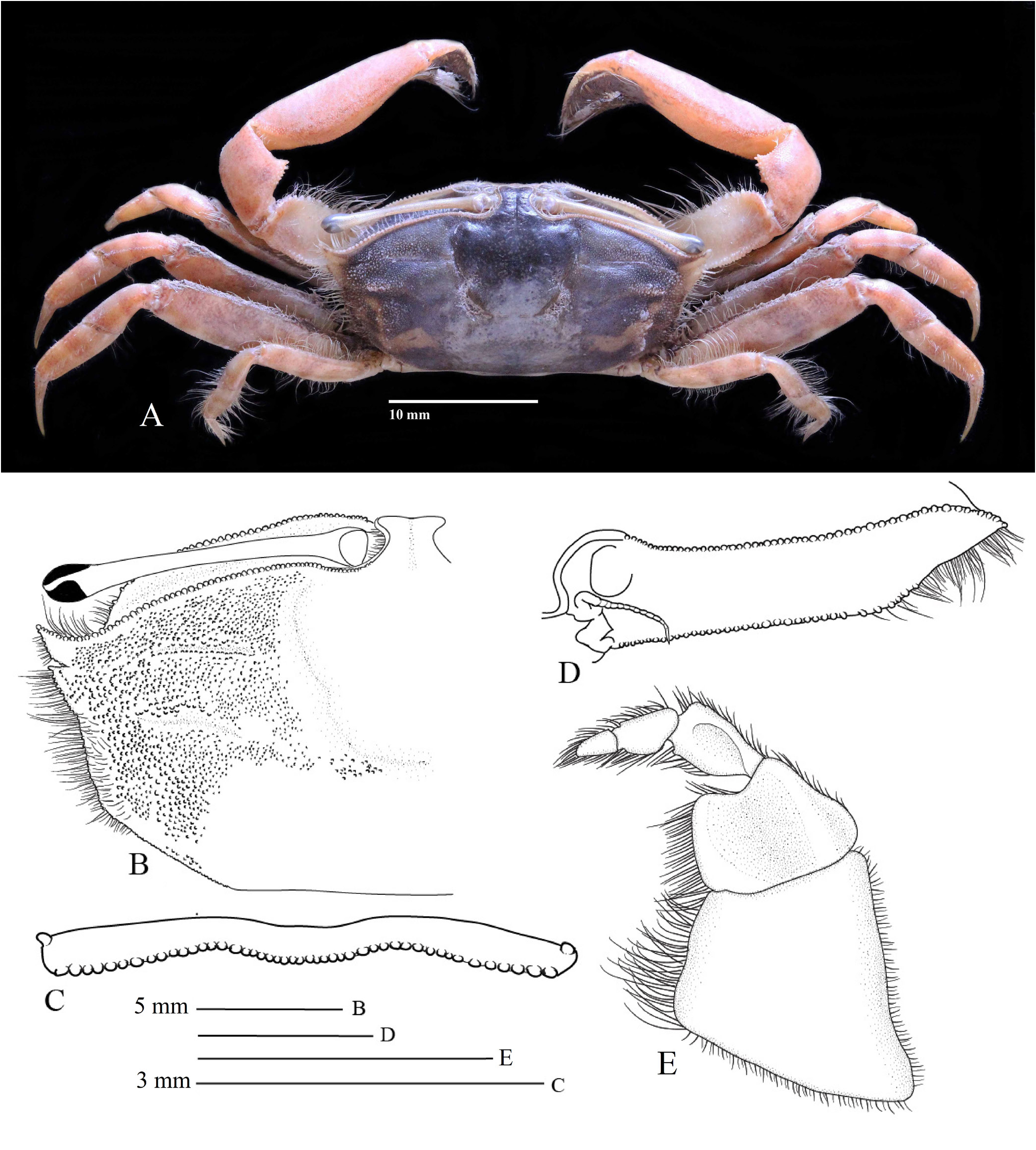
( Figs. 6–10 View Fig View Fig View Fig View Fig View Fig , 13A–C View Fig )
Material examined. Holotype: male (23.1 × 10.9 mm) ( MZB. Cru.5013), Liki Village, Sarmi District, Sarmi Municipality , Liki Island , Papua Province, 01º37′25.29″S, 138º44′26.54″E, 21 November 2018, coll. DC Murniati. GoogleMaps
Paratypes: 10 males (8.5 × 4.5 mm – 22.2 × 10.8 mm), 5 females (13.4 × 6.7 mm – 18.9 × 9.0 mm) ( MZB. Cru. 5014), Liki Village, Sarmi District , Sarmi Municipality , Liki Island , Papua Province, 01º37′25.29″S, 138º44′26.54″E, 21 November 2018, coll. DC Murniati GoogleMaps ; 2 males (18.0 × 9.7 mm, 23.2 × 12.3 mm), 1 female (20.3 × 10.8 mm) ( ZRC 2022.0913 View Materials ) Liki Village, Sarmi District, Sarmi Municipality , Liki Island , Papua Province, 01º37′25.29″S, 138º44′26.54″E, 21 November 2018, coll. DC Murniati. GoogleMaps
Comparative material examined. Macrophthalmus (Macrophthalmus) convexus Stimpson, 1858 , 1 male (20.1 × 10.3 mm) ( MZB. Cru. 1386), Sosobok, Halmahera Island , 10 April 1987, S Harminto ; 5 males (16.6 × 8.2 mm – 23.4 × 10.7 mm), 4 females (15.1 × 7.8 mm – 17.2 × 9.3 mm) ( MZB. Cru. 5060), Terima Bay, Sumber Klampok Village , Gerokgak District , Buleleng Municipality , Bali Province, 12 June 2020, H Kusumanegara, TEN Dian. Macrophthalmus (Macrophthalmus) parvimanus Guérin, 1834 , 2 males (14.6 × 8.1 mm – 24.0 × 12.4 mm), 3 females (12.5 × 6.9 mm – 2.2 × 11.1 mm) ( MZB. Cru. 4718), Saparua Island , Moluccas, S03º34′25.9″, E34º39′27.4″, 22 September 2016, DA Nugroho ; 1 male (13.5 × 8.0 mm) ( MZB. Cru. 3268), Eka’s Bay, East Lombok Municipality , Lombok Island , West Nusa Tenggara Province, 10 April 2018, DL Rahayu .
Diagnosis. Carapace rectangular, broad, about 1.9 times as broad as long. Front narrow, less than 0.2 times external orbital angles width. Lateral margins with two anterolateral teeth including external orbital angle. Chelipeds long, subequal. Palm long; inner surface of palm with microscopic granules, distally covered by mat of setae. Fixed finger short, deflexed; cutting margin with one low, subquadrate tooth submedially. Dactylus directed downward, cutting margin with one large, squarish tooth. Pleon moderately narrow, 1.2 times as long as wide. P4 longest. P5 shortest, margins with long setae. G1 stout, sligthly curved subproximally; mesial surface slightly concave; apical portion truncated, deeply depressed medially, with long setae; chitinous process very short, C-shaped or crescent-shaped. Female vulva rounded; protruded, with transverse groove medially, concave portion facing median portion of sternum; outer margin circular.
Description. Carapace rectangular, broad, about 1.9 times as broad as long, greatest width across external orbital angles ( Fig. 6A View Fig ). Regions on dorsal surface clearly defined, furrow on gastric region distinct; hepatic region with two distinct transverse ridges, anterior ridge straight on deep furrow, posterior ridge curved on bulging area; epigastric and mesogastric regions smooth; mesobranchial region covered by granules, larger laterally; scattered short setae on hepatic and branchial regions. Front narrow, less than 0.2 times external orbital angles width, constricted at bases of eyestalks ( Fig. 6A View Fig ), with deep longitudinal groove medially ( Fig. 6B View Fig ); frontal margin slightly concave medially, with microscopic tubercles. Supra-orbital margin moderately curved, regularly granular, granules smallest near base of eyestalk ( Fig. 6B, D View Fig ). Infra-orbital margin strongly granular, with larger granules in middle portion and smaller near base of eyestalk and lateral portion, shape of each granule similar to those in supra-orbital margin ( Fig. 6D View Fig ). Lateral margins of carapace oblique, strongly divergent anteriorly, with two anterolateral teeth including external orbital angle, dense long setae along margin; external orbital angle pointed anterolaterally, triangular, with acute angle, one distinct granule on its tip and one row of lateral granules; second anterolateral tooth triangular, separated by V-shaped incision from external orbital angle, apex pointed outward; distinctly smaller than preceding tooth, with granules on lateral margin, last granule adjacent to lateral margin; lateral margin straight, with several tubercles; posterior margin straight ( Fig. 6B View Fig ).
Epistome with posterior margin bearing regular row of tubercles, posteromedian margin distinctly convex, posterolateral margin slightly convex, anterior margin curved and without tuberculation ( Fig. 6C View Fig ).
Eyestalks slender, long, 0.4 times as long as carapace width; cornea not reaching beyond external orbital angle ( Fig. 6B View Fig ).
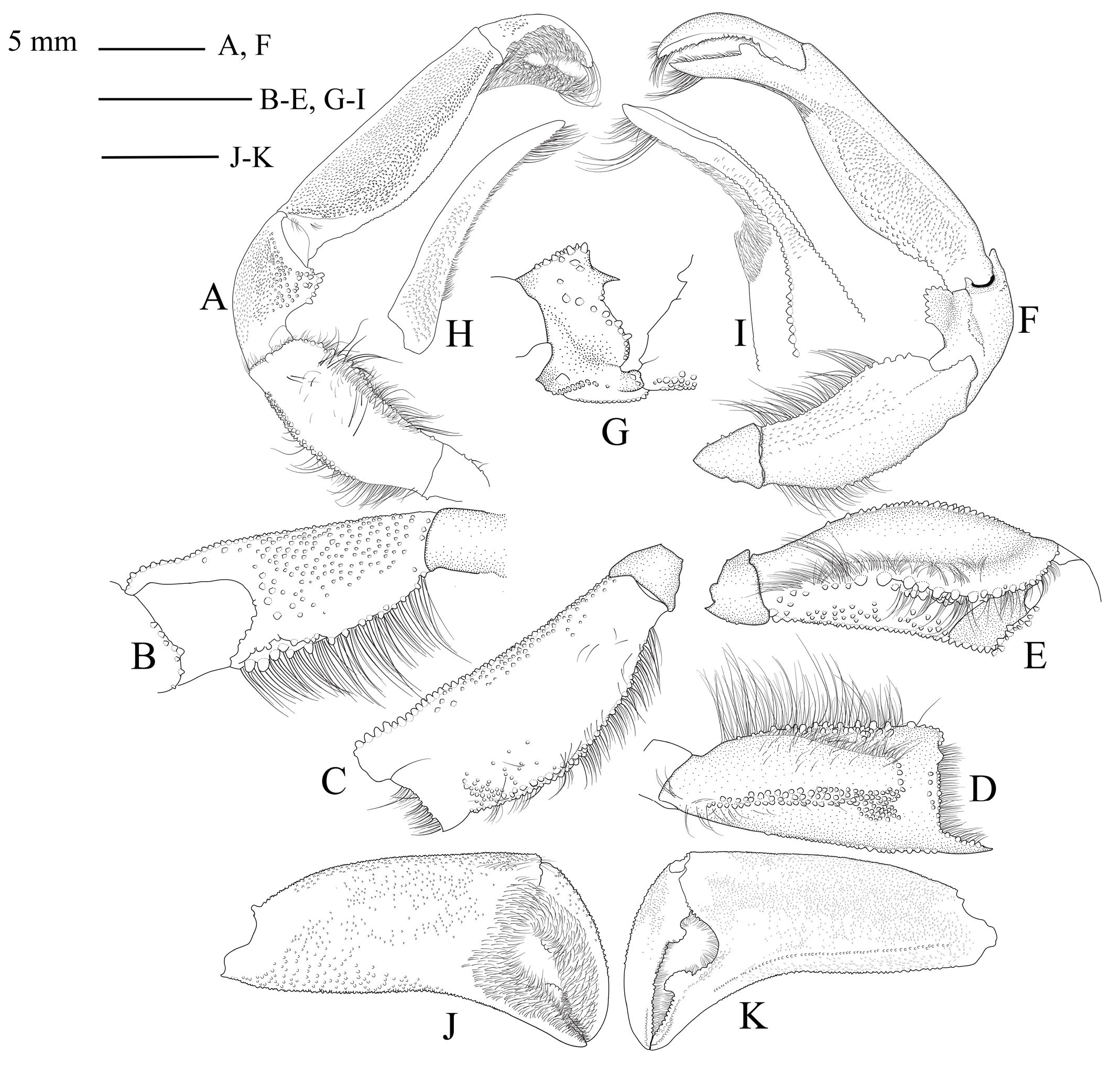
Third maxillipeds leaving large gap in between them even when closed. Ischium nearly subtrapezoidal; mesial margin with long setae, setae longer posteriorly; lateral margin nearly straight; lateral and posterior margins with short setae; outer surface smooth. Merus quadrate, smaller than ischium, about 0.5 times as long as ischium laterally; outer surface with wide depression near mesial margin, narrow depression near lateral margin; mesial margin with long and short setae, setae shorter anteriorly; lateral margin slightly curved, arched anteriorly, anterior margin distinctly concave, lined with long setae; anterolateral margin with scattered short setae, posterolateral margin without setae. Carpus trihedral, mesial and lateral margins with dense short setae. Propodus longer than dactylus, margins with dense short setae. Dactylus tubular, margins with dense setae ( Fig. 6E View Fig ).
Male chelipeds (P1) long, subequal. Merus with cross-section triangular, upper surface bearing scattered short setae, without granules ( Fig. 7A View Fig ); lower surface granular mainly on lower half, granules larger distally, densest near lower margin ( Fig. 7B View Fig ); outer surface coarse, with granules laterally, sparse setae near upper margin, median portion glabrous ( Fig. 7C View Fig ); upper margin distinctly tubercular, tubercles larger medially, no spine near articulation with carpus, with sparse long setae ( Fig. 7D View Fig ); lower margin evenly tubercular ( Fig. 7E View Fig ); outer margin with two rows of granules proximal half length, continuous with three row of granules and denser near distal portion, distal portion without granules except near distal margin ( Fig. 7E View Fig ). Carpus short, outer surface rectangular, with microscopic granules, without setae ( Fig. 7A View Fig ); inner surface with single curved row of granules ( Fig. 7G View Fig ); upper and lower margins distinctly tubercular ( Fig 7A, F View Fig ), tuberculation on upper margin larger than that of lower margin ( Fig. 7A View Fig ). Palm long, 2.2 times as long as high, 2.6 times as long as fixed finger ( Fig. 7J–K View Fig ); inner surface with microscopic granules on upper and median portions, larger granules on lower portion, distal margin to inner surface of fingers covered by mat of setae, without spine near articulation with carpus ( Fig. 7J View Fig ); outer surface with microscopic granules covered almost entirely, longitudinal row of microscopic granules from proximal portion of palm to fixed finger along lower margin ( Fig. 7K View Fig ); upper margin slightly arched, tubercular; lower margin straight, tubercular, tuberculation smaller distally. Fixed finger short, deflexed; cutting margin with one large, subquadrate submedial tooth, followed distally by denticulate teeth; lower margin tubercular 1/2 length; outer surface with row of granules parallel to cutting margin, one row of setae present half-distally parallel to cutting margin; tips of fingers spoon-shaped ( Fig. 7J–K View Fig ). Dactylus directed downward, cutting margin with one large, squarish, subproximal tooth, distally followed by single row of denticulate teeth; upper margin granular 3/4 length, one short row of long setae present distally; outer surface with microscopic granules on proximal portion, one row of setae present half-distally parallel to cutting margin ( Fig. 7K View Fig ).
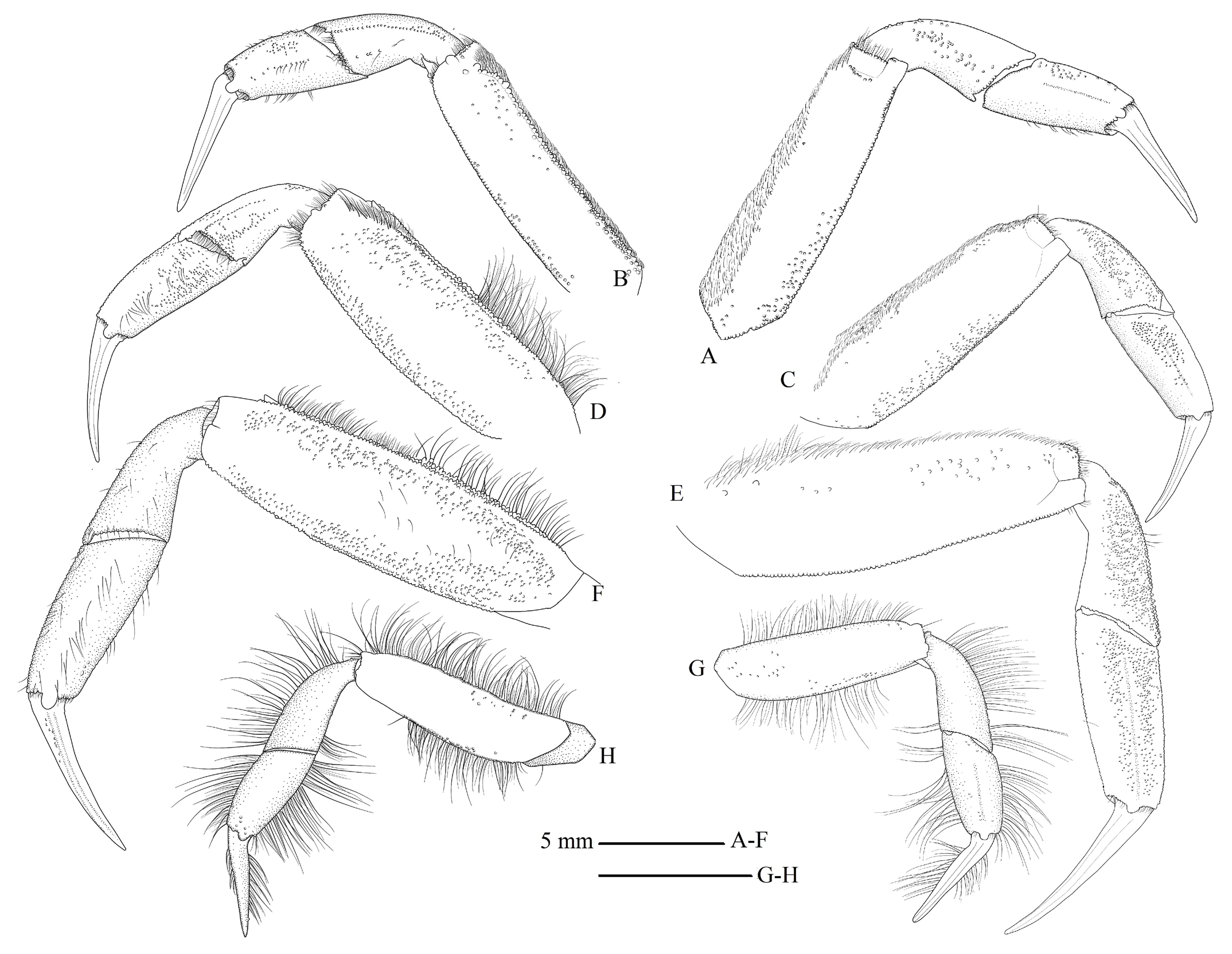
Ambulatory legs (P2–P5) medium sized. P2 shorter than P3. Merus 3.5 times as long as wide, with anterior surface nearly smooth, bearing dense setae near upper margin, densest setae proximally, setation extending to 3/4 length of anterior surface, microscopic granules near lower margin ( Fig. 8A View Fig ); posterior surface with sparse microscopic granules laterally ( Fig. 8B View Fig ); upper margin narrow, with one row of tubercles forming ridge structure, dense short setae extending whole length of margin ( Fig. 9A View Fig ); lower margin wide, slightly convex, with microscopic granules distributed irregularly, without setation ( Fig. 9E View Fig ). Carpus slightly shorter than propodus; anterior surface with irregular microscopic granules on median portion, lateral portion smooth ( Fig. 8A View Fig ); posterior surface with one row of microscopic granules near upper margin, irregular granules near lower margin, sparse setae on median portion ( Fig. 8B View Fig ); upper margin tubercular ( Fig. 9A View Fig ); lower margin nearly smooth ( Fig. 9E View Fig ). Propodus with anterior surface bearing one narrow groove, microscopic granules distributed near upper margin ( Fig. 8A View Fig ); posterior surface with one row of microscopic granules medially, with setae present along granulation, other granulation distributed between median granulation and upper margin ( Fig. 8B View Fig ); upper margin narrow, with one distinct row of tubercles ( Fig. 9A View Fig ); lower margin wide, nearly smooth, with sparse granules on distal portion ( Fig. 9E View Fig ). Dactylus slightly shorter than propodus, with one row of setae on upper margin, tuberculation rarely present on each margin and surface.
P3 longer than P2. Merus 3.2 times as long as wide, anterior surface with microscopic granules near lower margin, row of dense short setae on upper portion continuing to upper margin, setation extending whole length, sparse granules on upper portion within row of setae, median portion glabrous; posterior surfaces with dense microscopic granules near upper and lower margins, more granular than that of anterior surface, median portion without granules ( Fig. 8C–D View Fig ); upper margin with one row of tubercles extending whole length, tuberculation nearly forming one ridge, dense short setae and sparse long setae in one row, setation extending whole length ( Fig. 9B View Fig ); lower margin wide, with dense microscopic granules distributed almost whole length ( Fig. 9F View Fig ). Carpus slightly shorter than propodus; anterior surface with microscopic granules distributed evenly except near lower margin ( Fig. 8C View Fig ); posterior surface with sparse microscopic granules on median portion, one row of granules parallel to upper margin, sparse granules between row of granules and upper margin, single row of short setae on articulation with propodus ( Fig. 8D View Fig ); upper margin with sparse granules ( Fig. 9B View Fig ); lower margin wide, with microscopic granules distributed irregularly ( Fig. 9F View Fig ). Propodus with anterior surface bearing one narrow furrow, microscopic granules medially ( Fig. 8C View Fig ); posterior surface with microscopic granules, densest proximally, more granular than that of anterior surface and one row of spaced long setae medially ( Fig. 8D View Fig ); upper margin narrow, tubercular on whole length; lower margin wide, covered with microscopic tubercles ( Fig. 9B, F View Fig ). Dactylus with anterior and posterior surfaces smooth ( Fig. 8C–D View Fig ), upper margin smooth, lower margin with sparse tubercles and setae ( Fig. 9B, F View Fig ).
P4 longest. Merus long, 3.4 times as long as wide; anterior surface nearly smooth, without setae medially, with setae proximo-laterally, sparse granules distributed unevenly on upper half portion ( Fig. 8E View Fig ); posterior surface with dense granules near upper and lower margins, granulation densest proximally, sparse short setae medially, median portion smooth ( Fig. 8F View Fig ); upper margin distinctly narrow, with one row of tubercles, one subdistal tooth, and dense setae around tuberculation ( Fig. 9C View Fig ); lower margin wide, with microscopic granules, granulation more spaced than that of merus of P3, without setae ( Fig. 9G View Fig ). Carpus shorter than propodus; anterior surface with microscopic granules median to upper portion, more granular than that of P3, smooth near lower margin ( Fig. 8E View Fig ); posterior surface smooth, with sparse long setae ( Fig. 8F View Fig ); upper margin narrow, with one row of tubercles ( Fig. 9C View Fig ); lower margin wide, sparsely tubercular ( Fig. 9G View Fig ).
Propodus with anterior surface bearing one narrow furrow medially, covered by microscopic granules except on lower portion ( Fig. 8E View Fig ); posterior surface nearly smooth, covered with one row of spaced long setae ( Fig. 8F View Fig ); upper margin narrow, with microscopic tubercles distributed irregularly and sparse long setae; lower margin narrow, with one row of tubercles and sparse long setae ( Fig. 9C, G View Fig ). Dactylus with anterior surface smooth ( Fig. 8E View Fig ); posterior surface with sparse granules ( Fig. 8F View Fig ); upper margin smooth ( Fig. 9C View Fig ); sparse tubercles and setae on lower margin ( Fig. 9G View Fig ).
P5 shortest. Merus 3.1 times as long as wide, with anterior and posterior surfaces nearly smooth, sparse microscopic granules distributed unevenly ( Fig. 8G–H View Fig ); upper margin narrow, with one row of microscopic tubercles and one row of long setae extending whole length of upper margin; lower margin wide, with one row of microscopic tubercles, one row of long setae extending 2/3 length of lower margin ( Fig. 9D, H View Fig ). Carpus almost as long as propodus; anterior and posterior surfaces smooth ( Fig. 8G–H View Fig ); upper margin narrow, with one row of microscopic tubercles and one row of long setae extending whole length ( Fig. 9D View Fig ); lower margin with long setae near distal portion, without tubercles ( Fig. 9H View Fig ). Propodus with anterior and posterior surfaces nearly smooth ( Fig. 8G–H View Fig ); upper and lower margins narrow, with one row of microscopic tubercles and one row of long setae extending whole length ( Fig. 9D, H View Fig ). Datcylus with anterior and posterior surfaces smooth ( Fig. 8G–H View Fig ); upper margin with one row of short setae medially; lower margin with one row of long setae extending whole length of lower margin ( Fig. 9D, H View Fig ).
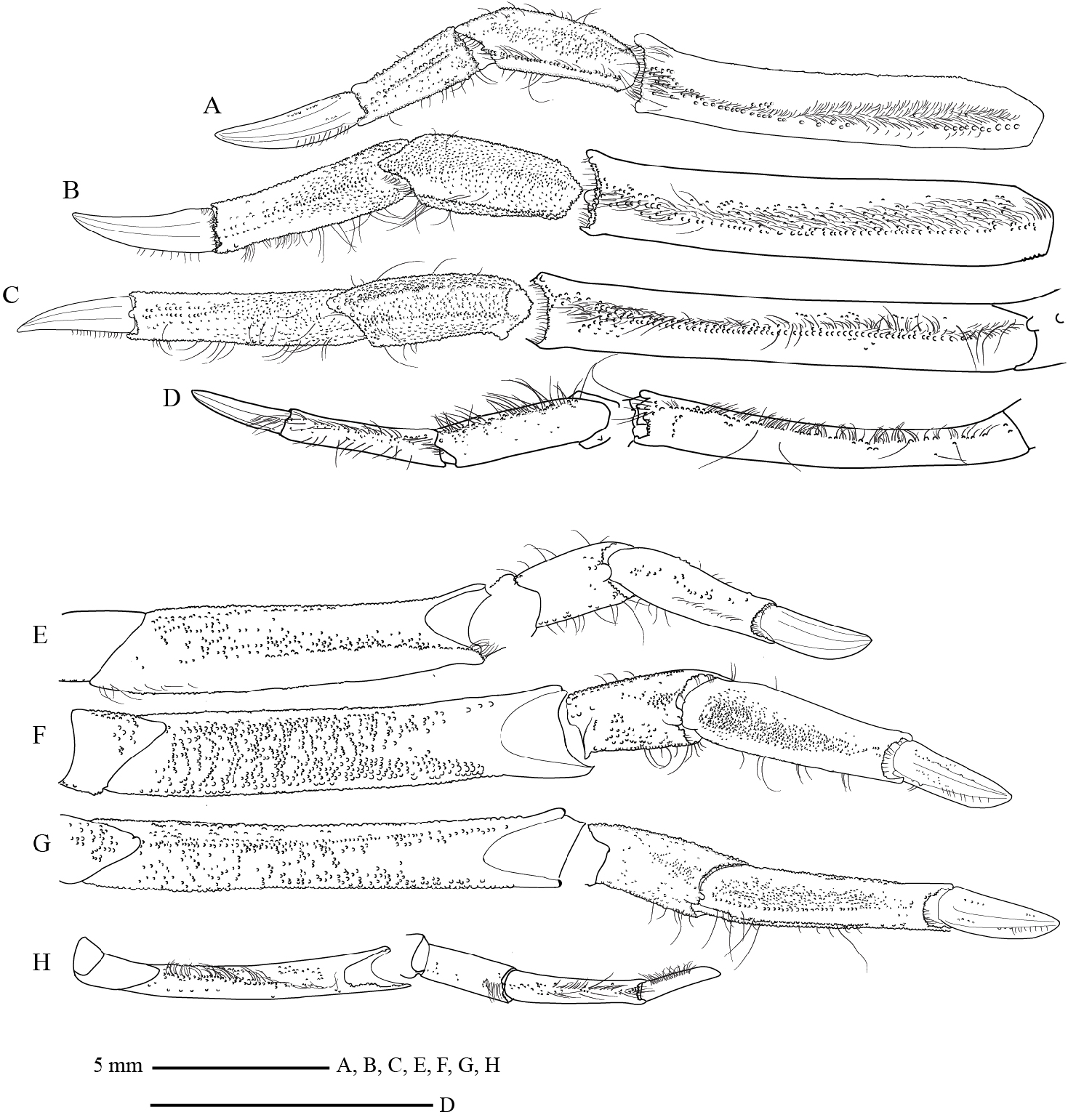
Male pleon moderately narrow, 1.2 times as long as wide; first and third somites wider than other somites. First somite 13.7 times as wide as long, anterior margin concave, posterior margin convex. Second somite shortest among all somites, wider than fourth somite. Third and fourth somites trapezoidal in shape. Third somite as wide as first somite, 5.1 times as wide as long, anterior and posterior margins slightly straight.
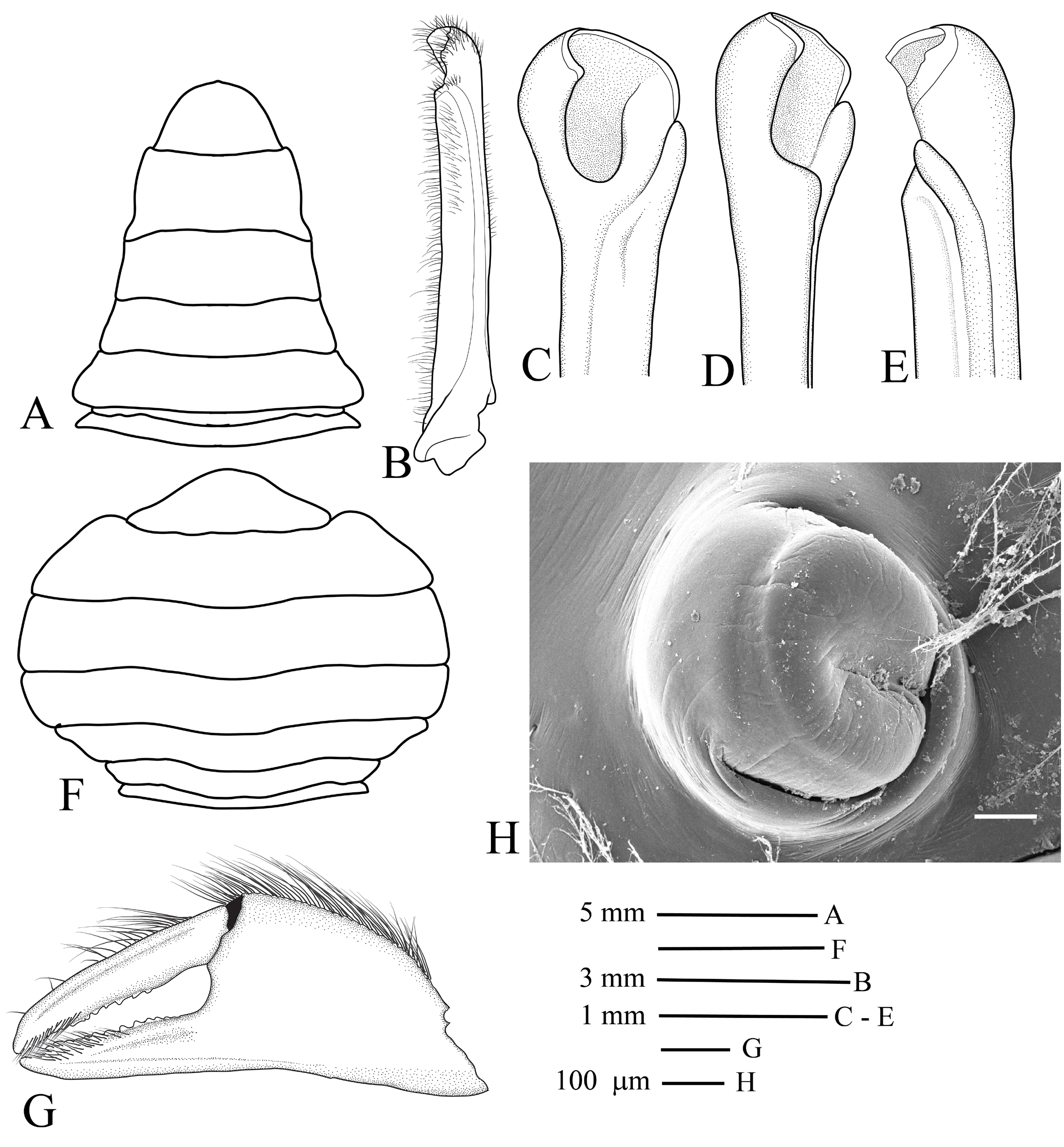
Fourth somite as long as third somite, narrower than third somite, 15.7 times as wide as long. Fifth somite 2.8 times as wide as long, longer and narrower than fourth somite, anterior and posterior margins slightly convex. Sixth somite longest, 2.4 times as wide as long, posterolateral margins weakly constricted, anterior and posterior margins concave. Telson 1.7 times as wide as long, slightly shorter than sixth somite, apically rounded ( Fig. 10A View Fig ).
G1 moderately long, stout, slightly curved subproximally ( Fig. 10B View Fig ); mesial surface slightly concave; dense short setae at outer margin of stem; apical portion with long setae, truncated, deeply depressed medially; apical chitinous process very short, C-shaped or crescent shape, directed distolaterally ( Fig. 10C–E View Fig ).
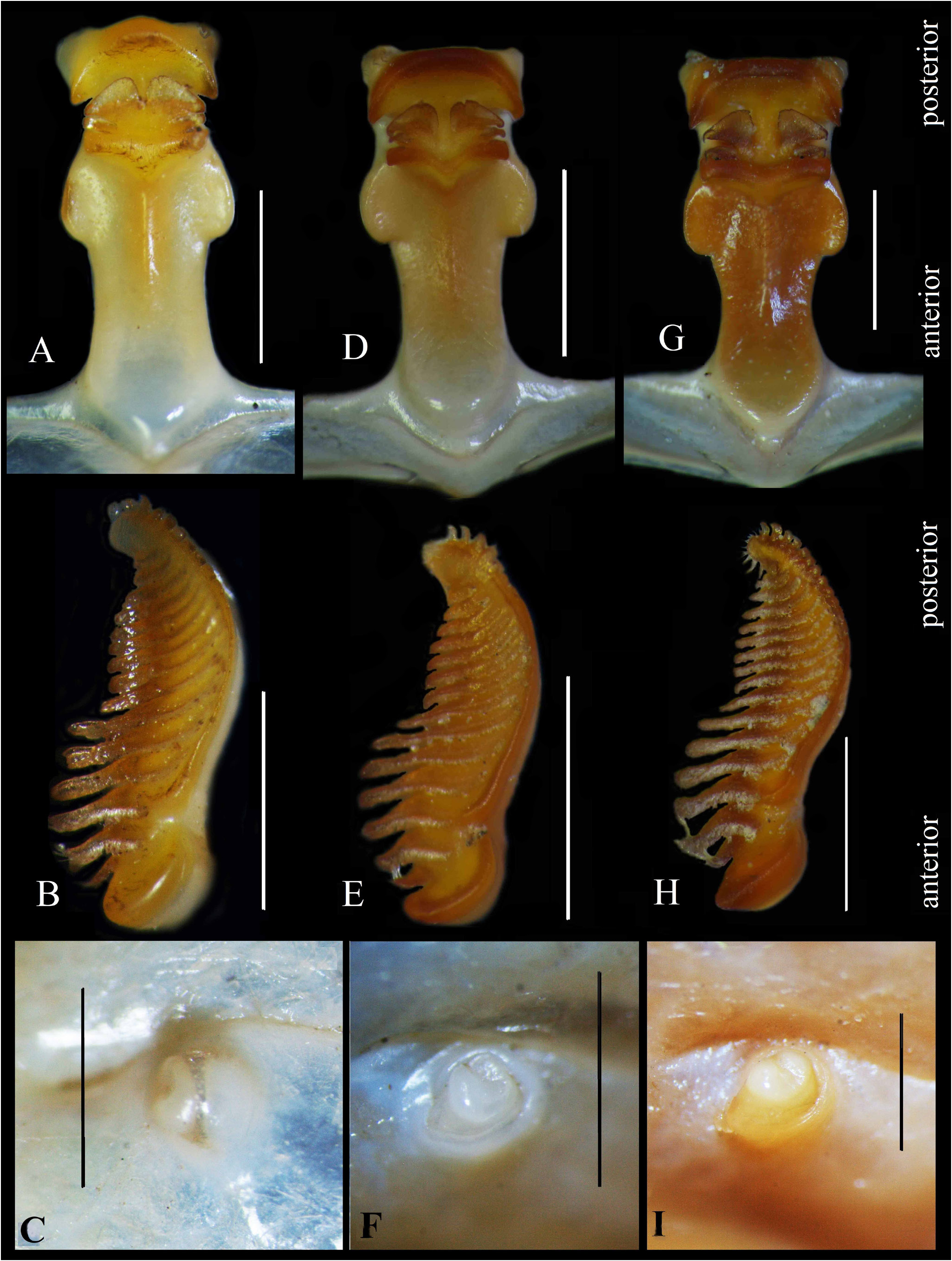
Gastric mill with median tooth plate 2.5 times as long as wide, consisting of two pairs of teeth, gap between two posterior teeth narrow ( Fig. 13A View Fig ); posterior margin of propyloric ossicle with narrow and acute shape; upper portion of anterior trunk shaped triangularly. Lateral tooth plate with 15–16 comb shape teeth, anterior tooth short and slender ( Fig. 13B View Fig ).
Female chelipeds small, equal. Palm convex medially, concave in lower-half portion; outer surface granular on upper half, smooth on lower half; upper and lower margins tubercular, upper margin with long setae ( Fig. 10G View Fig ); inner surface smooth, with row of long setae along upper margin. Fixed finger as long as dactylus; cutting margin serrated; outer surface with inferior ridge parallel to lower margin; inner surface smooth, with single row of long setae medially; distally spoon-shaped, margin chitinous ( Fig. 10G View Fig ). Dactylus as long as palm, convex; cutting margin serrated; upper margin tuberculate only on proximal portion; inner surface smooth, with single row of long setae along upper margin; distally spoon-shaped, accessorised with margined chitinous process.
Female pleon broad, subcircular, 0.8 as long as wide. First somite much narrower and shorter than other somites, 21 times as wide as long. Second somite 10.4 times as wide as long, with short and shallow furrow parallel to midposterior margin. Third somite 9.7 times as wide as long, as long as fourth somite. Fourth somite 7.5 times as wide as long, longer than third somite, as wide as fifth somite. Fifth somite six times as wide as long, slightly longer than fourth somite; as long as sixth somite. Sixth somite 5.3 times as wide as long, nearly as wide as third somite. Telson three times as wide as long; anterolateral margins slightly concave; posterolateral angles rounded, shallowly embedded in distal margin of sixth somite ( Fig. 10F View Fig ).
Vulva rounded; slightly protruded, with transverse groove medially, concave portion facing median portion of sternum; outer margin circular ( Fig. 10H View Fig ).
Etymology. The specific epithet is derived from the Nusa Manggala Expedition, and manggala is used here as a noun in apposition.
Remarks. Macrophthalmus (Macrophthalmus) manggala , new species, is most closely related morphologically to species of the Macrophthalmus (Macrophthalmus) convexus group sensu Barnes (2010). They share several diagnostic characters: the corneas are not positioned beyond the external orbital angle; the cornea is without a projection on its tip; the carapace is very broad (twice as broad as long); a small external orbital angle is followed with the second lateral tooth which has a larger and broader base; the shape of the second anterolateral tooth is flatter than that of the external orbital angle; the front is narrow, male chelipeds are elongate and the fixed finger is deflexed; the external orbital angle is large, flat, and forwardly curved; no spine is present on inner surface of the palm of the male cheliped.
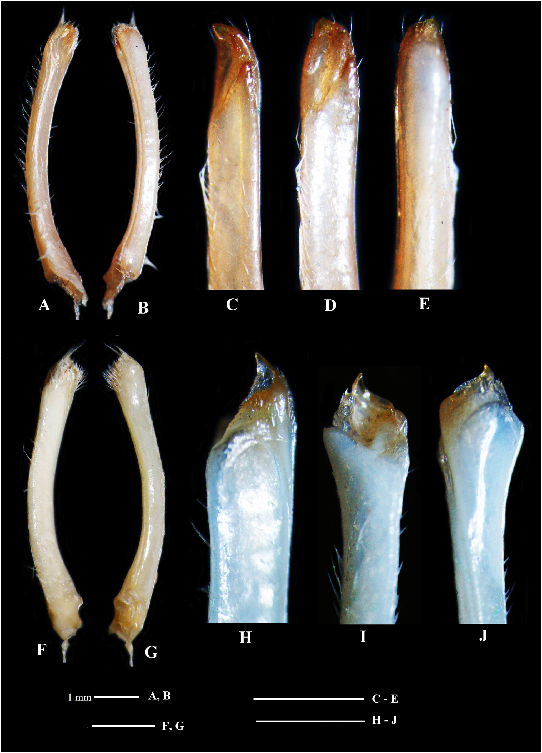
The M. (M.) convexus View in CoL group consists of M. (M.) consobrinus Nobili, 1906 View in CoL , M. (M.) convexus Stimpson, 1858 View in CoL and M. (M.) parvimanus Guérin, 1832 View in CoL ( Poupin, 1997; Barnes, 2010). Among the three species, M. (M.) manggala View in CoL , new species, most closely resembles M. (M.) convexus View in CoL morphologically in having large male chelipeds with deflexed fixed fingers and a differentiated tooth on the cutting margin of the fixed finger on each cheliped. The new species, however, differs from M. (M.) convexus View in CoL in the morphology of the male cheliped and G1 as listed in Table 1 View Table 1 . The new species clearly has a narrow tooth on the fixed finger of the male cheliped ( Fig. 7K View Fig ), and a C-shaped process on its truncated apical portion of the G1 ( Fig. 10C–E View Fig ). In contrast, M. (M.) convexus View in CoL has a wide tooth on the fixed finger of the male cheliped ( Fig. 11E View Fig ), and an arched or pointed process on its apical portion of the G1 ( Fig. 12A–E View Fig ; Komai et al., 1995: fig. 3O–Q).
Distinct differences exist in the morphology of the gastric mill between the new species M. (M.) manggala View in CoL and M. (M.) convexus View in CoL . The median tooth plate of the gastric mill of M. (M.) convexus View in CoL is shorter than that of the new species, which is 2.1 as long as wide, the posterior margin of the propyloric ossicle is wide and nearly flat, and the gap between the pair of posterior teeth is wider than that of the new species ( Fig. 13D View Fig ). The lateral tooth plate of the gastric mill of M. (M.) convexus View in CoL has 13–14 teeth which are arranged in a comb shape ( Fig. 13E View Fig ). On the other hand, that of the new species has 15–16 teeth ( Fig. 13B View Fig ). The anterior tooth of the lateral tooth plate of M. (M.) convexus View in CoL is short and stout ( Fig. 13E View Fig ); meanwhile, in the new species, the tooth is short and slender ( Fig. 13B View Fig ). Naderloo et al. (2010) used morphological characters of the median and lateral teeth plates of the gastric mills to distinguish closely related species.
The new species, M. (M.) manggala , is easily distinguished from the other two species of the M. (M.) convexus group, i.e., M. (M.) parvimanus and M. (M.) consobrinus . The latter two species have small and feeble chelipeds with straight fixed fingers in the males ( Komai et al., 1995: fig. 7; Poupin, 1997: fig. 7; MNHN, 2008; Barnes 2010: in key), whereas the new species has a pair of long and robust chelipeds in the males ( Komai et al., 1995: fig. 3) (see Table 1 View Table 1 for other minor differences between the new species and M. (M.) parvimanus and M. (M.) consobrinus ).
Poupin (1997) examined the morphology of the G1 of the three species of the M. (M.) convexus group, i.e., M. (M.) consobrinus , M. (M.) convexus and M. (M.) parvimanus , from different regions of the Indo-West Pacific region. He found two types of G 1 in these species: a G1 with a truncated apical portion (the typical form) and a G1 with an arched or pointed apical portion (Indo-Malaysian form). In M. (M.) convexus (type locality: Ryukyu Islands), specimens collected from the Western and Central Pacific have generally the typical form G1, and specimens collected from the Indo-Malaysian area have the Indo-Malaysian form G1. In M. (M.) parvimanus (type locality: Mauritius Island), specimens collected from the western Indian Ocean have the typical form G1, and specimens collected from the Indo-Malaysian area have the Indo-Malaysian form G1. Despite the geographical difference in G1 morphology, Poupin (1997) concluded that all populations of M. (M.) convexus he examined belonged to a single species, and he likewise considered the populations of M. (M.) parvimanus to be all conspecific, as judged from other morphological characteristics. The Indo-Malaysian form is not given with a specific status because the specimens from Hong Kong and the Philippines, belonging to the fifth group, are morphologically different from those of the IndoMalaysian. The specimen of M. (M.) convexus from Hong Kong has the similar G1 with M. (M.) parvimanus from Tanzania but distinctly different from M. (M.) convexus from Japan and Indonesia ( Poupin, 1997: fig. 3A–E).
In the present study, we examined the G1 of M. (M.) convexus from Halmahera Island and M. (M.) parvimanus from Saparua Island, both in the Maluku Islands, Indonesia, for comparison with the G1 of the new species. The G1 of the specimens of M. (M.) convexus ( Fig. 12A–E View Fig ) and M. (M.) parvimanus ( Fig. 12F–G View Fig ) agree well with that of the Indo-Malaysian form of M. (M.) convexus ( Poupin, 1997: fig. D’) and M. (M.) parvimanus ( Poupin, 1997: fig. 3B), respectively. Our specimens of M. (M.) parvimanus from Saparua and Lombok Islands ( Fig. 12H–J View Fig ) have the G1 with the pointed apex, similar to that from Thailand redescribed by Komai (1995: fig. 7N–P).
There is further minor variation in the G1 morphology within the Indo-Malaysian form of M. (M.) convexus ; the apical portion is either arched (like a human nail) or pointed ( Poupin, 1997: figs. 3D, D’). Specimens of M. (M.) convexus from Thailand that were illustrated by Komai et al. (1995: fig. 3O–Q) have the G1 with the pointed apex. Our samples of M. (M.) convexus from Halmahera Island, on the other hand, have the G1 with the arched apex ( Fig. 12A–E View Fig ). In contrast, the apical portion of the G1 of M. (M.) manggala is truncated, similar to that of M. (M.) convexus from Japan, which is the truncated (typical form) ( Fig. 10C–E View Fig ; Poupin, 1997: fig. 3C).
In females, the new species differs from M. (M.) convexus and M. (M.) parvimanus in morphology of the vulva; there is no transverse groove on the vulva of M. (M.) convexus and M. (M.) parvimanus ( Komai et al., 1995, figs. 3G, 7G; Fig. 13F, I View Fig ), but there is a median transverse groove on the vulva of M. (M.) manggala ( Fig. 13C View Fig ).
The new species is also morphologically related to species of the M. (M.) brevis group sensu Barnes (2010). However, the M. (M.) convexus group, now including the new species, distinctly differs from the M. (M.) brevis group in having a large, flat, forwardly curved external orbital angle, and no spine on inner surface of the palm in the male cheliped.
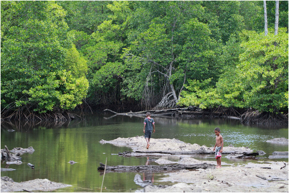
Habitat. This species inhabits muddy substrates. The habitat is submerged by sea water even at low tide ( Fig. 14 View Fig ).
Behavior. This species lives in a narrow burrow in a habitat similar to that of Gelasimus jocelynae on the sampling site but prefers fine mud substrates instead of sandy mud. Unlike G. jocelynae , this species rarely uses its chelipeds for waving display behaviour, and they are mostly used for feeding and burrowing.

| MZB |
Museum Zoologicum Bogoriense |
No known copyright restrictions apply. See Agosti, D., Egloff, W., 2009. Taxonomic information exchange and copyright: the Plazi approach. BMC Research Notes 2009, 2:53 for further explanation.
|
Kingdom |
|
|
Phylum |
|
|
Class |
|
|
Order |
|
|
Family |
|
|
Genus |
Macrophthalmus (Macrophthalmus) manggala
| Murniati, Dewi Citra, Asakura, Akira, Nugroho, Dharma Arif, Hernawan, Udhi Eko & Dharmawan, Wayan Eka 2022 |
(M.) manggala
| Murniati & Asakura & Nugroho & Hernawan & Dharmawan 2022 |
T. liki
| Murniati & Asakura & Nugroho & Hernawan & Dharmawan 2022 |
T. liki
| Murniati & Asakura & Nugroho & Hernawan & Dharmawan 2022 |
(M.) manggala
| Murniati & Asakura & Nugroho & Hernawan & Dharmawan 2022 |
Tmethypocoelis koelbeli
| Davie 1990 |
T. odontodactylus
| Davie 1990 |
T. koelbeli
| Davie 1990 |
T. odontodactylus
| Davie 1990 |
(M.) consobrinus
| Nobili 1906 |
M. (M.) convexus
| Stimpson 1858 |
M. (M.) convexus
| Stimpson 1858 |
M. (M.) convexus
| Stimpson 1858 |
M. (M.) convexus
| Stimpson 1858 |
M. (M.) convexus
| Stimpson 1858 |
M. (M.) convexus
| Stimpson 1858 |
M. (M.) convexus
| Stimpson 1858 |
M. (M.) convexus
| Stimpson 1858 |
(M.) parvimanus Guérin, 1832
| Guerin 1832 |