
Zephronia linkouzi, Chen & Zheng & Jiang, 2023
|
publication ID |
https://doi.org/ 10.11646/zootaxa.5257.1.6 |
|
publication LSID |
lsid:zoobank.org:pub:E18CE51E-CCD2-4E12-B480-930C79D8B745 |
|
DOI |
https://doi.org/10.5281/zenodo.7765913 |
|
persistent identifier |
https://treatment.plazi.org/id/03B087C8-0704-4405-3ACF-18CCDFAECD3F |
|
treatment provided by |
Plazi |
|
scientific name |
Zephronia linkouzi |
| status |
sp. nov. |
Zephronia linkouzi sp. nov.
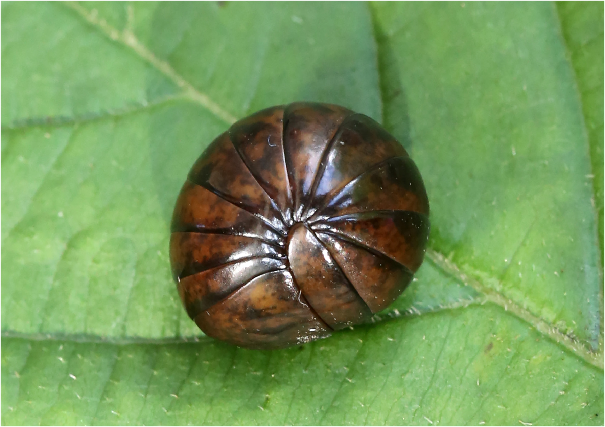
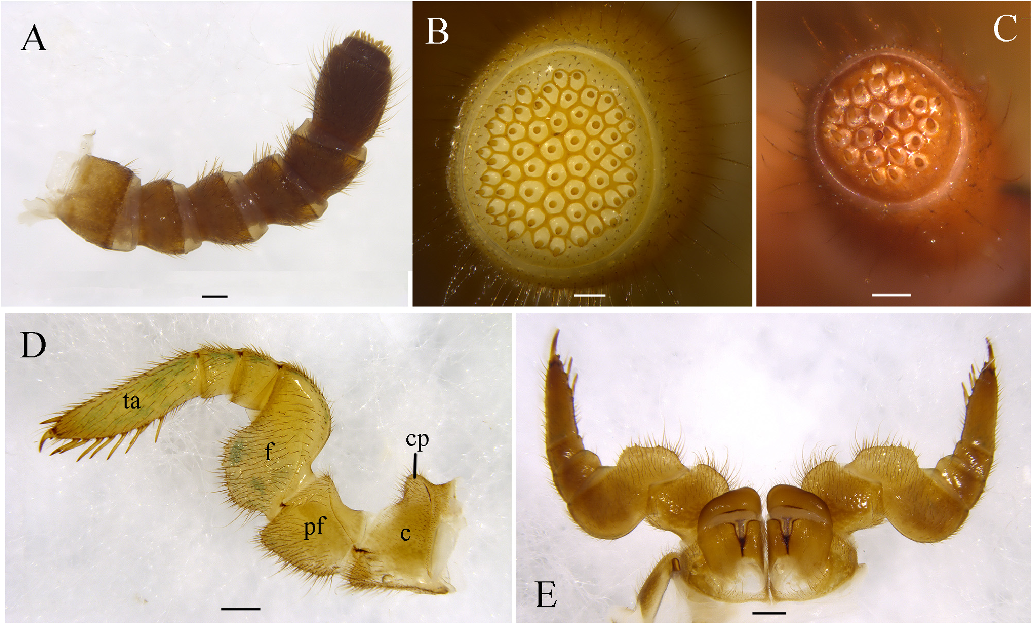
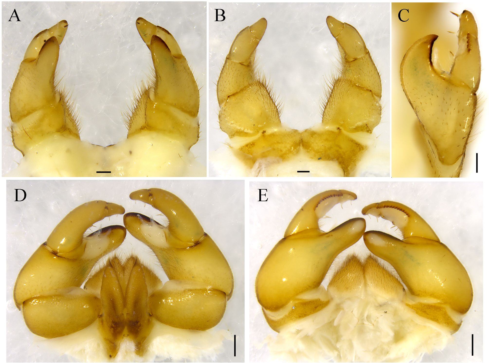
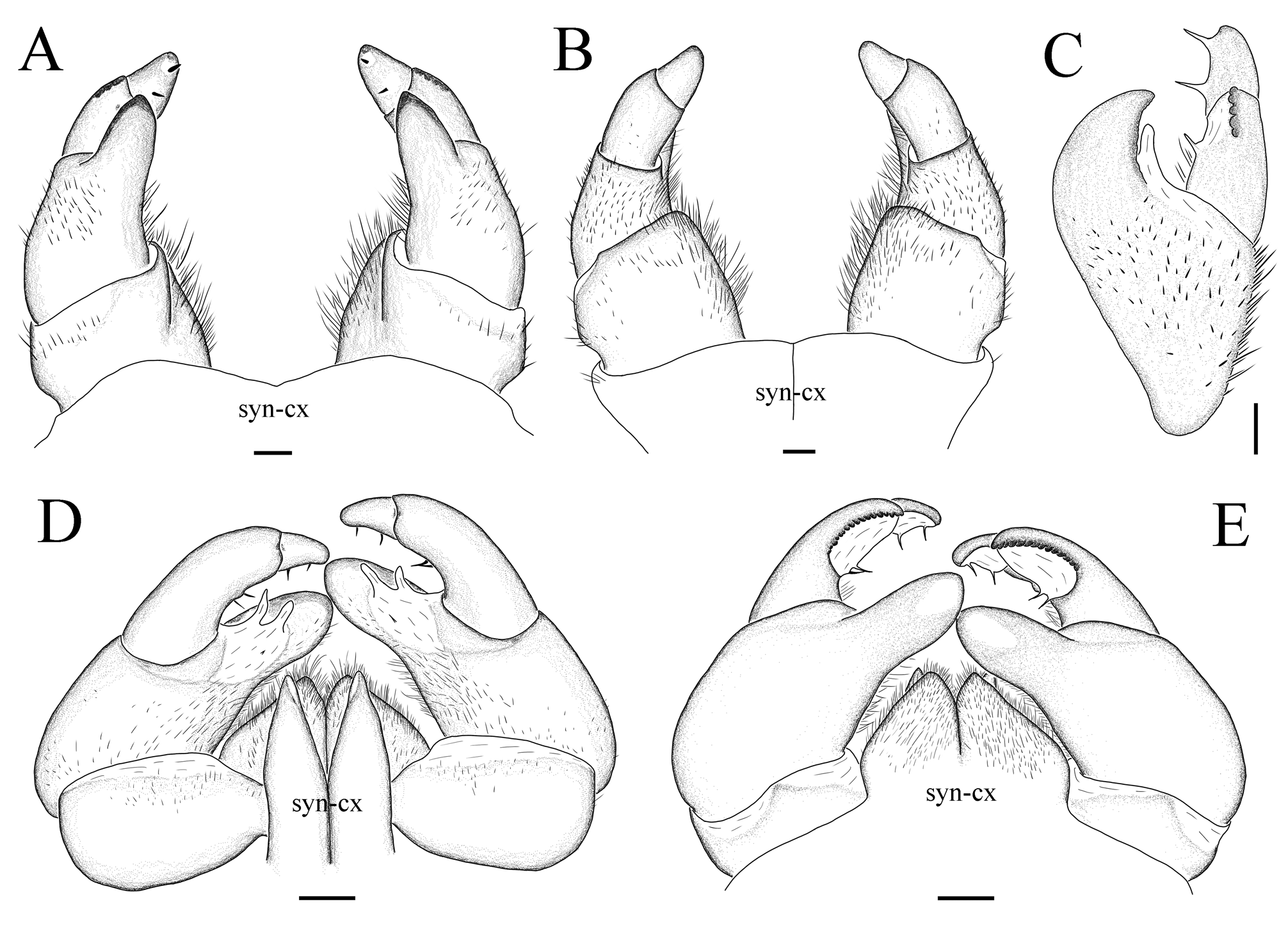
Figs 1–4 View FIGURE 1 View FIGURE 2 View FIGURE 3 View FIGURE 4
Type materials. Holotype male: China, Chongqing, Wuxi County, Yintiaoling National Nature Reserve, Linkouzi , 31°28’19.47” N, 109°52’58.34” E, alt. 1680 m, 17 August 2022, X.K. Jiang & H.M. Chen leg. GoogleMaps Paratypes: 6 males and 2 females, same data as holotype GoogleMaps ; 1 female, same locality, 18 August 2022, X.K. Jiang & H.M. Chen leg. GoogleMaps
Diagnosis. This species is very similar to Zephronia hui Liu & Wesener, 2022 from Jiangkou County, Guizhou Province, China ( Zhao et al. 2022), but it differs from the latter by the antennae slender ( Fig. 2A View FIGURE 2 ) (short and stout in Z. hui ), the process of telopoditomere 2 of the anterior telopod obviously shorter than the length of telopoditomeres 3 and 4 ( Figs 3C View FIGURE 3 , 4C View FIGURE 4 ) (subequal in Z. hui ).
Etymology. This species is named after the type locality, Linkouzi, noun in apposition.
Description. Male body length ca. 19–31 mm. Width of thoracic shield = 8.0–11.0 mm, of tergite 8 = 8.8–12.0 mm. Height of thoracic shield = 4.3–6.0 mm, of tergite 8 = 6.0–8.2. Holotype 31 mm long, 11.0 mm (thoracic shield) wide, 6.0 mm (thoracic shield) high, 12.0 (tergite 8) wide, 8.2 (tergite 8) high. Female body length ca. 32–34 mm long. Width, of thoracic shield = 10.8–11.0 mm, of tergite 8 = 11.9–12.0 mm. Height, of thoracic shield = 6.4–6.9 mm, of tergite 8 = 8.3–8.9 mm. Body generally brown with irregular dark marks scattered on body surface ( Fig. 1 View FIGURE 1 ).
Eyes with ca. 55 ocelli. Antennae short and thick. Last antennomere obviously longer than other antennomeres, lengths of antennomeres: 1=2=3=4=5<<6 ( Fig. 2A View FIGURE 2 ). Apical disc with 39–55 apical cones (male), 24–27 (female) ( Fig. 2B, C View FIGURE 2 ). Organ of Tömösváry located inside antennal groove. Palpi sensory cones located in a single field. Head surface setose. Structure of gnathochilarium typical. Sensory cones of palpi all located in single field. Mandibles not examined. First stigmatic plate widely rounded, apex slightly curved anteriorly. Posterior-lateral margin of laterotergite 1 strongly projecting into a sharp tip. Laterotergite 2 with a broad, stout, much shorter projection. Collum glabrous except for marginal setae. Thoracic shield grooves deep, anterior margin thickened. Surface glabrous like tergites, setae only in grooves. Tergites surface glabrous. Tips of paratergites of midbody tergites projecting posteriorly. Inner section of endotergum lacking any spines or setae. Middle area with a single row of small, elliptical, cuticular impressions. Distance between impressions twice as wide as their diameter. Apically, 2 dense rows of short marginal bristles, tips of longest setae barely protruding beyond midpoint towards tergal margin. Anal shield massive, well-rounded, shiny and glabrous. Locking carina weakly developed. Leg 1 and 2 with 3 or 4 ventral spines. Leg pairs 4–21 ( Fig. 2D View FIGURE 2 ) with 8–10 ventral spines and single apical spine. Coxal process weakly developed and well-rounded ( Fig. 2D View FIGURE 2 ), absent from leg pairs 1 and 2. Tarsus 3.3 times longer than wide. Femur with toothed ridge of medium length.
Male gonopore covered with a small, inconspicuous plate. Telopoditomere 1 of anterior telopods ( Figs 3A, B View FIGURE 3 ; 4A, B View FIGURE 4 ) stout, slightly wider than long. Telopoditomere 2 as long as telopoditomere 3 in anterior view. Process of telopoditomere 2 originated posteriorly, broader than telopoditomere 3, tapering apically, curved, and protruding as high as basal part of telopoditomere 4. Inner margin towards movable finger with one large, membranous lobe. Posterior surface with a row of 5 crenulate and sclerotized teeth ( Figs 3C View FIGURE 3 , 4C View FIGURE 4 ). Telopoditomere 4 short, with at inner margin two long spines ( Figs 3A–C View FIGURE 3 , 4A–C View FIGURE 4 ). Mesial and lateral parts of telopoditomere 1 and anterior and lateral parts of telopoditomere 2 covered by setae. Other parts glabrous. Telopoditomere 1 of posterior telopods cylindrical, twice as long as wide. telopoditomere 2 stout ( Figs 3D, E View FIGURE 3 ; 4D, E View FIGURE 4 ). Immovable finger (process of telopoditomere 2) slightly shorter than movable finger, consisting of telopoditomeres 3 and 4. Margin towards movable finger with two large, membranous lobes. Tip of immovable finger with a white spot in posterior view. Telopoditomere 3 elongated, 3 times longer than telopoditomere 4. Margin near immovable finger with a large membranous lobe and two slender spine, posterior surface with a row of 15 crenulate and sclerotized teeth. Telopoditomere 4 with at inner margin two long spines and a single membranous lobe. Entire telopod with few setae, mostly at anterior margins of telopoditomere 1 and inner margins of telopoditomere 2 ( Figs 3D, E View FIGURE 3 ; 4D, E View FIGURE 4 ).
Female vulva ( Fig. 2E View FIGURE 2 ) large, covering 2/3 of coxa, located at mesal margin, extending mesally to basal third of prefemur length. Operculum rounded. Subanal plate large and wide, not subdivided.
Distribution. Known only from the type locality.
No known copyright restrictions apply. See Agosti, D., Egloff, W., 2009. Taxonomic information exchange and copyright: the Plazi approach. BMC Research Notes 2009, 2:53 for further explanation.
|
Kingdom |
|
|
Phylum |
|
|
Class |
|
|
Order |
|
|
Family |
|
|
Genus |