
Hymedesmia (Hymedesmia) noramaloneae, Goodwin & Berman & Hendry, 2019
|
publication ID |
https://doi.org/10.11646/zootaxa.4658.3.3 |
|
publication LSID |
lsid:zoobank.org:pub:D926CCEC-56EF-4E9A-98BE-CEB4D4D3D60A |
|
DOI |
https://doi.org/10.5281/zenodo.5584974 |
|
persistent identifier |
https://treatment.plazi.org/id/03B087ED-FFDA-FF8C-FF59-F9D3FBACDD79 |
|
treatment provided by |
Plazi |
|
scientific name |
Hymedesmia (Hymedesmia) noramaloneae |
| status |
sp. nov. |
Hymedesmia (Hymedesmia) noramaloneae View in CoL sp. nov.
( Figure 9 View FIGURE 9 , Table 6 View TABLE 6 )
lsid:zoobank.org:act: D3D74DBA-258E-4FAB-8E5F-343DD3FB562B
Type material. Holotype: BELUM. Mc 2015.701 Vieugue Island (65°38.758’S, 65° 12.540’W), depth 10–22 m; collected by C. Goodwin and E. Priestley, 23/02/2015. GoogleMaps
Paratype: BELUM.Mc2015.722and BELUM. Mc 2015.726 Port Charcot, Booth Island (65°03.853’S, 64° 01.868’W), depth 6–16 m; collected by C. Goodwin and E. Priestley, 23/02/2015 GoogleMaps .
Other specimens: BELUM. Mc 2015.601 Grotto Island, Verdansky Base (Site 1) (65°14.615’S, 64° 15.019’W), depth 14–24 m. GoogleMaps BELUM. Mc 2015.734 Port Charcot, Booth Island (65°03.853’S, 64° 01.868’W), depth 6–16 m. GoogleMaps
Diagnosis. Hymedesmia (Hymedesmia) with tornote ectosomal spicules (245–358 µm long) and two categories of acanthostyles (267–633 and 137–208 µm long), the larger of which are only spined for their basal third.
Etymology. Named after Nora Malone, daughter of Jade Berman, who was born a few months after the expedition.
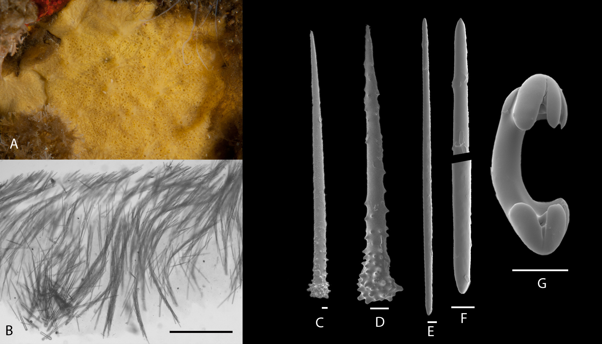
External morphology. In situ appearance ( Figure 9A View FIGURE 9 ): Very thin pale yellow encrusting sponge forming patches up to 20 cm in diameter. Surface covered with an irregular pattern of veins and pores sieves—pore sieves do not have rims so are less distinct than in many other Hymedesmia species.
Preserved appearance. Firm white crust, 1 mm thick.
Skeleton ( Figure 9B View FIGURE 9 ): Hymedesmoid with a basal layer of primary and secondary acanthostyles and ascending columns (5–15 spicules thick) of tornotes. Chelae sparsely scattered throughout tissue.
Spicules (for measurements of all specimens see Table 6 View TABLE 6 ).
Primary acanthostyles ( Figure 9C View FIGURE 9 ). With a tylote head. The head and up to the lower 1/3 of the shaft are spined with small conical spines but the majority of the shaft is smooth. The shaft is often slightly curved.
Secondary acanthostyles ( Figure 9D View FIGURE 9 ). With a tylote head. Entirely spined with small conical spines but these are densest at the head and become slightly sparser towards the tip.
Ectosomal tornotes ( Figure 9E, F View FIGURE 9 ): Anisotornotes, sometimes with one slightly rounded end so style-like in form, others with two pointed ends.
Arcuate chelae ( Figure 9G View FIGURE 9 ): Normal arcuate chelae with a slight bend in the shaft and three, fairly short, rounded alae on each end.
Remarks. Of the 13 species of Hymedesmia (Hymedesmia) recorded from the region (see Goodwin et al. 2012) three possess two categories of acanthostyles and have tornotes as ectosomal spicules: H. antarctica Boury-Esnault & Van Beveren, 1982 , H. mariondufresni Boury-Esnault & Van Beveren 1982 , and H. barnesi Goodwin, Brewin & Brickle 2012 . H. barnesi can be distinguished as it has smaller primary acanthostyles (272–392 µm) and is bright orange when living. Boury-Esnault & Van Beveren (1982) note that the primary difference between H. antarctica and H. mariondufresni is that the tornotes of the latter are shorter than the acanthostyles, whilst the reverse is true for the former. On this basis our specimen would be closer to H. mariondufresni , the tornotes of our species are certainly much shorter than those in H. antarctica (422–593 µm). However, the primary acanthostyles of H. mariondufresni are smaller and thinner than those in our specimens (243–512 by 13–25 µm) and only smooth at the end of the shaft, whereas the primary acanthostyles of our specimens are normally only spined for the basal third.
Distribution. Currently only known from the type and holotype localities.
| BELUM |
Ulster Museum, Belfast |
No known copyright restrictions apply. See Agosti, D., Egloff, W., 2009. Taxonomic information exchange and copyright: the Plazi approach. BMC Research Notes 2009, 2:53 for further explanation.
|
Kingdom |
|
|
Phylum |
|
|
Class |
|
|
Order |
|
|
Family |
|
|
Genus |
|
|
SubGenus |
Hymedesmia |