
Sinocaulus truncatus, Jin, Zhenyu, Ślipiński, Adam & Wang, Wenkai, 2015
|
publication ID |
https://doi.org/ 10.11646/zootaxa.3974.4.9 |
|
publication LSID |
lsid:zoobank.org:pub:30EC4B9F-8C2A-41BA-A16E-FD6AEE80A19B |
|
DOI |
https://doi.org/10.5281/zenodo.6098068 |
|
persistent identifier |
https://treatment.plazi.org/id/03B087F2-F667-FF9E-148A-FBB675D4FEF0 |
|
treatment provided by |
Plazi |
|
scientific name |
Sinocaulus truncatus |
| status |
sp. nov. |
Sinocaulus truncatus sp. n.
( Figures 1–27 View FIGURES 1 – 8 View FIGURES 20 – 26 View FIGURE 27 )
Etymology. The species name refers to the truncate phallobase of the aedeagus.
Diagnosis. This species is most similar to S. omiensis , both having the strongly protuberant eyes, but the phallobase of the latter species is acute at base and ventral lobes are obviously shorter than dorsal lobe.
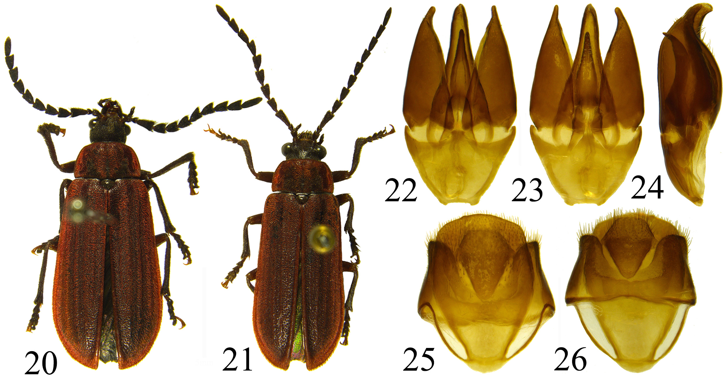
Description. Body length 13.0– 14.6 mm, width 4.9–5.9 mm, females usually larger than males ( Figs. 20–21 View FIGURES 20 – 26 ). Body narrowly elongate oval, 2.4–2.6 times as long as wide, sides weakly convex, head, pronotal and elytral setae uniformly red, venter covered by brown dense pubescence.
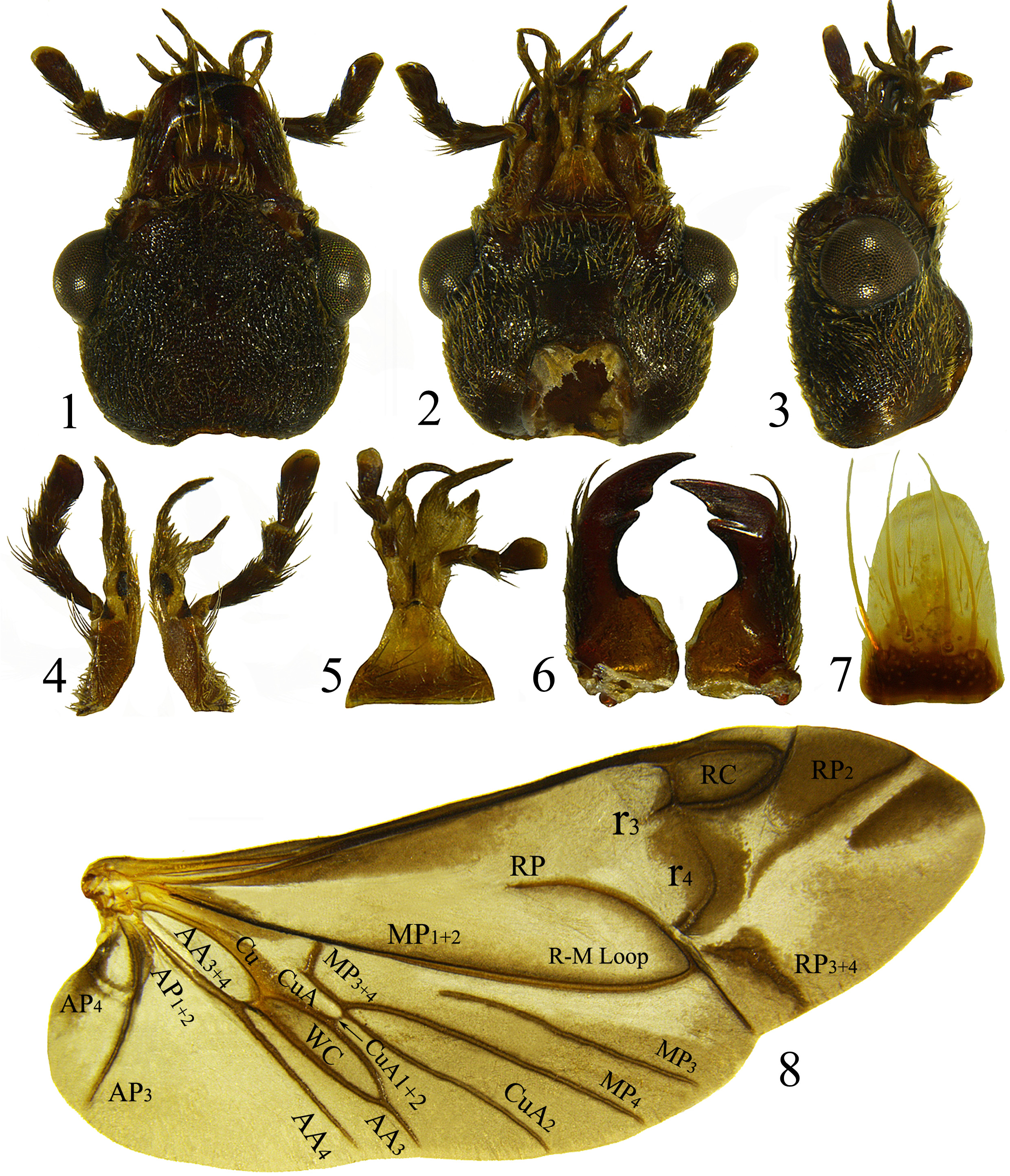
Head ( Figs. 1–3 View FIGURES 1 – 8 ) subquadrate. Eyes large, entire, finely facetted, strongly protuberant and circular in crosssection. Frontoclypeal suture distinct, straight; clypeus short, arcuate anteriorly. Occiput without longitudinal impression or endocarina. Labrum ( Fig. 7 View FIGURES 1 – 8 ) elongate, distinctly sclerotised and setose at base, prominent and membranous anteriorly, rounded at apex. Antenna (Fig. 18) serrate, reaching mid-length of elytra; scape slender, about 2.0–2.2 times as long as pedicel; antennomere 3 about 2.2–2.4 times as long as broad, 1.4–1.5 times as long as antennomere 4, terminal antennomere obtuse apically and longer than penultimate. Mandibles ( Fig. 6 View FIGURES 1 – 8 ) moderately long, broad at base, curved apically with sharp apical tooth; incisor edge with 2 teeth; mola blunt, indistinct, prostheca membranous. Maxillary ( Fig. 4 View FIGURES 1 – 8 ) lobes well developed and membranous apically; galea with two slender lobes; lacinia less prominent and densely setose; terminal maxillary palpomere weakly expanded apically. Mentum large, trapezoidal, obtuse anteriorly; ligula membranous, deeply bilobed, bearing long setose process on each side.
Prothorax (Fig. 9) trapezoidal, transverse, 0.5–0.6 times as long as wide; sides distinctly narrowing anteriorly; lateral carinae complete, simple with upturned margin but without distinct bead; anterior and posterior angles obtuse; posterior edge tri-emarginate and smooth; disc convex with median impressed line and distinctly explanate lateral margins; punctation hardly visible under dense pubescence.
Prosternum (Fig. 10) in front of coxa as long as longitudinal mid-coxal diameter; prosternal process entirely separating procoxae, slightly elevated, very narrow and pointed apically. Notosternal suture complete. Procoxae strongly projecting below prosternum. Procoxal cavities strongly transverse, narrowly separated and broadly open externally and internally; protrochantin exposed. Scutellar shield (Fig. 11) distinctly elevated, truncate anteriorly, distinctly pointed posteriorly, wider than long; densely setose.
Elytra taken together 1.9–2.0 times as long as wide and 4.7–4.9 times as long as pronotum; weakly convex, sides weakly expanding to about apical fourth and then abruptly narrowing to apex, apices blunt; lateral margins narrow with distinct bead, entirely visible from above. Alternate elytral intervals convex and setose, intervals with dense and coarse punctures not forming rows; scutellary striole absent. Epipleuron broad and mostly horizontal, distinctly setose.
FIGURES 9–19. Sinocaulus truncatus sp. n. (9–11, 13–19) female; (12) male: (9) prothorax; (10) prosternum; (11) mesoscutum; (12–13) abdomen; (14) pterothorax ventral; (15) fore leg; (16) mid leg; (17) hind leg; (18) antenna; (19) ovipositor.
Mesoventrite (Fig. 14) broad anteriorly, separated from mesanepisterna by complete sutures; anterior edge medially on the same plane as metaventrite, forming weak elevation laterally bordered by shallowly declined and oblique procoxal rests. Mesoventral process very narrow, emarginate apically; meso-metaventral junction monocondylic. Mesocoxal cavities separated by 0.1–0.2 coxal diameter; open laterally, closed by mesepimeron.
Mesocoxae weakly projecting, mesotrochantin exposed. Metaventrite wider than long, moderately convex; discrimen complete; transverse, katepisternal suture complete; exposed portion of metanepisternum long and broad; metepimeron exposed. Metacoxae contiguous, extending laterally to meet elytra; metacoxal plates poorly developed and well visible in mesal part only.
Hind wing ( Fig. 8 View FIGURES 1 – 8 ) about 2.1–2.3 times as long as wide; radial cell 1.8–1.9 times as long as wide, not forming equilateral triangle, pigmented, inner posterior angle obtuse; cross-vein r3 short and oblique; apical field about 0.1– 0.2 times total wing length with pigment patches around apical folds and r4, plus RP vein remnants; medial field with 5 free veins and MP3 complete; wedge cell long, apically acute; anal lobe well-developed, anal notch absent; AP divided, with posterior branch meeting basal wing margin.
Legs (Figs. 15–17) slender, similar in shape, covered with setae; femur elongate and straight, about as long as tibia in fore- and mid-legs, while shorter than tibia in hind legs; tibia with external dorsal side spinose; spurs paired, serrate. Tarsi 5–5– 5 in both sexes; tarsomere 1 longer than 2; divided membranous ventral lobes present on tarsomeres 1–4 but the first one comparatively smaller and not entirely divided; claws simple; empodium absent.
Abdomen (Fig. 12–13) with five ventrites, ventrites 1 and 2 of similar lengths, connate; ventrites uniformly, densely pubescent without glabrous spots on each side; apex of ventrite 5 sexually dimorphic, truncate in males (Fig. 12) and broadly rounded in females (Fig. 13); intercoxal process indistinct, metacoxal cavities not delimited on ventrite 1. Sternite IX ( Fig. 25 View FIGURES 20 – 26 ) apically trapezoidal and broadly rounded at base, bearing uniformly short setae in middle and apical part; posterior edge of tergite IX emarginate; tergite X slightly shorter than tergite IX; apex of tergite X trapezoidal ( Fig. 26 View FIGURES 20 – 26 ).
Male genitalia ( Figs. 22–24 View FIGURES 20 – 26 ) trilobate, symmetrical; phallobase without struts, with median endocarina and base truncate; parameres articulated, obviously longer than phallobase, apices slender and narrowly rounded, apical third with sparse setae and not upturned in inner margin of apical part. Penis with longer dorsal and shorter ventral lobes, both lobes slender and narrowly obtuse at apex; anterior edge of penis with short paired struts.
Female genitalia weakly sclerotised with vagina and bursa copulatrix not clearly separated; bursa copulatrix without sclerites; spermatheca small and not sclerotised. Ovipositor (Fig. 19) short; paraprocts entirely sclerotised without baculi, about as long as gonocoxites; proctiger absent; gonocoxites entirely sclerotised, ventral to paraprocts, triangular and strongly bent with prominent apices densely setose dorsally, without baculi; gonostyli absent.
Types. Holotype (♂): China-Guizhou: Guiyang, Daozhen, Yangxisha, 31-v-2004, Yang Yu (NKUM).
Paratypes (2♀): China-Guizhou: Guizhou province, Suiyang, Kuankuoshui Reserve, 840-1200, 9-vi-2010, Kai Dang (1♀, MHBU); Guizhou province, Suiyang, Kuankuoshui Reserve, Xiangshuwan Village, 840-1200m, 9- vi-2010, Kai Dang (1♀, MHBU).
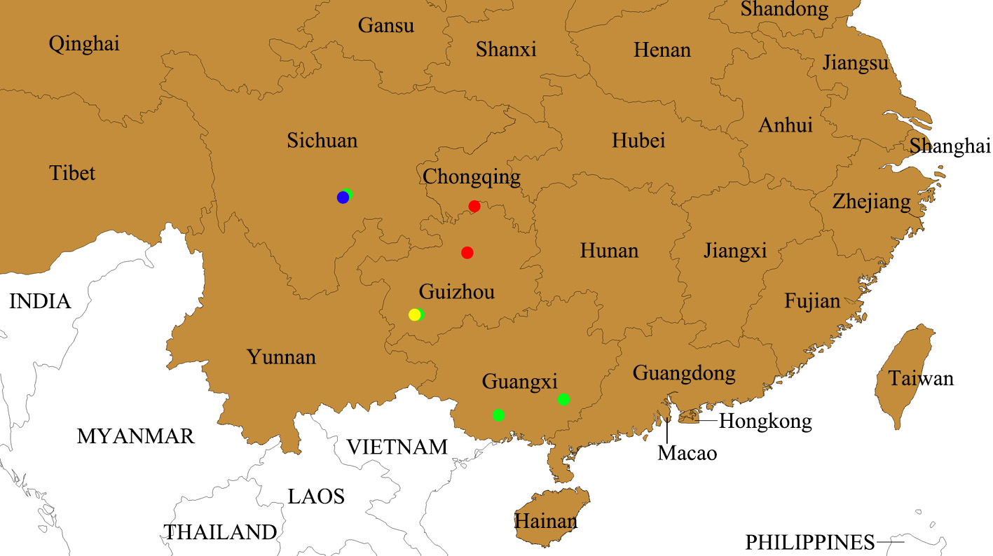
Distribution. China: Guizhou ( Fig. 27 View FIGURE 27 ).
No known copyright restrictions apply. See Agosti, D., Egloff, W., 2009. Taxonomic information exchange and copyright: the Plazi approach. BMC Research Notes 2009, 2:53 for further explanation.