
Mummucina titschacki Roewer, 1934
|
publication ID |
https://doi.org/ 10.11646/zootaxa.3884.4.2 |
|
publication LSID |
lsid:zoobank.org:pub:DD461E43-28C7-4842-85CC-885286DAA9E3 |
|
DOI |
https://doi.org/10.5281/zenodo.6123404 |
|
persistent identifier |
https://treatment.plazi.org/id/03B0924E-490D-FFA5-FF64-FEA79DA4C8EA |
|
treatment provided by |
Plazi |
|
scientific name |
Mummucina titschacki Roewer, 1934 |
| status |
|
Mummucina titschacki Roewer, 1934 View in CoL
( Figures 1–43 View FIGURES 1 – 5 View FIGURES 6 – 9 View FIGURES 10 – 13 View FIGURES 14 – 17 View FIGURES 18 – 21 View FIGURES 22 – 25 View FIGURES 26 – 31 View FIGURES 32 – 35 View FIGURES 36 – 39 View FIGURES 40 – 43 , Table 1)
Mummucina titschacki Roewer, 1934: 589 View in CoL , fig. 334h.
Mummucina titschacki: Kraus 1966: 182 View in CoL , fig. 1; Muma 1971: 10; Muma 1976: 24; Harvey 2003: 290, 291; Martins et al. 2004: 2371; González-Reyes & Corronca 2013: 538, 539.
Mummicina titschacki (as Mummicinatitschacki [sic]): Weidner 1959: 110 [genus name is a lapsus calami].
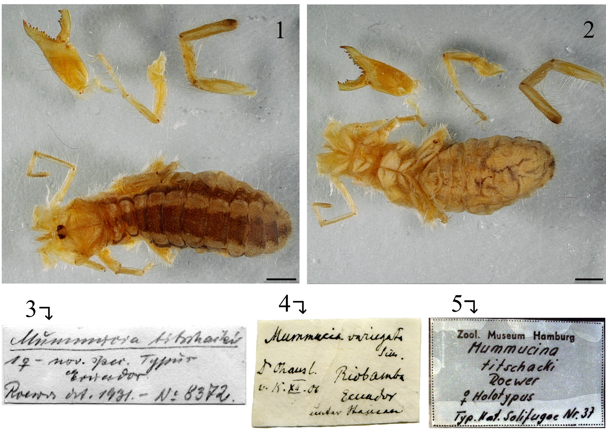
Type material. Shared between two collections. FEMALE HOLOTYPE: body and some appendages housed in the ZMH. Labels verbatim: “ Mummucina titschacki / 1♀ - nov. spci. Typus / Ecuador / Roewer det. 1931. - Nº 8372 ” (C. F. Roewer’s hand-writing). “ Mummucia variegata Gerv. / Dr. Ohaus I. / v. [=vom] 15.XII.06 / Riobamba / Ecuador / unter Steinen [historical German script Sütterlin meaning´under stones/rocks´]” (K. Kraepelin’s handwriting). “ Zool. Museum Hamburg / Mummucina titschacki Roewer / ♀ holotypus / Typ. Nat. Solifugae Nr. 37 ” ( ZMH collection label). Examined by photos. FEMALE HOLOTYPE: one leg III, one leg IV, a pedipalp and the left chelicera in a microscope slide housed in the Senckenberg Museum, Frankfurt ( SMF) (catalogue number 6821- 71). Not examined.
Remarks. The depository of the holotype of Mummucina titschacki was not specified in the original description ( Roewer 1934). Weidner (1959) mentioned that the specimen was in the collection of the Hamburg Zoological State Institute and Museum (currently ZMH, Germany).
Kraus (1966: fig. 1) illustrated the right chelicera of a female Mummucina titschacki . Although this author did not mention the material on which the illustration was based, it was most likely the holotype since it was the only specimen reported in literature and no new locality records were made in that contribution. Afterwards, Muma (1976: 24) stated that it was unknown where the holotype of this species was deposited. This belief propagated and was neither confirmed nor contradicted in subsequent works ( Harvey 2003; Martins et al. 2004; Rocha & Carvalho 2006; Carvalho et al. 2010; González-Reyes & Corronca 2013).
Recently, the author established contact with Dr. Andreas Wessel and Dr. Hieronymus Dastych of the ZMH, who managed to locate the holotype in their collection. As shown in Figs. 1–2 View FIGURES 1 – 5 , the specimen has some appendages detached and others missing (see also “ type material”). Likewise, the colour of the specimen has faded, particularly in the propeltidium and chelicerae. Still, it provides information about the species’ morphology and especially on diagnostic features of the chelicerae. The specimen is accompanied by two hand-written labels, one by C. F. Roewer and the other by K. Kraepelin ( Figs. 3–4 View FIGURES 1 – 5 ); the latter reads “ Mummucia variegata ”, which was the only mummuciid species described during Kraepelin’s lifetime.
Material examined. ECUADOR: Chimborazo, Road 35th, 3 km N of Riobamba, 1 km before San Andrés, 100 m from “Cantera (quarry) San Andrés”, 3000 m elev., 1.5990417° S 78.6973167° W, manual capture and pitfall traps (12:00 to 15:00 hs), 22–23 March 2014; R. Botero Trujillo: 31 males, 3 females, 4 juveniles ( MACN, 80% ethanol; 2 males and 1 female used for SEM); 5 males, 1 female ( PUCE, 80% ethanol); 2 males, 2 juveniles ( AMNH, 80% ethanol); 2 males, 2 juveniles (MCN, 80% ethanol); 18 males, 2 juveniles ( MACN, 96% ethanol).
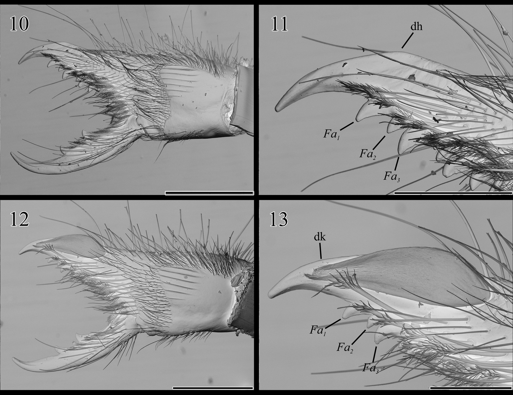
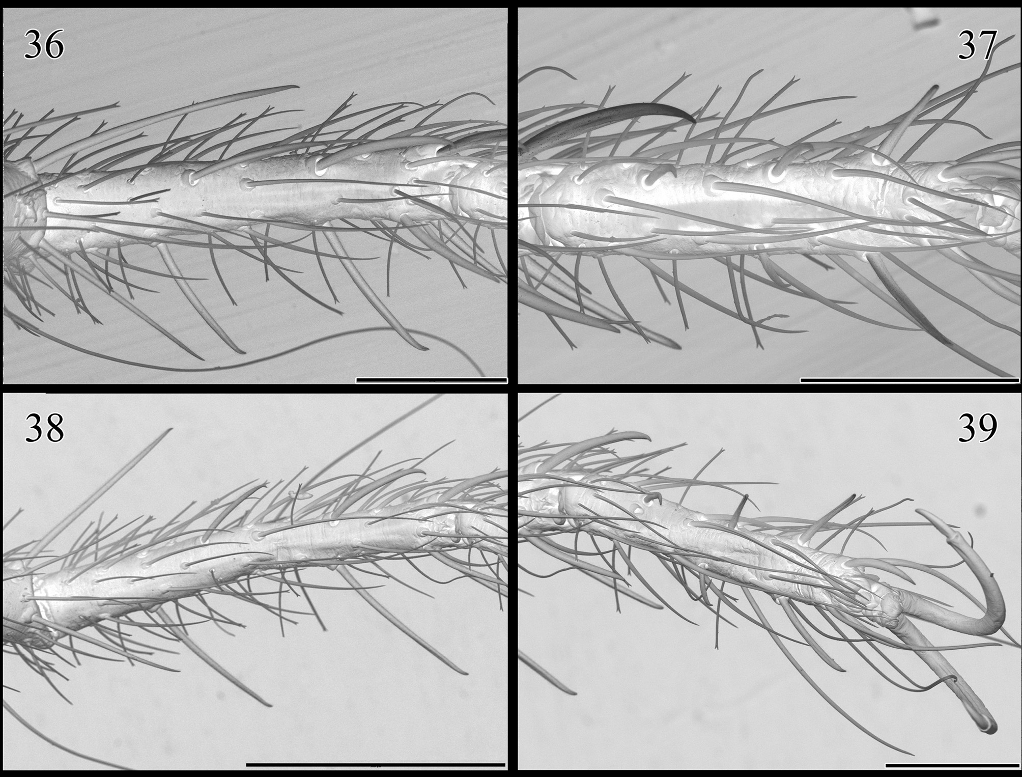
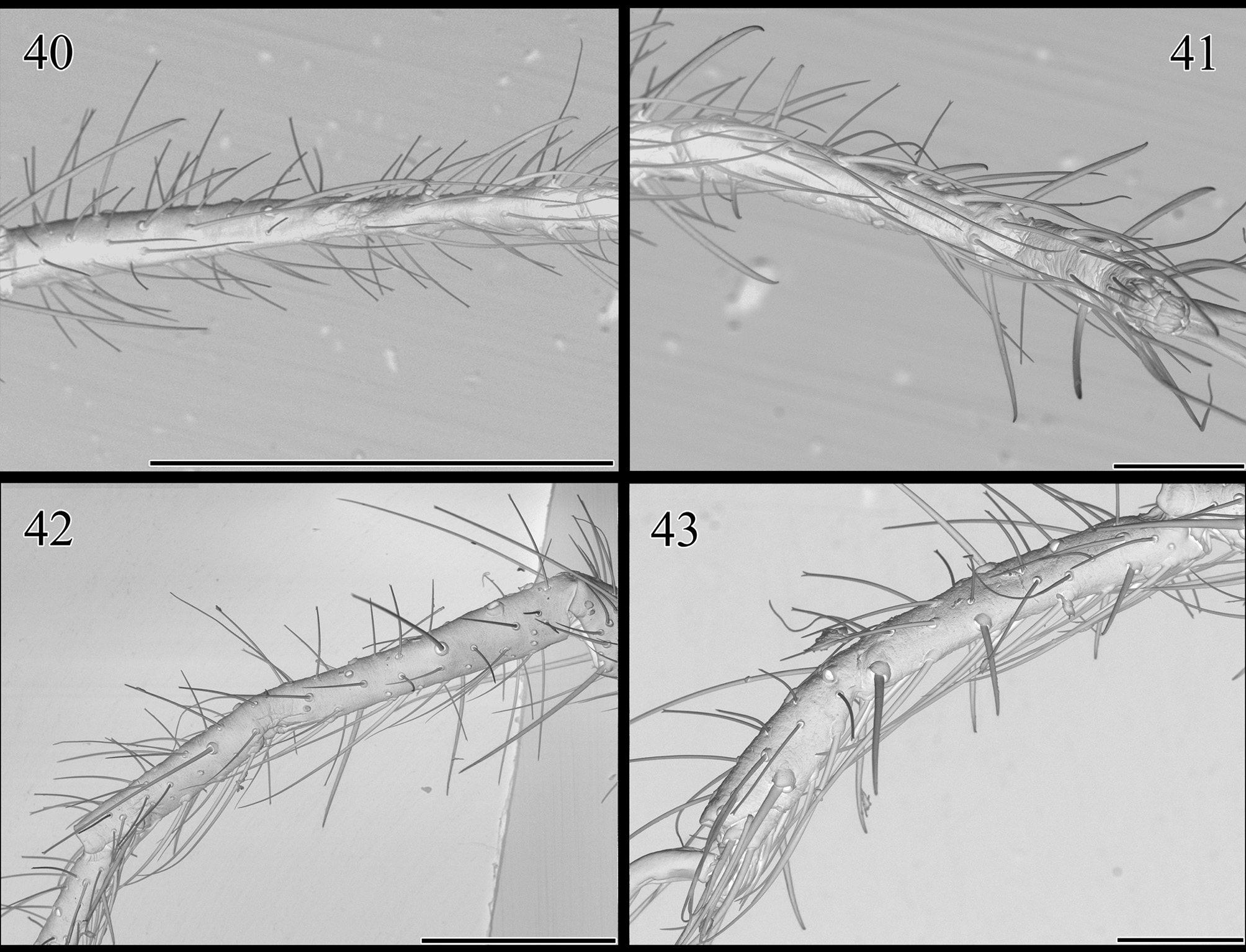
Revised diagnosis. A member of Mummuciidae as defined by Maury (1984, 1998), characterized by the following features. Cheliceral fixed finger with weak dorsal hump, at level of Fa2 tooth in female and of Fp in male, preceded anteriorly by a more or less straight portion ( Figs. 16–17 View FIGURES 14 – 17 ). Male and female cheliceral fixed finger with three anterior teeth (Fa1–3), contiguous; Fa2 tooth noticeably reduced in comparison to Fa1 and Fa3, which are approximately subequal in size ( Figs. 11, 13 View FIGURES 10 – 13 ). Flagellum thin, with dorsal margin keel-shaped, slightly visible in ectal aspect; apex of the flagellum surpassing the level of Fa1 tooth, almost reaching the finger tip ( Figs. 13 View FIGURES 10 – 13 , 16 View FIGURES 14 – 17 ). Male and female with 1.2.2/2.2 (5 prolateral, 4 retrolateral) spiniform setae on telotarsi of legs II–III and 2.2.2-2/ 2.2 on that of leg IV ( Figs. 37, 39 View FIGURES 36 – 39 , 41, 43 View FIGURES 40 – 43 ).
Description of male. Meristic data in Table 1.
TABLE 1. Meristic data for Mummucina titschacki Roewer, 1934 . Above, measurements in millimeters for one male and one female (MACN). L = length; W = width; H = height. 1Measured from the anterior margin of propeltidium to posterior end of abdomen. 2Measured at widest point. 3Measured in ectal aspect parallel to dorsal surface, from the basal condyle (estimated below the lateral lobe) to the fixed finger tip. 4Sum of individual segment lengths. 5Measurement excludes claws. Below, variability report obtained for some cheliceral teeth counts.
MALE FEMALE
Total body L (w/o chelicerae):1 7.00 9.37
Propeltidium: L: 1.25 1.63
W:2 1.43 2.29
Chelicera: L:3 1.73 3.04
W:2 0.60 1.01
H: 0.58 1.04
Pedipalp total L:4 4.56 5.52
Femur L: 1.65 1.95 MEASUREMENTS Leg I total L (w/o claws):4 Tibia Tibia Metatarsus L W::2 + tarsus L: 3.62 1.31 0.29 1.60 1.60 1.97 0.44 4.17
Femur L: 1.00 1.12
Tibia L: 1.15 1.37
Metatarsus L: 0.79 0.92
Tarsus L:5 0.67 0.77
Leg IV total L (w/o claws):4 6.06 6.89
Femur L: 1.98 2.29
Tibia L: 1.77 2.05
Metatarsus L: 1.42 1.58
Tarsus L: 5 0.88 0.96
MALES FEMALES
Fixed finger: Ectal anterior teeth: (n = 60) (n = 7)
60 with 3 7 with 3
Ectal intermediate teeth: (n = 61) (n = 8)
45 with 1 8 with 2 REPORT Ectal fondal teeth: (49 16 n = with with 61) 2 5 (6 n with = 8) 6 VARIABILITY Mesal fondal teeth: (1 1 58 10 n with with = with with 61 7 6) 3 4 (8 2 n with with = 8) 7 3
2 with 2
1 with 4
Movable finger: Intermediate teeth: (n = 61) (n = 8)
55 with 1 5 with 2
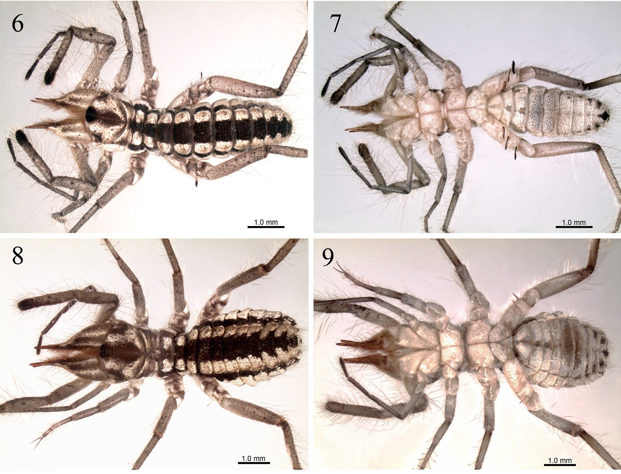
6 with 2 3 with 1 Color. As in Figs. 6–7 View FIGURES 6 – 9 . Propeltidium base color whitish, with brown-to-purplish areas in median region, in the posterior border, and in the suture line with the lateral lobes. Ocular tubercle black. Chelicerae with a pattern of whitish and brownish-to-chestnut areas on dorsal and ectal surfaces of manus; fingers chestnut to reddish. Meso-, metapeltidium and abdominal tergites, all crossed medially by a (broad) brown longitudinal band, and dorsolaterally by a (less broad) whitish band on each side. The whitish bands extend into part of the lateral pleural membranes, creating a very thin whitish border on them. Contiguous to this border, pleural membranes have a brown longitudinal band (thinner than others) which at the level of the last four-to-five abdominal segments bears some whitish spots. The rest (and largest portion) of the membranes is entirely whitish and spotless. Ventral aspect of prosoma and abdomen predominantly whitish, except for faint brown mottling on post-spiracular sternites IV–VI and black anus. Pedipalps and legs whitish brown, without any remarkable coloration feature other than the tarsi of pedipalp and leg I brown-to-purplish. Malleoli white basally and brownish distally.
Prosoma. Propeltidium slightly wider than long ( Fig. 6 View FIGURES 6 – 9 ); with long, medium-sized and short bifurcated setae, the former of which are bilaterally symmetrical; anterior margin convex, with ocular tubercle elevated; complete median longitudinal furrow present; lateral lobes separated from the propeltidium by an incomplete lateral groove. Peltidium narrow, with row of bifurcated setae of variable size. Parapeltidium smooth. Meso- and metapeltidium wider than long, with bifurcated setae of variable size. Coxae densely covered with bifurcated setae; those of pedipalps and legs I–III with some bilaterally paired longer bifurcate setae, and one or two long that are non-bifid. Sternum glabrous ( Figs. 18–21 View FIGURES 18 – 21 ).
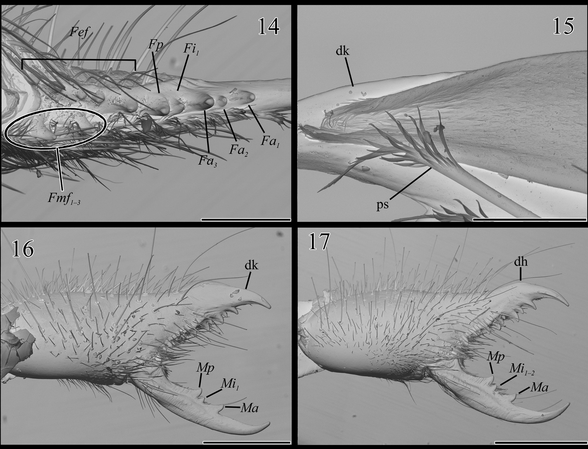
Chelicerae. Ectal and dorsal surfaces with abundant bifurcated setae of variable size, some of them bilaterally symmetrical ( Figs. 12 View FIGURES 10 – 13 , 16 View FIGURES 14 – 17 ). Mesal surface with array of setal types ( Figs. 12–13 View FIGURES 10 – 13 ), which starting from the cutting edge of fixed finger are as follows: first row is made up of plumose setae bordering the teeth line, which reaches the base of the fixed finger; second row of similar plumose setae, but more extended basally reaching the condyle of the movable finger; third row of few thick blunt setae in front of the stridulatory apparatus, and others thinner and placed more apically; following, a carpet-like area of barbed bristles covers the most distal third of the manus, this area is twice higher than wide, and inclined so that dorsal section is slightly advanced to the front. From the base of the chelicerae to the setal area on mesal surface, there is a completely glabrous region, approximately wider than high; on its dorso-frontal quarter is the stridulatory apparatus of six parallel ridges ( Fig. 12 View FIGURES 10 – 13 ). Movable finger with a first mesal row of plumose setae bordering the teeth line on basal half, followed by a row of blunt setae, and another row of barbed bristles. Dentition as in Figs. 13 View FIGURES 10 – 13 , 14, 16 View FIGURES 14 – 17 : Movable finger with one anterior (Ma), one or two intermediate (Mi1, Mi2), and one principal tooth (Mp), arranged in increasing size: Mi1, Mi2, Ma = Mp; all teeth close together in the medial region of the finger. Fixed finger with three anterior teeth (Fa1, Fa2, Fa3; contiguous), one or two intermediate teeth (Fi1, Fi2), and one principal tooth (Fp), arranged in increasing size: Fi1, Fa2 = Fi2, Fa1, Fa3 = Fp; ectal fondal teeth (Fef) five to seven in number, row uninterrupted and teeth variable in size; mesal fondal teeth (Fmf) most frequently three or two in number, arranged in increasing size Fmf3, Fmf2, Fmf1. Fixed finger with dorsal keel starting close to the base of the flagellum and ending close to the finger tip, which protects the flagellum on ectal aspect ( Figs. 15–16 View FIGURES 14 – 17 ); this keel has a weak dorsal hump at level of Fp tooth, preceded anteriorly by a more or less straight portion; ventral border (cutting edge) of the mucron without keel, shaped in a rather straight line with respect to the teeth line and curved at the tip ( Fig. 16 View FIGURES 14 – 17 ). Movable finger mucron predominantly straight, with moderate elevation on basal half of dorsal border (cutting edge) and the tip curved against the fixed finger.
Flagellum. A thin translucent membrane, slightly visible in ectal aspect, with dorsal margin keel-shaped and ventral margin almost straight. Mesal surface with abundant spicules on anterior half which become more prominent as they approach the apex. Attachment ring of the flagellum approximately at the level of the second ectal fondal tooth. Apex of the flagellum surpassing the level of Fa1 tooth, almost reaching the finger tip ( Figs. 12–13 View FIGURES 10 – 13 , 15–16 View FIGURES 14 – 17 ).
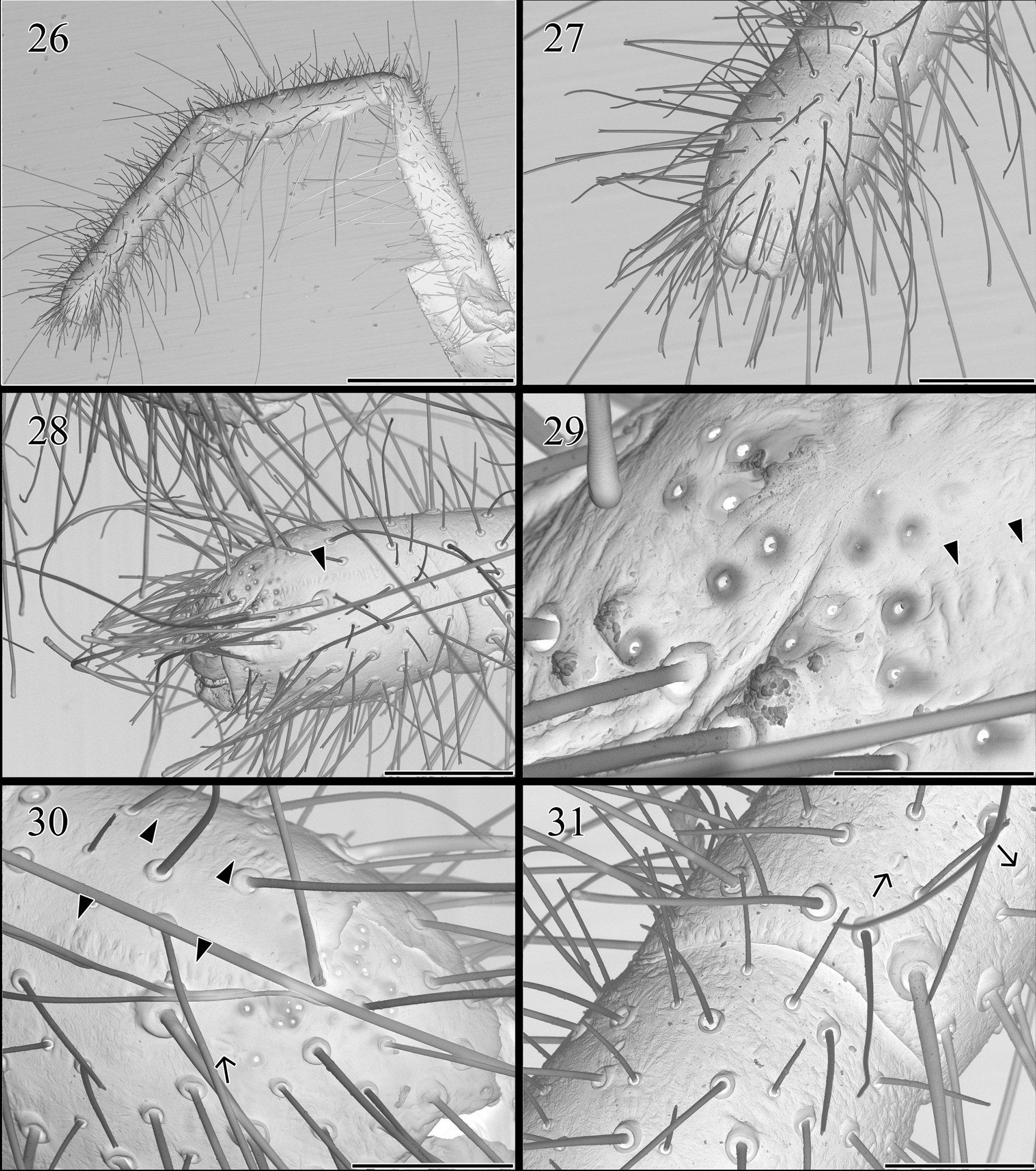
Pedipalps. All segments coated with bifurcated setae of different sizes, from very minute to even longer than the tibia ( Fig. 26 View FIGURES 26 – 31 ); clubbed setae present only on tarsus and metatarsus. Tarsus with a dorsal pore area on distal third ( Figs. 28–30 View FIGURES 26 – 31 ); each pore is defined by an elevated border and bears a seta inside (i.e., sensilla ampullacea sensu Bauchhenss 1983). The pore area is spatially associated to a couple of dorsal longitudinal areas, reticular in appearance, which end near the suture line with the metatarsus ( Figs. 28–30 View FIGURES 26 – 31 ). All segments with slit sensilla randomly distributed ( Figs. 30–31 View FIGURES 26 – 31 ).
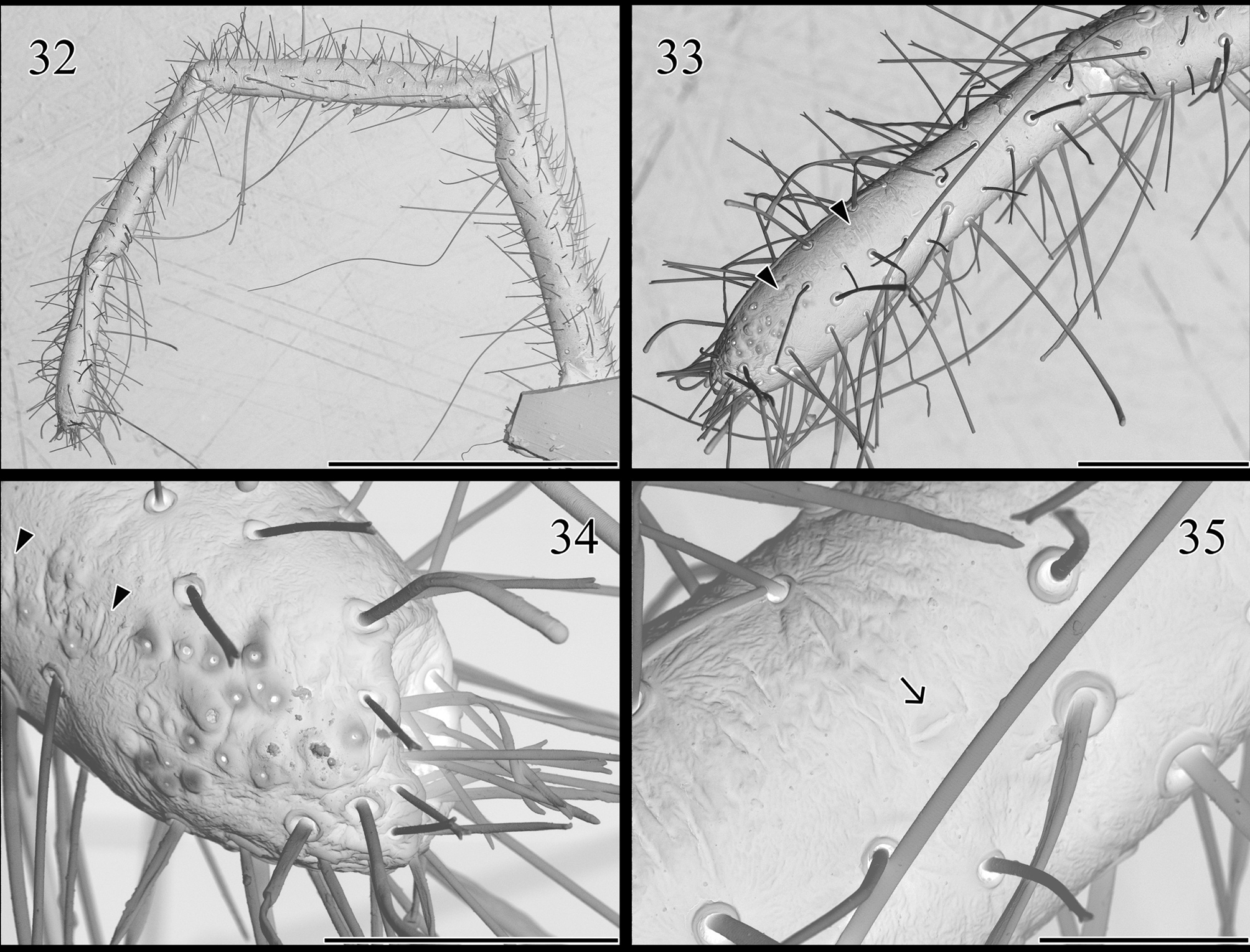
Leg I. Similar to pedipalp in the types of setae that are present, their density and their distribution in the segments; without claws or spiniform setae ( Fig. 32 View FIGURES 32 – 35 ). Tarsus with a dorsal pore area on distal fifth, similar to that described above and also associated to the dorsal longitudinal reticular areas, which end about the middle of the tarsus ( Figs. 33–34 View FIGURES 32 – 35 ). Slit sensilla present at least on tarsus and metatarsus, but fewer than in pedipalps ( Fig. 35 View FIGURES 32 – 35 ).
Walking legs. Covered with abundant bifurcated setae of medium to small size, and a few longer setae (e.g., Fig. 38 View FIGURES 36 – 39 ). Legs II and III: tibia with distal pair of ventrolateral spiniform setae; metatarsus with 2.2.3 ventrolateral spiniform setae ( Figs. 36, 38 View FIGURES 36 – 39 ); tarsus bi-segmented with 1.2.2/2.2 ventrolateral spiniform setae (unpaired seta of tarsus is on prolateral aspect) ( Figs. 37, 39 View FIGURES 36 – 39 ). Leg IV: Tibia with distal pair of ventrolateral spiniform setae; metatarsus with 1.1.1.2 ventral spiniform setae (unpaired setae are on prolateral aspect) ( Figs. 40, 42 View FIGURES 40 – 43 ); tarsus bisegmented with pseudosegmentation on basal segment, and 2.2.2-2/2.2 ventral spiniform setae ( Figs. 41, 43 View FIGURES 40 – 43 ).
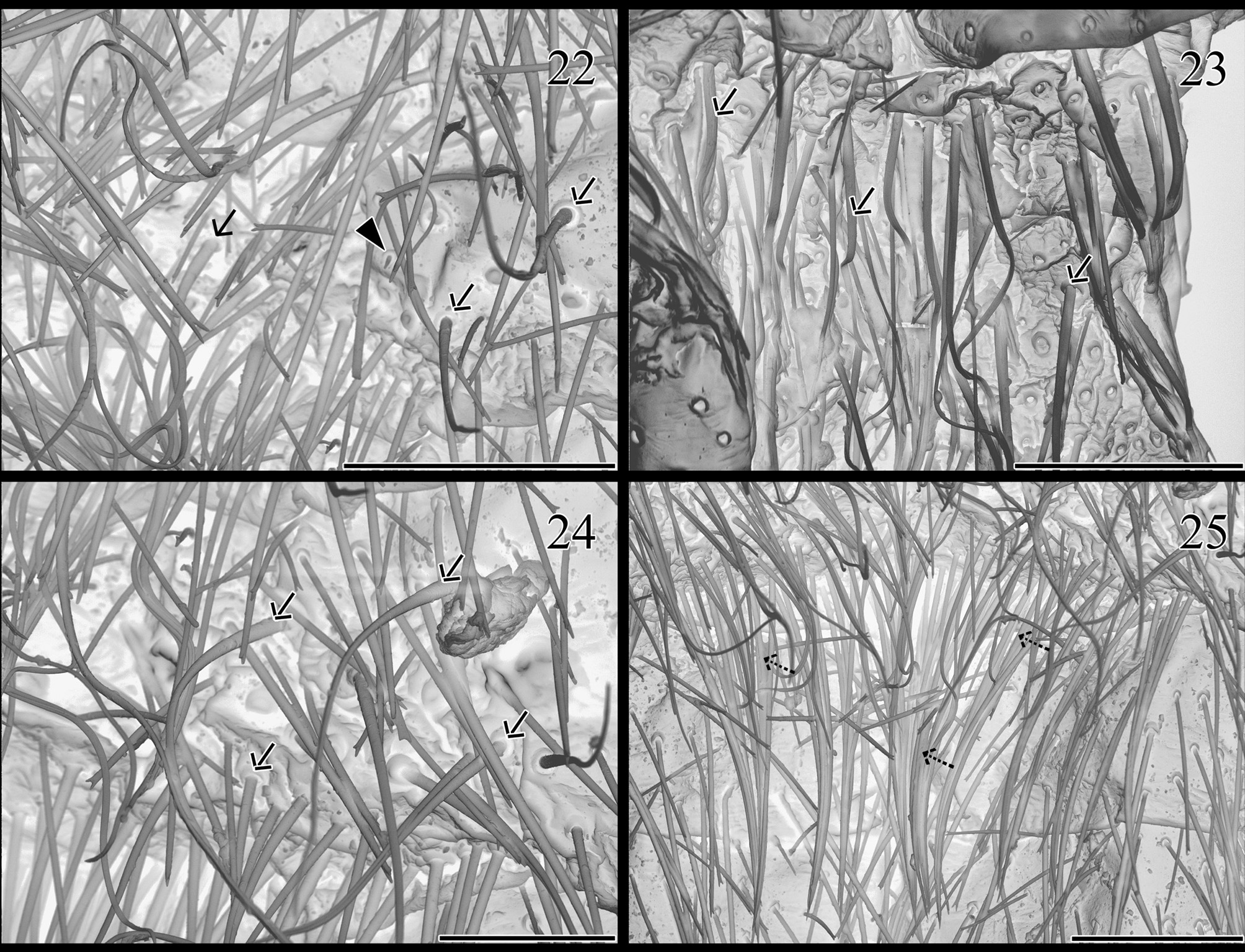
Opisthosoma. Tergites with some bifurcated setae of medium size, some of which are longer and bilaterally symmetrical. Sternites with several bifurcated setae (e.g., Figs. 22, 25 View FIGURES 22 – 25 ). Two pairs of microsetae, similar to those reported by Iuri et al. (2014), are placed in the posterior half of genital plate; one of these microsetae is also present on each side of post-spiracular sternite I ( Fig. 22 View FIGURES 22 – 25 ) (these could not be seen in other sternites due to dense setation). Posterior half of post-spiracular sternites I and II with many filiform ctenidia, not remarkably thicker than setae but with broad insertion socket ( Figs. 22–24 View FIGURES 22 – 25 ); each side of spiracular sternite II with one to three ctenidia of similar appearance and position; post-spiracular sternite II with an additional row of densely-packed “specialized setae” (ctenidia?) along posterior margin ( Fig. 25 View FIGURES 22 – 25 ).
Female. Meristic data in Table 1.
Similar to male but larger and more robust. Color as in Figs. 8–9 View FIGURES 6 – 9 . Propeltidium wider than long ( Fig. 8 View FIGURES 6 – 9 ). Chelicerae with a dorsal hump on the fixed finger at level of Fa2 tooth, preceded anteriorly by a portion of the finger with a straight border ( Figs. 11 View FIGURES 10 – 13 , 17 View FIGURES 14 – 17 ). Mesal surface of the chelicerae with denser setation ( Fig. 10 View FIGURES 10 – 13 ). Abdomen apparently without ctenidia, but post-spiracular sternite II with row of densely-packed “specialized setae” along posterior margin.
Variability. Characters listed in the diagnoses were confirmed to be constant in all the specimens. The pattern of spiniform setae on legs was analysed in 20 specimens among which it was invariable. Teeth counts were performed on both chelicerae of 31 males and four females, and data are presented in Table 1. Noteworthy, there is a male with an abnormal chelicera in which Fa1 and Fa2 teeth are fused on its base. Another male lacks Fa1 tooth in one chelicera, while other teeth are visibly worn suggesting that it might have worn completely. One specimen has an additional minute tooth in one chelicera between Fmf1 and Fmf2 teeth; this is likely an anomalous condition since that additional tooth was not present in any other specimen. In the specimens with only two mesal fondal teeth, it is Fmf3 which is absent.
No known copyright restrictions apply. See Agosti, D., Egloff, W., 2009. Taxonomic information exchange and copyright: the Plazi approach. BMC Research Notes 2009, 2:53 for further explanation.
|
Kingdom |
|
|
Phylum |
|
|
Class |
|
|
Order |
|
|
Family |
|
|
Genus |
Mummucina titschacki Roewer, 1934
| Botero-Trujillo, Ricardo 2014 |
Mummucina titschacki:
| Gonzalez-Reyes 2013: 538 |
| Martins 2004: 2371 |
| Harvey 2003: 290 |
| Muma 1976: 24 |
| Muma 1971: 10 |
| Kraus 1966: 182 |
Mummicina titschacki
| Weidner 1959: 110 |
Mummucina titschacki
| Roewer 1934: 589 |