
Halesus nurag, Malicky, 1974
|
publication ID |
https://doi.org/ 10.11646/zootaxa.4425.3.8 |
|
publication LSID |
lsid:zoobank.org:pub:AAEC95A7-62ED-4ADB-BD8F-558E5AB1365A |
|
DOI |
https://doi.org/10.5281/zenodo.5957431 |
|
persistent identifier |
https://treatment.plazi.org/id/03B0F41C-FF85-FFAC-2093-FD3708F7FA12 |
|
treatment provided by |
Plazi |
|
scientific name |
Halesus nurag |
| status |
|
Description of the fifth instar larva of Halesus nurag View in CoL
Biometry. Body length of final instar larva ranging from 19.0 to 22.5 mm, head width from 1.89 to 1.92 mm (n = 2).
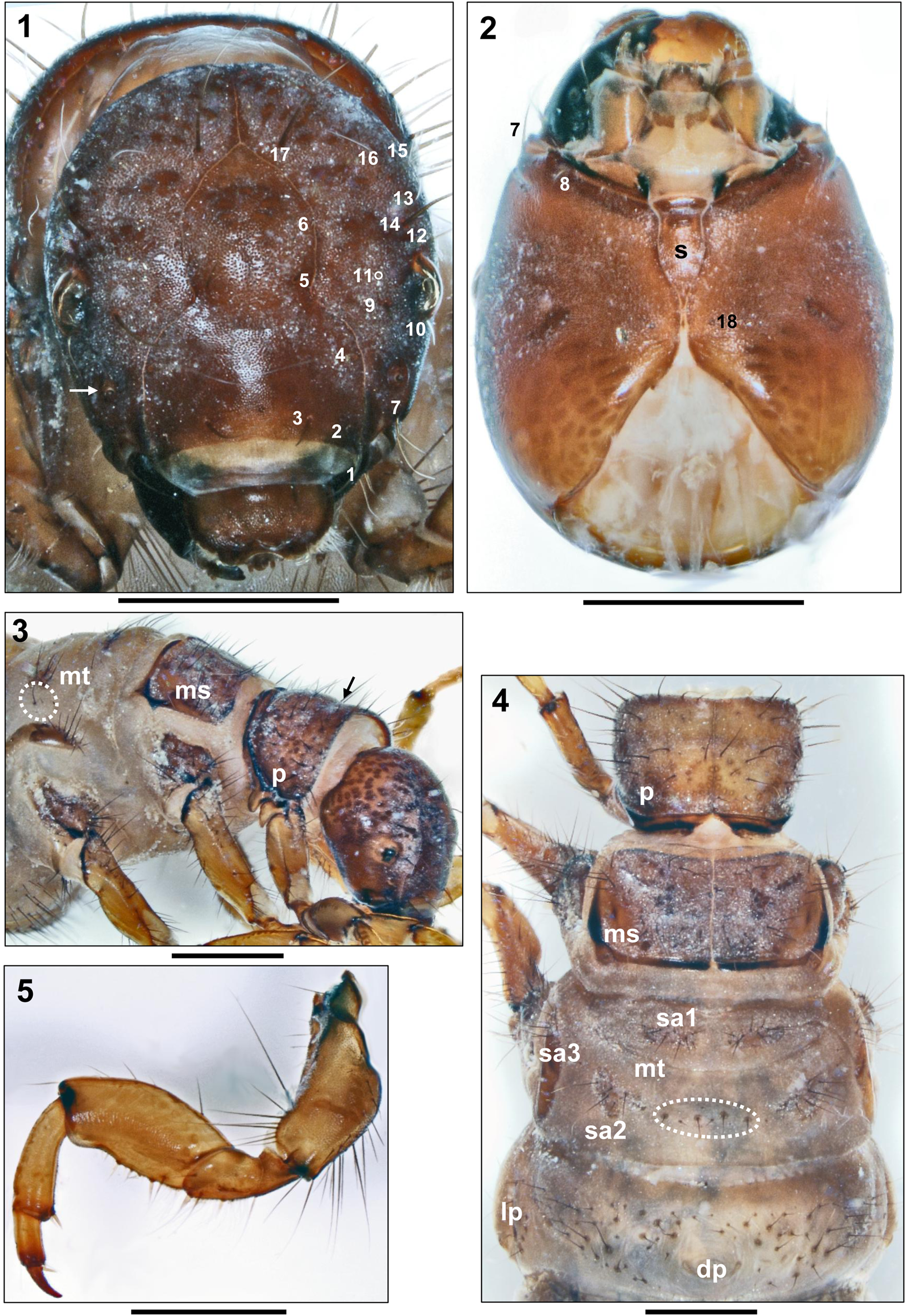
Head. Head capsule elongate, hypognathous, with dark brown coloration frontally and laterally, fading to orange brown posterolaterally and ventrally. With elliptical, dark brown muscle attachment spots on frontoclypeus and parietalia, strongly contrasting in color on posterior sections of parietalia ( Figs 1–3 View FIGURES 1–5 ). Large areas of head surface covered by spinules ( Fig. 1 View FIGURES 1–5 ). Whitish ring present around each eye ( Fig. 3 View FIGURES 1–5 ); eyes slightly protruding ( Fig. 1 View FIGURES 1–5 ). Head capsule with complete set of 18 pairs of primary setae ( Figs 1, 2 View FIGURES 1–5 ). Frontoclypeus bell-shaped, with deep central constriction ( Fig. 1 View FIGURES 1–5 ). Antennal bases roundish, situated halfway between eyes and anterior head margin ( Fig. 1 View FIGURES 1–5 , white arrow). Antennae short, each consisting of 1 short cylindrical base and 1 short flagellum. At each parietal, 10 dorsal and 2 ventral primary setae present, with setae #9 and 14 long and conspicuous ( Fig. 1 View FIGURES 1–5 ). Frontoclypeus with 6 pairs of primary setae, 3 of them along anterior border (#1–3). Labrum dark brown, with setal brush and primary setae #1–3 at anterolateral margins and primary setae #4–6 on dorsal area ( Fig. 1 View FIGURES 1–5 ). Ventral apotome wedge-shaped, orange brown with dark brown anterior border; postgenal suture approximately 18% of apotome length ( Fig. 2 s View FIGURES 1–5 ). Mandibles black, each with 5 terminal teeth along its edge (3 of them visible in Fig. 2 View FIGURES 1–5 ); in addition, ridges present in central concavity ( Fig. 2 View FIGURES 1–5 ).
Thorax. Pronotum ( Fig. 4 p View FIGURES 1–5 ) completely covered by two thick sclerites meeting in a straight mid-dorsal ecdysial line; pronotal sclerites light brown, with strongly-contrasting dark brown ovoid muscle attachment spots; surface finely granulated ( Figs 3, 4 View FIGURES 1–5 ). Its posterior and posterolateral margins thickened and bent dorsad, thereby creating semicircular groove with black stripes ( Fig. 3 View FIGURES 1–5 ). With earlike posterolateral projection ( Fig. 3 View FIGURES 1–5 ). Pronotal transverse groove at end of anterior 3rd distinct and with dark furrow ( Figs 3 View FIGURES 1–5 arrow, 4). Along anterior border three setal rows present: (1) dense fringe of short, curved, fine, yellow short setae; (2) widely-spaced, continuous row of intermediate curved, pale setae; and (3) widely-spaced, continuous row of long, straight, dark setae ( Figs 3, 4 View FIGURES 1–5 ). In total, 55–62 dark setae of varying lengths distributed over each pronotal half. Prosternal horn present ( Fig. 8 h View FIGURES 6–12 ). Median brown central prosternite conspicuous, outlined like a flying bird’s silhouette: slightly pointed at anterior center, with winglike lateral sections and fan-shaped posterior extension ( Fig. 8 s View FIGURES 6–12 ). Mesonotum ( Fig. 4 View FIGURES 1–5 ms) completely covered by 2 light brown sclerites meeting in straight mid-dorsal ecdysial line; with dark brown muscle attachment spots. Their posterolateral and posteromedian margins strongly sclerotized and with black margins ( Fig. 4 View FIGURES 1–5 ). Counts for mesonotal setae on each sclerite are as follows (nomenclature sensu Wiggins 1996): anterior setal group sa 1: 5–7, posterior group sa 2: 10–12, lateral group sa 3: 12–16. Metanotum ( Fig. 4 View FIGURES 1–5 mt) partially covered by 3 pairs of light brown sclerites with dark brown muscle attachment spots. Anterior metanotal sclerites (sclerites of setal area 1, or sa 1 sensu Wiggins 1996) narrow and transversally elongate; their intermediate separation distinctly larger than length of each of them ( Fig 4 View FIGURES 1–5 sa 1); with 12–16 setae per sclerite. Posterior metanotal sclerites (sclerites of setal area 2, or sa 2 sensu Wiggins 1996) broadly triangular, with 12–15 setae per sclerite and dense group of 8– 10 setae between them ( Fig. 4 View FIGURES 1–5 , dotted oval). Lateral metanotal sclerites (sclerites of setal area 3, or sa 3 sensu Wiggins 1996) narrow, crescent-shaped, each with 12–15 setae concentrated at anterior third of sclerite ( Fig. 3 View FIGURES 1–5 ). With small groups of 4–6 setae between each lateral (sa 3) and posteromedian sclerite (sa 2) ( Fig. 3 View FIGURES 1–5 , dotted oval). Pleurae light brown, with black pleural suture; epimera of 2nd and 3rd legs with digiform ventral process bearing one or more setae. Legs light brown, with dark brown muscle attachment spots and numerous setae on coxae, trochanters, and femora ( Figs. 5–7 View FIGURES 1–5 View FIGURES 6–12 ); tibiae and tarsi with only small number of setae. Femora of 2nd and 3rd legs with several proximodorsal setae ( Figs 6, 7 View FIGURES 6–12 arrows). Coxa, femur, and tibia of each foreleg much wider than those of mid- and hind legs Fig. 5 View FIGURES 1–5 ). Additional setae lacking on anterior and posterior faces of mid- and hind femora ( Figs 6, 7 View FIGURES 6–12 ). Ventral trochanteral brush at distal section of each trochanter present on all legs. Proximal section of all trochanters with only one primary seta each. Rows of minute spines present along ventral edges of femora; pairs of ventral-edge setae pale on fore femora, but dark on mid- and hind femora. Tibiae of all legs with 2 pale, stout subapical spines, tarsi with 2 long subapical setae; tarsal claws sickle-shaped, with stout basal spines ( Figs 5–7 View FIGURES 1–5 View FIGURES 6–12 ).
Abdomen. Abdominal segment I with 1 dorsal ( Fig. 4 View FIGURES 1–5 dp) and 2 lateral fleshy protuberances ( Figs 4 View FIGURES 1–5 lp, 9). Dorsal setal areas sa 1, sa 2, and sa 3 (sensu Wiggins 1996) fused, thereby creating continuous transverse row of 65– 75 setae on distinct medium brown basal sclerites anterior to dorsal protuberance ( Figs. 4 View FIGURES 1–5 , 9 View FIGURES 6–12 ); without setae posterior to dorsal protuberance ( Fig. 4 View FIGURES 1–5 ). Lateral protuberance with large brown, smooth posterior sclerite without setae, but with 1–3 holes, usually 2 ( Fig. 9 View FIGURES 6–12 , dotted oval). Lateral protuberance setae consisting of dorsal group of 12–16 setae and single ventral seta. On abdominal sternum I, setal areas sa 1 and sa 2 fused, creating continuous central field of approximately 110 setae, about half of them with medium-sized, brown basal sclerites widely separated from each other; setal areas sa 3 situated ventral of lateral protuberances and separated from sa 1 and sa 2, consisting of 15–20 setae, about half of them with medium-sized, brown basal sclerites ( Fig. 10 View FIGURES 6–12 ). Abdominal segments II–VII with 2 dorsal setae each. On abdominal dorsum VIII, number of posterodorsal setae (pds) typically 10, with 6 long and remainder short ( Fig. 12 View FIGURES 6–12 pds). Only 1 posterolateral seta ( Figs 11, 12 View FIGURES 6–12 , black arrows) and 2 tiny ventral setae present on each half of abdominal dorsum IX. Light brown abdominal tergite IX semicircular, with medium brown muscle attachment spots ( Fig. 12 View FIGURES 6–12 ); along its posterior border, 7–9 long and several shorter setae present, 1 of these long setae having position of central intermediate seta. Anal prolegs of limnephilid type, yellowish brown, with medium brown muscle attachment spots. Lateral sclerite ( Fig. 11 View FIGURES 6–12 ls) with 5 dark dorsal and row of 5 dark ventral setae, 3 of latter very strong and prominent. Ventral sole plate ( Fig. 11 View FIGURES 6–12 vsp) with black dorsal stripe and single anterior seta. Anal claw basal sclerite Fig. 11 View FIGURES 6–12 ac) with 3 tiny pale ventral setae and 3 darker dorsal setae; anal claw dark brown, with 1 small dorsal accessory hook ( Fig. 11 View FIGURES 6–12 ).
All gills single filaments ( Fig. 13 View FIGURES 13–19 ). Dorsal gills present at most from segment II (presegmental position) to segment VII (presegmental position). Ventral gills ranging from segment II (presegmental) to segment VII (postegmental). Lateral gills present from segment II (presegmental) to segment IV (postsegmental position). Lateral fringe extending from anterior border of abdominal segment III ( Fig. 14 View FIGURES 13–19 lf) to end of abdominal segment VIII. With large oval chloride epithelia on abdominal sterna II–VII ( Fig. 13 View FIGURES 13–19 , dotted oval). Abdominal segments II– VII with 3–7 forked lamellae immediately dorsal of lateral fringe ( Fig. 14 View FIGURES 13–19 fl).
Case. Larval case 27.5–33.0 mm long (n = 2), very slightly curved and almost untapered (width at anterior opening 7.0– 7.5 mm and at posterior opening 5.3–7.8 mm). Cases consist of mix of mostly detrital particles of unequal size arranged longitudinally and sometimes few sand grains; typically also including fibers, conifer needles or longitudinal detritus protruding from posterior end ( Fig. 15 View FIGURES 13–19 ).


No known copyright restrictions apply. See Agosti, D., Egloff, W., 2009. Taxonomic information exchange and copyright: the Plazi approach. BMC Research Notes 2009, 2:53 for further explanation.