
Heterofragilia hirsuta Nakamura & Child, 1991
Bamber, Roger N., 2004, with description of three new species, Zootaxa 458, pp. 1-12 : 5-6
|
publication ID |
https://doi.org/ 10.5281/zenodo.157995 |
|
DOI |
https://doi.org/10.5281/zenodo.6271528 |
|
persistent identifier |
https://treatment.plazi.org/id/03B12173-FFE6-6D35-8465-FD5BFB5C5402 |
|
treatment provided by |
Plazi |
|
scientific name |
Heterofragilia hirsuta Nakamura & Child, 1991 |
| status |
|
Heterofragilia hirsuta Nakamura & Child, 1991 ( Fig 2 View FIGURE 2 )
Nakamura & Child, 1991, 21–24; fig. 9.
Remarks: The genus Heterofragilia Hedgpeth, 1943 is known from five species, H. fimbriata Hedgpeth, 1943 , the type species, from one specimen collected off Martinique at 870.5 m; H. amica Stock, 1954 from two specimens from 165 to 203 m off Kyushu, Japan, ( Stock 1954; Utinomi 1955); H. major Stock, 1986 , from one specimen from St Vincent, W. Indies, at 1053–1540 m; H. brevicauda Stock, 1991 , from off New Caledonia, W. Pacific at 650–680 m, and H. hirsuta , two males and a female from Sagami Bay, Japan at 454– 495 m. All known specimens of the species other than H. hirsuta were female.
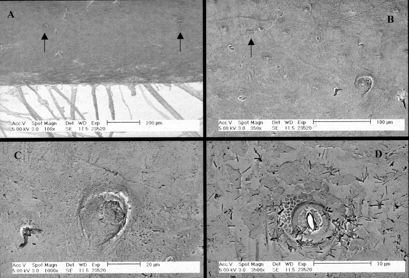
While these species are generally quite similar, the present specimens agree with H. hirsuta as described by Nakamura & Child (1991), the only species for which a male has been described previously. In the male, the oviger articles 7 to 10 form a strigilis, bearing stout but simple ventral spines, numbering 10, 6, 6 and 6 per article respectively in the present material (9, 5, 4, 6 in the holotype). The male genital pore is situated on a ventrodistal spur on the second coxa of legs 3 and 4 only. The cement gland is evident as a whitened swelling along the ventral twothirds of the femur of the fourth leg only. Nakamura and Child (1991) describe the cement gland opening through ‘many tiny slitlike pores in the integument surface’ (without a figure). SEM observation of the fourth right leg of the present male specimen revealed that, on the ventral proximal margin of the naked integument covering the cement gland, there were two small (20 m diameter) capped pores ( Fig. 2 View FIGURE 2 A, 2C) within a field of very small (5 m diameter) secretory pores ( Figs. 2 View FIGURE 2 B, 2D) which were scattered over much of the integument surface. These two cement gland pores were not evident under light microscopy.
The present specimens are from slightly deeper and further south than the types.
No known copyright restrictions apply. See Agosti, D., Egloff, W., 2009. Taxonomic information exchange and copyright: the Plazi approach. BMC Research Notes 2009, 2:53 for further explanation.