
Cacopsylla ( Hepatopsylla ) cinereosignata, Luo, Xinyu, Li, Fasheng, Ma, Yanfang & Cai, Wanzhi, 2012
|
publication ID |
https://doi.org/10.5281/zenodo.213975 |
|
publication LSID |
lsid:zoobank.org:pub:2C43EA7B-94F7-4133-9070-21AC4A8AB734 |
|
DOI |
https://doi.org/10.5281/zenodo.3504159 |
|
persistent identifier |
https://treatment.plazi.org/id/03B1723D-FFE7-FF87-FF60-FB1C501171F1 |
|
treatment provided by |
Plazi |
|
scientific name |
Cacopsylla ( Hepatopsylla ) cinereosignata |
| status |
sp. nov. |
Cacopsylla ( Hepatopsylla) cinereosignata View in CoL sp. n.
( Figs 33–43 View FIGURES 33 – 40 View FIGURES 41 – 43 )
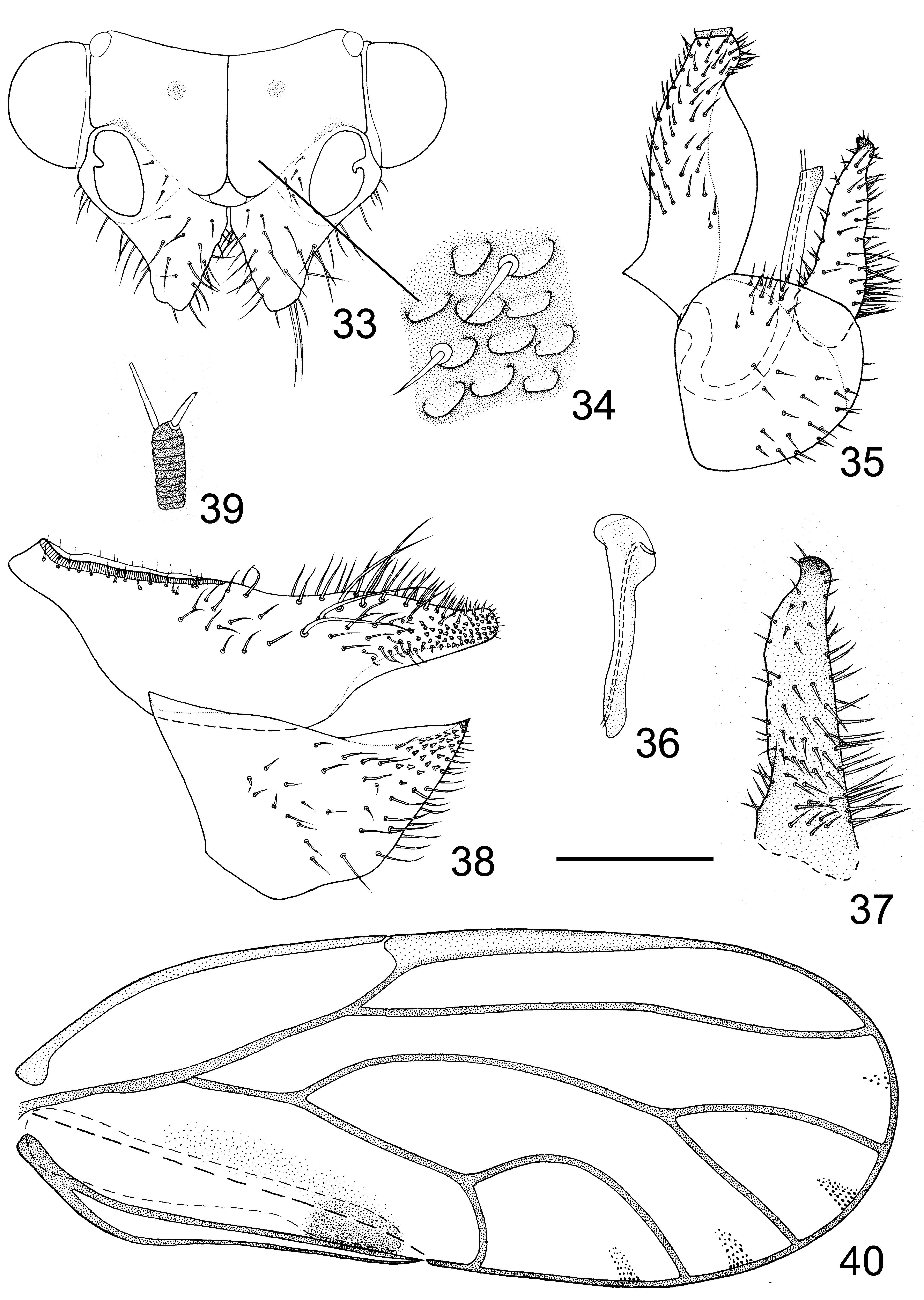
Adult. Coloration: Body brown. Vertex light brown, fore angles and mid of the posterior margin yellow; discal foveae black. Genal process yellowish brown, darker apically. Antennae dark brown, with black apices on segments III–VI and segments VII–X entirely black. Ocelli orange; compound eyes red. Thoracic terga yellow to brown, with black stripes, median stripe in mesoscutum brown. Legs yellow, dorsal surface of femora irregularly blackened, apical tarsal segment dark brown. Fore wing ( Fig. 40 View FIGURES 33 – 40 ) transparent, veins black, C+Sc and pterostigma light yellow; marking near apex of claval suture light black, with margin obscure; centre of cell cu2 with one obscure light grey oblong marking, successive with the marking near apex of claval suture. Abdominal segments blackish orange to entirely black. Male terminalia black. Female proctiger black in the basal half, yellow in the apical half.
Structures: Body glabrous and relatively slender. Head moderately inclined from longitudinal body axis, narrower than mesoscutum. The section of border between vertex and gena above the antennal fossa relatively concave, suppressing the long and narrow area of vertex above it, forming a shallow gap nearly parallel with lateral margin of vertex ( Fig. 33 View FIGURES 33 – 40 ). Discal foveae deep. Vertex ( Fig. 34 View FIGURES 33 – 40 ) finely sculptured with microscopic setae and scaly micro structures that are near elliptical, relatively prominent, and moderately spaced from each other. Genal processes ( Fig. 33 View FIGURES 33 – 40 ) cone-shaped, slightly shorter than vertex along median suture and relatively widely divergent, covered with long setae; apex relatively acute and slightly raised upward. Antenna long and relatively thick, terminal setae ( Fig. 39 View FIGURES 33 – 40 ) not as long as each other, the longer one about twice as long as the shorter one, and about as long as antennal segment X. Metatibia with well developed basal spine, apical spurs arranged in (1+3+1). Fore wing ( Fig. 40 View FIGURES 33 – 40 ) oblong oval, widest at apical 1/4; pterostigma relatively long, ending in the apical 1/3 of cell r1; cell cu1 relatively larger, turning of vein Cu1a relatively smooth; surface spinules present only in cell cu2, with the field seemingly guided along the claval suture; 4 sets of radular spinules present in cells r2, m1, m2 and cu1, in r2 less developed and varies in size among individuals.
Male terminalia relatively small. Proctiger ( Fig. 35 View FIGURES 33 – 40 ) simple and slightly arched, covered with short setae. Paramere ( Fig. 35 & 37 View FIGURES 33 – 40 ) lamellar and slender, anterior margin expanded into one thin and narrow extension, posterior margin slightly sinuate; apex tooth-shaped, moderately inflexed and projecting cephalad; erect setae present in both inner and outer surface, in anterior margin rather short and sparse, in basal half of inner surface and posterior margin especially long and dense. Apical dilatation ( Fig. 36 View FIGURES 33 – 40 ) of aedeagus rounded in the tip without forming a hook; sclerotised end tube of ductus ejaculatorius moderately curved apically and slightly projecting beyond the dorsal margin of apical dilatation. Subgenital plate ( Fig. 35 View FIGURES 33 – 40 ) near triangular in profile, with several setae that vary in length in the dorsal margin and dense short setae in the ventral surface.
Female terminalia ( Fig. 38 View FIGURES 33 – 40 ) short. Proctiger convex dorsally, with a shallow transverse gap in the mid, lying cephalad to the convex; setae of varied length present in dorsal-lateral surface, longest at top of the convex; apex blunt; laterally and apex of the apical 2/3 of apical part covered with moderately dense peg setae, the involved field completely surrounded by fields of short setae. Ventral surface of subgenital plate sparsely covered with short setae and peg setae.
5th instar nymph. Coloration: For specimens preserved in absolute ethanol and not dissected. Body general color yellow. Sclerites yellow to dark brown. Compound eyes crimson. Apical 2/3 of antennal segment 7 black.
Structures: Body oblong oval. Dorsal aspect ( Fig. 41 View FIGURES 41 – 43 ) unevenly covered with capitate setae that vary in length in general. Ventral surface of abdomen ( Fig. 41 View FIGURES 41 – 43 ) covered with simple setae that are longer laterally. Micro spinules present in both dorsal and ventral aspects, fields as shown in Fig. 41 View FIGURES 41 – 43 ; in dorsal aspect short, lamellar and multicuspid; in ventral aspect long, spinous and unicuspid. Ocular seta ( Fig. 41 View FIGURES 41 – 43 ) capitate and relatively long. A short capitate seta ( Fig. 41 View FIGURES 41 – 43 ) present behind compound eye in submargin of dorsal surface. Antenna ( Fig. 41 View FIGURES 41 – 43 ) slender, 7-segmented, with a rhinarium on apices of segments 3 and 5, two rhinaria on segment 7. A sclerite with a spiracle present in ventral surface between praecoxa and mesocoxa, and a sclerite with a spiracle present in ventral surface between mesocoxa and metacoxa ( Fig. 41 View FIGURES 41 – 43 ). Fore wing pad with a line of 9–12 relatively short capitate setae along outer margin, hindwing pad with two capitate setae in distal angle ( Fig. 41 View FIGURES 41 – 43 ). Dorsal surface of mesotibia with 1 long and 1 relatively short capitate setae; dorsal surface of metatibia with 1 long capitate setae ( Fig. 41 View FIGURES 41 – 43 ). Tarsal arolium ( Fig. 43 View FIGURES 41 – 43 ) petiolate and fishtail, relatively narrow apically. 2+2 lateral free sclerites ( Fig. 41 View FIGURES 41 – 43 ) bearing a spiracle each present in ventral surface of abdomen. Caudal plate ( Fig. 42 View FIGURES 41 – 43 ) relatively large, with a pair of spiracle on bilateral sides of circum-anal pore ring. Outer circum-anal pore ring ( Fig. 42 View FIGURES 41 – 43 ) oval, with anterior margin sharply depressed, posterior margin weakly depressed and lateral margin not depressed. Inner circum-anal pore ring ( Fig. 42 View FIGURES 41 – 43 ) generally following the shape of the outer. Ventral surface of caudal plate ( Fig. 42 View FIGURES 41 – 43 ) with 2+2 simple setae right anterior to outer circum-anal pore ring, 2 simple setae within the suture, a hemicycle row of simple setae from around the spiracles to anterior of anal suture, a series of simple setae near anterior margin and a series of simple setae near posterior margin. Abdominal margin ( Fig. 41 View FIGURES 41 – 43 ) decorated with 7 pairs of long and 3 pairs of short capitate setae.
Material examined. Holotype: male, dry mounted, China, Gansu, Zhujiahetan, Sanshilipu, Hezheng, on Pyrus ussuriensis , 10.ix.2011, Ma Yanfang.
Paratypes: 8 male, 11 female with same data as holotype, along with numerous nymphs.
Non-paratypic specimens: Additional specimens from the same series are preserved in absolute ethanol.
Etymology. From Latin, cinereus = grey, signatus = marked. This species is named after its obscure light grey oblong marking in cell cu2.
Remarks. This species displays conspicuous intraspecific variation in the shape of cell cu1, which in some individuals is taller than in Fig. 40 View FIGURES 33 – 40 , exceeding the shape of that of C. chinensis summer form ( Fig. 28 View FIGURES 20 – 29 ).


No known copyright restrictions apply. See Agosti, D., Egloff, W., 2009. Taxonomic information exchange and copyright: the Plazi approach. BMC Research Notes 2009, 2:53 for further explanation.
|
Kingdom |
|
|
Phylum |
|
|
Class |
|
|
Order |
|
|
SuperFamily |
Psylloidea |
|
Family |
|
|
Genus |