
Echinoderes zacharyi, Kennedy & Sørensen & Landers, 2022
|
publication ID |
https://doi.org/ 10.1016/j.jcz.2022.09.002 |
|
publication LSID |
lsid:zoobank.org:pub:97E7ECBB-8B9B-4CD8-83B8-7930EC5C0D41 |
|
persistent identifier |
https://treatment.plazi.org/id/03B1878F-8218-FF70-FCB0-FB9393F80C04 |
|
treatment provided by |
Felipe |
|
scientific name |
Echinoderes zacharyi |
| status |
|
Genus Echinoderes View in CoL Clapar`ede, 1863
Species Echinoderes zacharyi sp. nov.
Zoobank codes:
urn:lsid:zoobank.org:act:57671314-AF1F–41FC-BCE7-0E80583670E3
urn:lsid:zoobank.org:pub:97E7ECBB-8B9B-4CD8-83B8-7930EC5C0D41 .
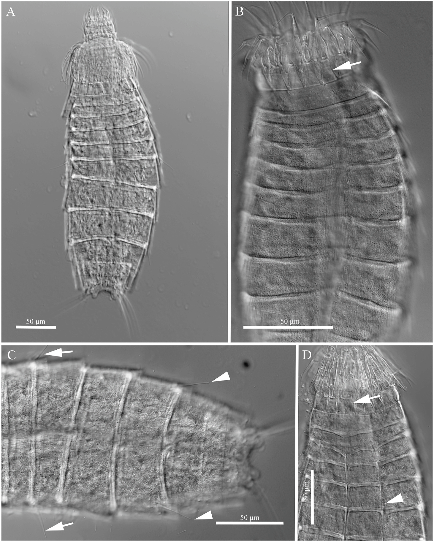
Figs. 2–6 View Fig View Fig View Fig View Fig View Fig .
Synonymy. Echinoderes coulli group – Landers et al. 2020: page 496; Table III.
Material. Holotype: Adult male collected at Station 09–2020 from sediment on March 9, 2020 at 15.4 m (30.2911 ◦ N, 88.0330 ◦ W), and deposited in the Natural History Museum of Denmark (Copenhagen) under catalogue number NHMD–1174539. Three paratype specimens from the nGOM ( NHMD –1174540 - 1174542) include adult females, one collected from the same site as the holotype ( NHMD –1174540), and two at station 00–2020 (30.2654 ◦ N, 88.0733 ◦ W), at a depth of 4.46 m ( NHMD –1174541 and 1174542). All type material was fixed in 5–10% buffered formalin and mounted with Fluoromount G ®. One non-type specimen ( SEM) was collected on March 16, 2018 in the nGOM at site 10–2018 (30.1354 ◦ N, 88.0768 ◦ W), at a depth of 16.3 m. Five additional non-type specimens were collected from the sGOM, which came from the two most-shallow locations that were sampled in Mexican waters (stations LT1 and LT2). This material includes four non-type specimens (1 SEM, 3 LM) collected on August 4, 2015 at site LT1 (18.8484 ◦ N, 92.0705 ◦ W) at 16m depth, and one non-type specimen (LM) collected on August 4, 2015 at site LT2 (19.0564 ◦ N, 92.1245 ◦ W) at 21m depth. GoogleMaps
Diagnosis: Echinoderes without acicular spines, except for lateral terminal ones. Midlateral and lateroventral tubes on segment 5; midlateral and lateral accessory tubes on segment 8. Large oval sublateral sieve plate on segment 9. Males with 3 sublateral penile spines, with the lateral-most spine twice as long as the others. Females without lateral terminal accessory spines. Segments 10 and 11 retractable into segment 9.
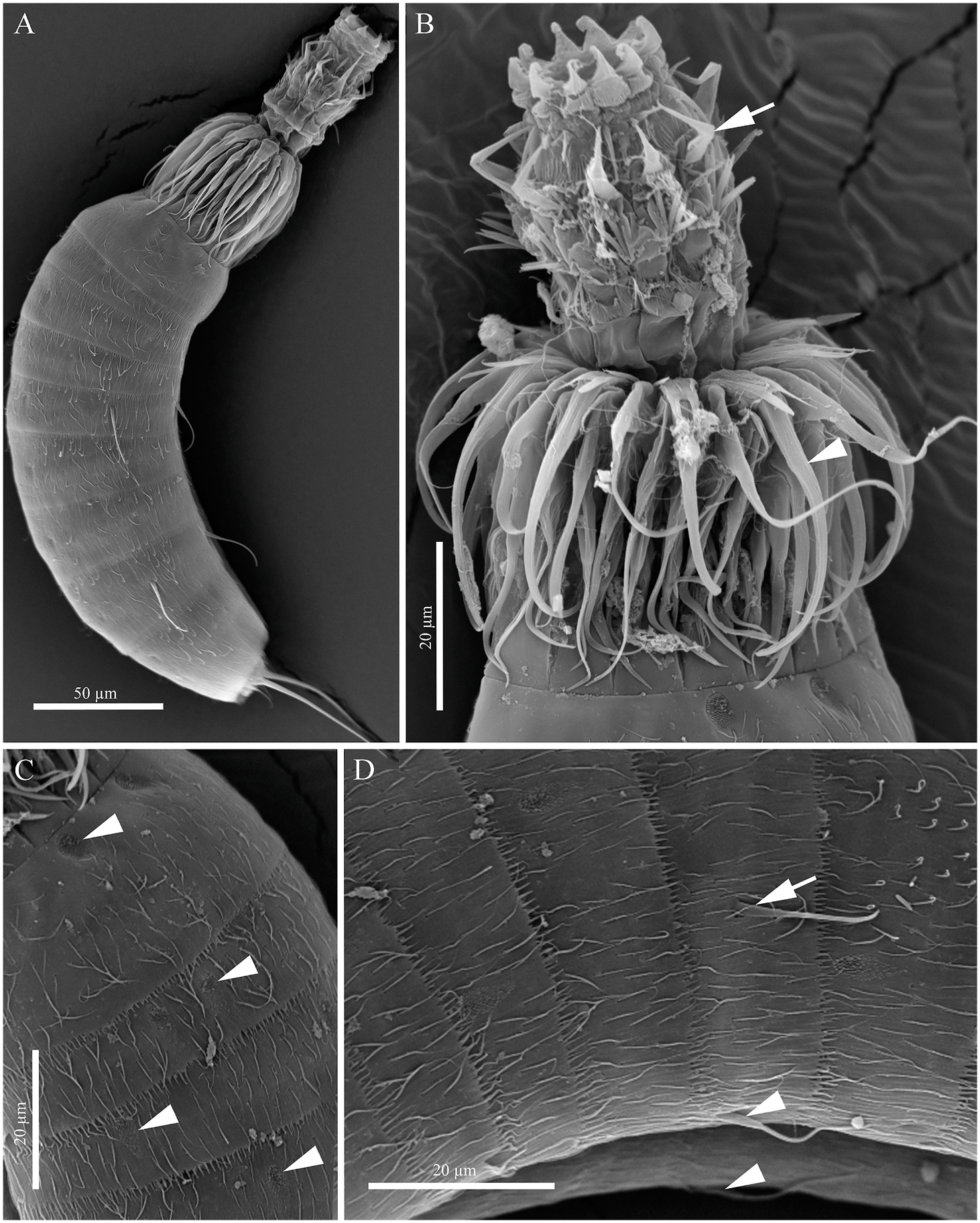
Description: Adults with head, neck, and trunk, with eleven trunk segments ( Figs. 2–6 View Fig View Fig View Fig View Fig View Fig ). Trunk length (segments 1–11) ranging from 214 to 265 μm, measured from anterior-most location on segment 1 to the posterior tip of the tergum (measured from animals with segments 10 and 11 retracted into segment 9). All specimens had segments 10 and 11 retracted into segment 9. Complete measurements are listed in Table 2. Location of glandular cell outlets, sensory spots, spines, and tubes are listed in Table 3.
The head has a retractable mouth cone and an introvert composed of scalids ( Figs 3A and B View Fig ; 5A, B View Fig ). The mouth cone has a ring of 9 outer oral styles, each with a proximal portion which articulates with an elongated distal tip, with the total length of the two approximately 17 μm. A fringe with spikes is located at the base of each outer oral style. The introvert sectors are defined by 10 primary spinoscalids in the first ring. The primary spinoscalids are composed of a basal sheath and a distal end piece. The distal end piece of each primary spinoscalid is elongated and extends posteriorly to segment 1. Six rings of spinoscalids are visible by SEM.
The neck has 16 placids ( Figs. 2A and B View Fig ; 3B,D View Fig ). The midventral placid is the largest, measuring 15 μm in length and 16 μm in width, with the remaining placids measuring 10 μm in width at their bases. Trichoscalid plates are narrow, but otherwise well developed ( Fig. 2 View Fig ).
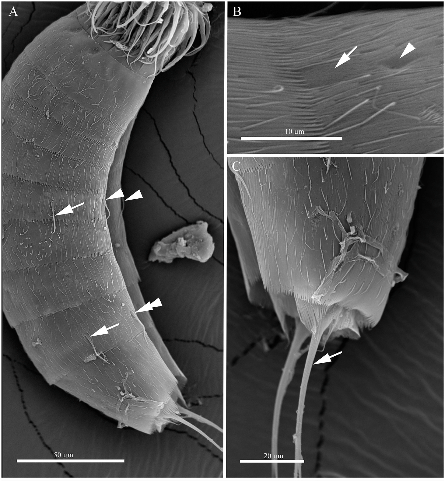
Segment 1 ( Figs. 2A and B View Fig ; 3A, B, D View Fig ; 5A, B, C View Fig ; 6A View Fig ) is a complete cuticular ring. Sensory spots are located at the anterior margin of the segment, in subdorsal and laterodorsal positions. Sensory spots are located in a circular indentation with fine, stubby hairs. Glandular cell outlets type 1 are located anteriorly in the segment, in the middorsal and lateroventral position. Cuticular hairs are evenly distributed. Pectinate fringe with delicate filiform extensions transitioning from longer extensions (3 μm) along the ventral surface to shorter extensions (1.2 μm) along the dorsal surface, with the transition occurring approximately at the midlateral position.
Segment 2 ( Figs. 2A and B View Fig ; 3A, B, D View Fig ; 5A, C, D View Fig ; 6A View Fig ) is a complete cuticular ring. Two pairs of sensory spots are located in tandem in laterodorsal positions, and a single pair is present in the ventromedial position. Sensory spots are round at the anterior end, and posteriorly form an oblong field of hairs that taper to a point, and have an approximate length of 6.6–8.0 μm. Glandular cell outlets not observed on this segment or subsequent segments. Cuticular hairs are evenly distributed. Pectinate fringe as on the preceding segment and on subsequent segments 3–6. Pachycyclus of the anterior segment of regular thickness.
Segment 3 ( Figs 2A and B View Fig ; 3A, B, D View Fig ; 5A, C, D View Fig ; 6A View Fig ) and remaining segments consist of one tergal and two sternal plates. Sensory spot shape on this and subsequent segments are consistent with segment 2; sensory spots located in the subdorsal and sublateral positions. Pachycyclus of the anterior segment disrupted along margins at the middorsal, midventral, and at the tergosternal junctions. Cuticular hairs evenly distributed across tergal and sternal plates of segment with a hairless midventral area on this and segments 4–10.
Segment 4 ( Figs 2A and B View Fig ; 3A, B, D View Fig ; 5A, C, D View Fig ; 6A View Fig ) with sensory spots located in the laterodorsal position. Pachycyclus as on preceding segment.
Segment 5 ( Figs 2A and B View Fig ; 3A–D View Fig ; 5A, D View Fig ; 6A View Fig ) with tubes in midlateral and lateroventral positions; tubes positioned medially in the segment. Tube morphology consists of a flattened basal portion that is wider than the distal portion (a cylindrical distal portion extends for approximately 2/3 of the length of the tube). Sensory spots located in subdorsal and ventromedial positions. Pachycyclus as on preceding segment.
Segment 6 ( Figs 2A and B View Fig ; 3A–D View Fig ; 4A View Fig ; 5A, D View Fig ; 6A View Fig ) with sensory spots in subdorsal, sublateral, and ventromedial positions. Pachycyclus as on preceding segment.
Segment 7 ( Figs 2A and B View Fig ; 3A–C View Fig ; 4A View Fig ; 5A View Fig ; 6A View Fig ) with sensory spots in subdorsal, midlateral, and ventrolateral positions. Pectinate fringe with delicate filiform extensions uniform in length across the posterior margin, approximately 1.8 μm. Pachycyclus as on preceding segment.
Segment 8 ( Figs 2A and B View Fig ; 3A–C View Fig ; 4A View Fig ; 5A View Fig ; 6A View Fig ) with tubes in the midlateral and lateral accessory positions; tubes positioned medially in the segment. Sensory spots present in subdorsal and ventrolateral positions. Pachycyclus and pectinate fringe as on preceding segment.
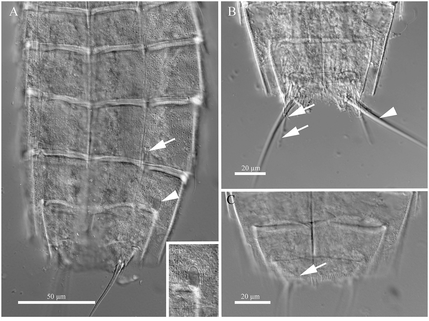
Segment 9 ( Fig. 2A–D View Fig ; 3A, C View Fig ; 4A–C View Fig ; 5A View Fig ; 6A–C View Fig ) with a pair of sensory spots in tandem in the subdorsal position. Additional sensory spots present in the sublateral and ventrolateral positions. Oval sieve plates positioned medially at the sublateral position; sieve plates measure 11 μm in length, corresponding to 18.7% of the total segment length. Sieve plate accompanied by a cuticular pore approximately 0.25 μm posterior to the plate. Pachycyclus, and pectinate fringe as on preceding segment.
Segment 10 ( Fig. 2A–D View Fig ; 4B, C View Fig ) nearly fully retracted into segment 9; sensory spots and glandular cell outlets not observed. Pectinate fringe with uniform filiform extensions.
Segment 11 ( Fig. 2A–D View Fig ; 3A, C View Fig ; 4A–C View Fig ; 5A View Fig ; 6A, C View Fig ) nearly fully retracted into segment 10; with lateral terminal spines that extend for 20–30% of the total trunk length. Sensory spots and glandular cell outlets not observed. Lateral terminal accessory spines not observed on females. Males with three pairs of sublateral penile spines, with one pair stout and extending well past the tergal and sternal posterior margins (when the last two segments are contracted into segment 9). Cuticular hairs sparsely distributed. Tergal extensions blunt and end in short narrowtipped hairs at the posterior margin. Posterior margin of the sternum with very short, rounded extensions on each side of the midline, with fine hairs on the edge.
Etymology. This species is named after the late Zachary J. Kennedy, brother of Madison C. Kennedy, first author of this publication.

No known copyright restrictions apply. See Agosti, D., Egloff, W., 2009. Taxonomic information exchange and copyright: the Plazi approach. BMC Research Notes 2009, 2:53 for further explanation.
|
Kingdom |
|
|
Phylum |
|
|
Class |
|
|
Order |
|
|
Family |
|
|
Genus |