
Meotipa picturata Simon, 1895
|
publication ID |
https://doi.org/ 10.11646/zootaxa.4344.3.9 |
|
publication LSID |
lsid:zoobank.org:pub:E93BAB87-ED61-4AA0-9F5B-DFDF84258978 |
|
DOI |
https://doi.org/10.5281/zenodo.6040715 |
|
persistent identifier |
https://treatment.plazi.org/id/03B1886A-FFC6-690A-FF02-66C39D38DB3A |
|
treatment provided by |
Plazi |
|
scientific name |
Meotipa picturata Simon, 1895 |
| status |
|
Meotipa picturata Simon, 1895 View in CoL
( Figs. 1A–J View FIGURE 1 , 2A–F View FIGURE 2 & 4A–D View FIGURE 4 )
Redescription. Deeleman-Reinhold (2009)
Material examined. 3♂ (ADSH 101671 A), 4♀ ( ADSH 101671 B) India, Kerala, Pathiramanal Island (9o37'07.11''N, 76o23'04.95''; 4 m. alt.) from foliage by hands ; 20 April. 2015, 9 June. 2015, 11 July. 2015 and 27 Nov. 2015; Jobi M.J, Pradeep M.S & Prajapati D.A.
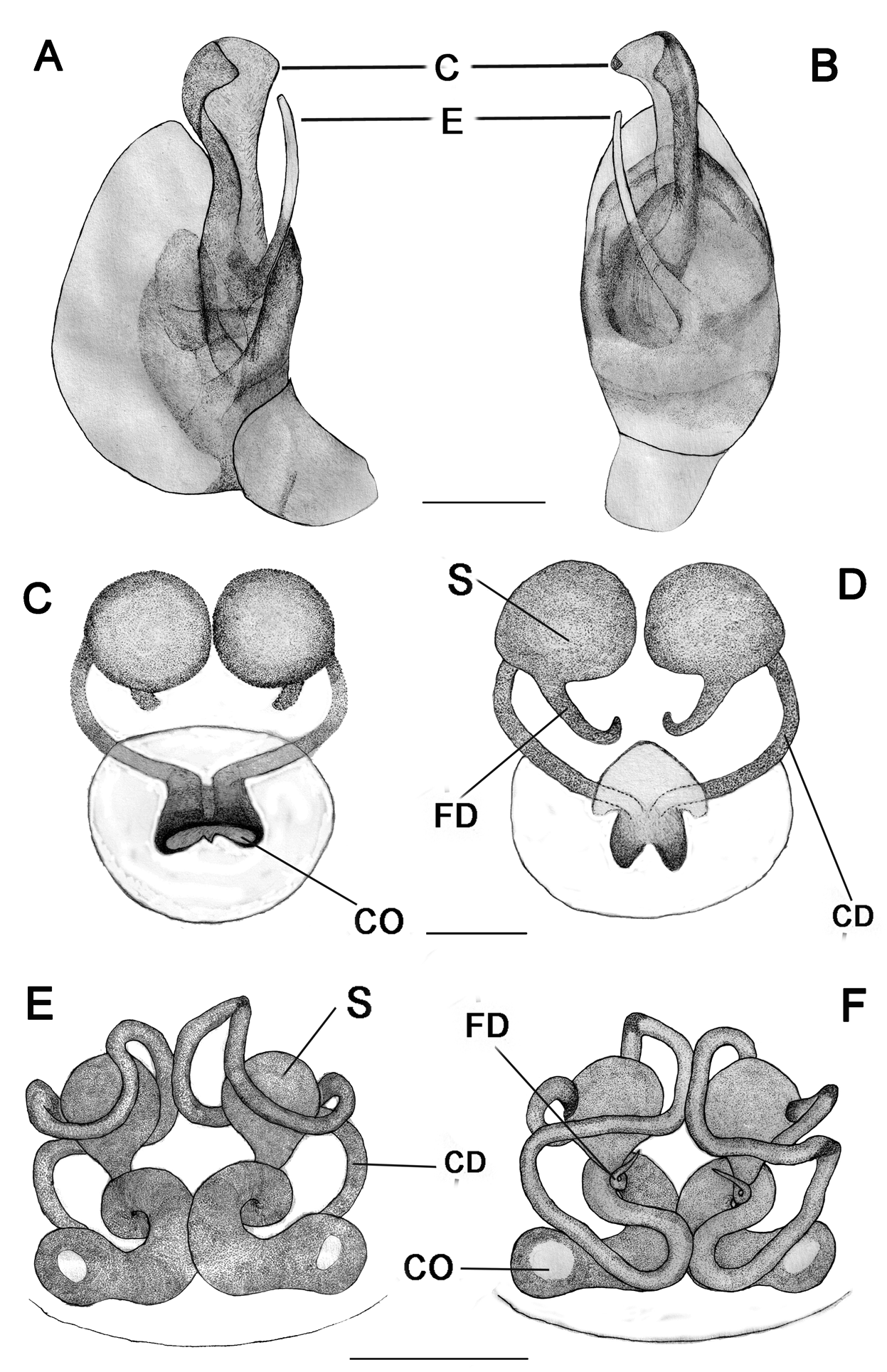
Diagnosis. M. picturata is more similar to M. thalerorum Deeleman-reinhold, 2009 with which shares characters such as absence of flattened abdominal spines ( Fig. 1A View FIGURE 1 ); abdomen with faint grey spots ( Fig. 1A & C View FIGURE 1 ); male palp with spoon shaped conductor ( Fig. 1G, H View FIGURE 1 & Fig. 4A, B View FIGURE 4 ); epigyne with deep round pit, in the middle of which a rod-shaped projection ( Fig. 1I View FIGURE 1 ). It can be distinguished from the former species by the transparent palp, broad conductor, flat with retrolaterally folded sides; embolus narrow, spine like with a blunt tip separated from the conductor ( Fig. 4A View FIGURE 4 ) (in M. thalerorum , conductor distally a membranous disc, distal edge partly aligned with chitinized arch. Embolus simple, straight, see in Deeleman-reinhold, 2009 Fig. 10 & 11); Fertilization duct basal to globular spermathecae, apically with a curve; widely placed copulatory duct ( Fig. 4C View FIGURE 4 ).
Description. Female in alcohol ( Figs. 1D–F, 1I –J View FIGURE 1 , 2A–F View FIGURE 2 , 4C–D View FIGURE 4 ): Prosoma front and back truncated; medially flat; Cephalic area elevated; clypeus broad, bulged out; All eyes nearly uniform in size, AME black, rest pearly white; Cephalothorax with reddish stripes, medially radiating streaks; Prosoma glabrous; Eye field, clypeus with few short hairs; Fovea broad, smooth with distinct depression and irregular ridges; Sternum rebordered, brownish with black patches; Maxillae with short hairs and without scopulae; labium short with small hairs; Pedipalp long; Tibia dorsally with lanceolate spines, long proximally, distally small with swelling, apicoventrally reddish; Tarsus with a pectinate claw; Opisthosoma triangular, very broad in lateral view, its dorsum provided with long alternate red and black stripes, caudal region knobbed extending downwardly towards spinnerets, provided with lanceolate spines. Venter transparent, some small patches posteriorly; Body length 5.60; Prosoma length 1.80, width (at middle) 1.80, height (at middle) 0.50; Opisthosoma length 3.80, width (at middle) 2.03, height (at middle) 2.80; Eye diameter: AME 0.097, ALE 0.095, PME 0.100, PLE 0.092; Eye interdistances: AME–AME 0.10, PME–PME 0.09, PLE–ALE contiguous; AME–ALE 0.04, PME–PLE 0.07, PME-AME 0.06; Clypeus height at AMEs 0.36, at ALEs 0.48; Measurements of legs & palp; Palp 2.08 [0.79, 0.27, 0.23, 0.79]; leg I 17.36 [6.20, 0.76, 3.40, 6.00, 1.00], II 11.13 [4.00, 0.50, 2.30, 3.60, 0.73], III 7.38 [2.70, 0.48, 1.50. 2.10, 0.60], IV 16.06 [6.00, 0.56, 2.80, 5.80, 0.90]; Leg formula 1423; Femur, patella & tibia with lanceolate spines distally; Tibia with reddish black spot distally; Leg segments except tibia with short, fine, dense hairs; Femur IV with a row of lanceolate spines distoventrally; Metatarsus I blackish distally; Epigyne ( Figs. 1I –J View FIGURE 1 & 4C–D View FIGURE 4 ): very small having a broad atrium medially with a rod or scape like structure narrowing apically and flattened; Copulatory openings depressed, contiguous, medially placed, deeply rest inside the atrium ( Fig. 4D View FIGURE 4 ); Copulatory ducts narrow, moderately long, tube like without convolution, having a single opening; Spermathecae globular ( Fig. 4D View FIGURE 4 ); Fertilization duct relatively long, basal to spermathecae, proximal part confronting with an apical curve ( Fig. 4C View FIGURE 4 ).
Males in alcohol ( Figs.1A–C, 1G–H View FIGURE 1 , 4A–B View FIGURE 4 ). Like the female, except by the following. Dwarf, nearly ¼th smaller than female, without characteristic lanceolate spines; Prosoma smooth, creamy, strongly truncated anteriorly, two short setae medially; Clypeus slightly bulged with a median longitudinal patch; Eye field wide, slightly elevated, some short hairs, eyes uniform sized, rose coloured appearance except bulged out black anterior medians; Fovea indistinct with longitudinal patch; Sternum heart shaped; maxillae, labium without scopulae; Body length 1.88; Prosoma length 0.82, width (at middle) 0.50, height (at middle) 0.36; Opisthosoma length 1.06, width (at middle) 0.48, height (at middle) 0.70; Eye diameter: AME 0.07, ALE 0.06, PME 0.06, PLE 0.06; Eye interdistance: AME–AME 0.04, PME–PME 0.05, PLE–ALE contiguous, AME–ALE 0.02, PME–PLE 0.01, PME- AME 0.02; Clypeus height at AMEs 0.07, at ALEs 0.12. Measurements of legs and palp: Palp 0.79 [0.23, 0.06, 0.17, 0.33]; Leg I 3.30 [1.30, 0.20, 0.10, 1.20, 0.50], II 2.65 [0.90, 0.10, 0.50, 0.75, 0.40], III 1.78 [0.60, 0.08, 0.30, 0.50, 0.30], IV 3.1 [1.10, 0.10, 0.70, 0.80, 0.40]; Leg formula 1423; Opisthosoma with irregular, alternate grey and white patches, slightly high, medially with indistinct humps; Caudal region lower, abruptly bent downwards, surface between caudal end and spinnerets concave with white patches; Legs with short, fine hairs, small grey spots ventrally; Palp ( Figs. 1G–H View FIGURE 1 & 4A–B View FIGURE 4 ): Simple, cymbium short with numerous setae; Tegulum small, without any apophysis (also in M. bituberculata and M. impatiens ); Conductor broad, flat with retrolaterally folded sides, originating distoretrolaterally directed at 12’O clock ( Fig. 4B View FIGURE 4 ); embolus long, narrow, spine like separated from the conductor, medially originate with a blunt tip, facing towards the apical part of the conductor ( Fig. 4A View FIGURE 4 ).
Variation: Spiders are notable for intra specific variation and it is proved that specimens collected from the same area show variation in their morphological and copulatory structures ( Malamel et. al, 2013). In our study also the different specimens collected from Pathiramanal Island show variation in the caudal region short to moderately long; white patches to yellow stripes on their opisthosoma ( Fig. 1 D–F View FIGURE 1 & Fig. 2A–F View FIGURE 2 ).
Distribution. India, Thailand, Indonesia. In India, known from Goa, Kerala and Tamil Nadu ( Fig 5 View FIGURE 5 ).


| ADSH |
Arachnology Division, Sacred Heart College |
No known copyright restrictions apply. See Agosti, D., Egloff, W., 2009. Taxonomic information exchange and copyright: the Plazi approach. BMC Research Notes 2009, 2:53 for further explanation.
|
Kingdom |
|
|
Phylum |
|
|
Class |
|
|
Order |
|
|
Family |
|
|
Genus |