
Protonemura alexidis, Vinçon & Launay & Reding, 2021
|
publication ID |
https://doi.org/10.11646/zootaxa.5061.3.2 |
|
publication LSID |
lsid:zoobank.org:pub:572805BF-0121-469D-8B0D-625DBEEE14EB |
|
DOI |
https://doi.org/10.5281/zenodo.5700083 |
|
persistent identifier |
https://treatment.plazi.org/id/CE147455-A56B-449A-8BBD-AEEE9E2B7C94 |
|
taxon LSID |
lsid:zoobank.org:act:CE147455-A56B-449A-8BBD-AEEE9E2B7C94 |
|
treatment provided by |
Plazi |
|
scientific name |
Protonemura alexidis |
| status |
sp. nov. |
Description of Protonemura alexidis View in CoL sp. n.
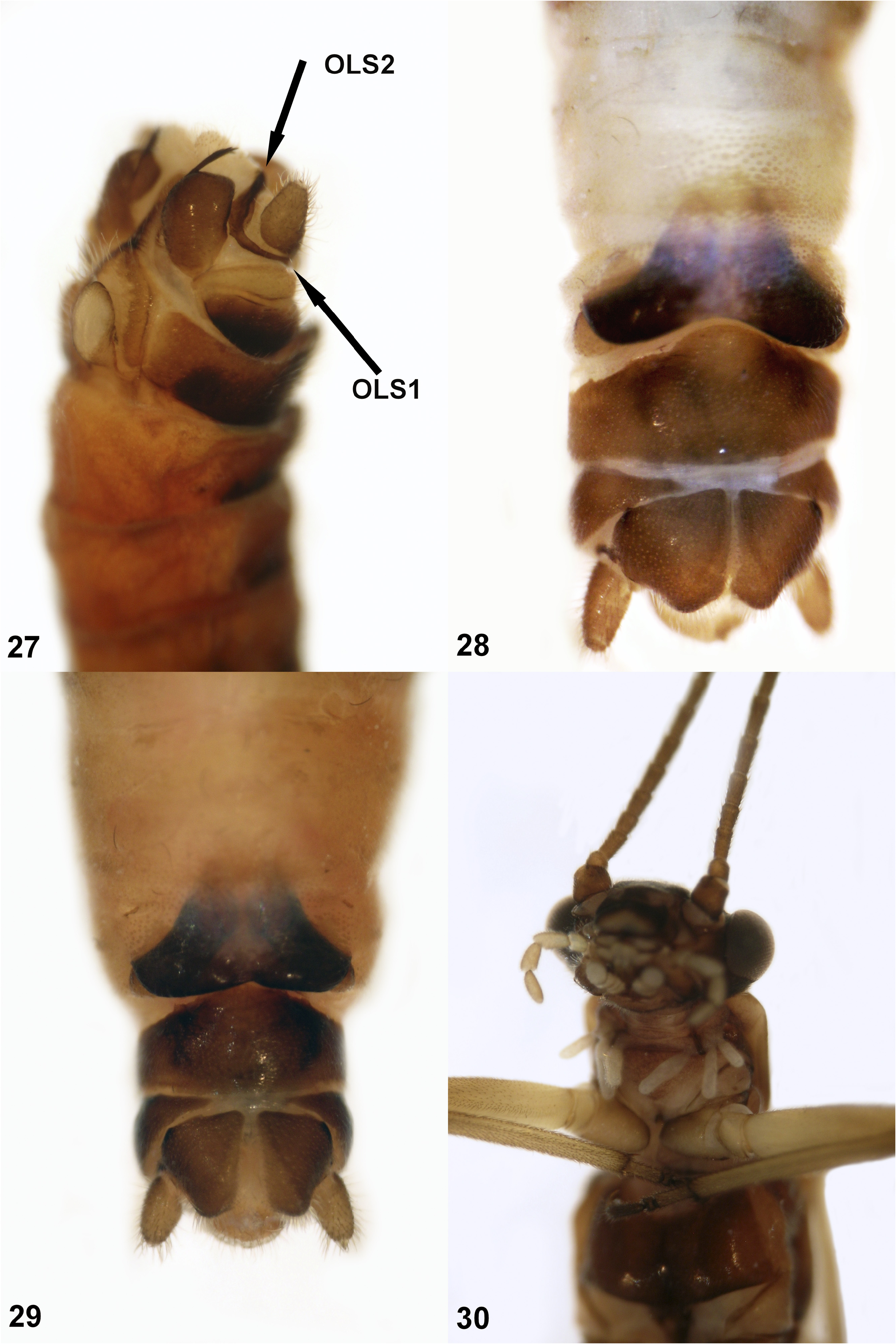
Males (Figs. 11–18). In dorsal view, tergites 8 and 7 with several rows of strong spines on each side of posterior margin, medially interrupted; tergite 6 with one to nine smaller spines in one or two rows on each side (Fig. 13). Hypoproct terminated by a finger-shaped expansion (Figs. 15, 17). Vesicle ovoid-shaped (Figs. 15, 17). Inner lobe of paraprocts thinly sclerotized and mostly hidden by hypoproct (Fig. 18). In ventral view, sclerotized base of median lobe of the paraprocts of adult males pea-shaped (Fig. 15) or sub-rectangular (Figs. 16–17). In lateral view, sclerotized base of median lobe of the paraprocts sub-rectangular (Figs. 16, 18); membranous field reduced, not extending over the length of the cercus (Figs. 15, 17). Insertion point of the sclerotized stem located medially on the side of the median lobe of the paraprocts (Figs. 15, 17). Sclerotized stem of the sclerotized median lobe of the paraprocts short, thick and slightly curved, not extending over the membranous field, and with several (three to four) strong apical spines (Figs. 11, 16, 18). Sclerite of the outer lobe of the paraprocts bifurcated, with a narrow basal branch turning around the cercus (= OLS1, Fig. 18), and a second, wider branch, located between the cercus and the membranous field of the median lobe (= OLS2, Fig. 18). Epiproct with a median widening (Figs. 11–12). Tip of epiproct swollen (Figs. 11–12). Epiproct with a U-shaped notch between the tip and the upper median part of the epiproct (Figs. 11–12). Tip of epiproct with two small protruding elongated spines pointing forward, in lateral (Figs. 11–12) and in dorsal (Fig. 14) views. Dorsal sclerites on each side of the epiproct bifurcated (Figs. 11–12); the dorsal branch of the sclerite is wide and straight, the ventral branch tapers out to a thin band near the ventral edge of the epiproct (Figs. 11–12). Ventral sclerite of the epiproct curved (Fig. 11), bearing a row of short spines in its middle section (Fig. 11).
Females (Figs. 19–22). Subgenital plate trapezoidal in shape, large and wide (Figs. 19–20). Lower edge of the subgenital plate rectilinear (Fig. 19) or slightly convex in its middle (Fig. 20). Vaginal lobes medium-sized, well visible at the lower edges of the subgenital plate (Figs. 21–22).
Larvae. Unknown.
Morphological affinities. Males. Adult males of Protonemura alexidis sp. n. are morphologically closest to those of P. risi and P. spinulosa . The epiproct of P. alexidis sp. n. bears two small protruding triangular spines pointing forward in lateral (Figs. 11–12) and dorsal (Fig. 14) views, as is also the case in P. spinulosa (Figs. 31–32; Vinçon & Ravizza 2005, fig. 4k; Despax 1929, fig. 3), whereas there are no such spines in P. risi (Figs. 23–25). The shape of the sclerotized base of the median lobe of the paraprocts of P. alexidis sp. n. (Figs. 15–17) is also similar to P. spinulosa (Figs. 33–34; Vinçon & Ravizza 2005, fig. 4i), but both species are easily separable by the presence of a trifurcated outer lobe sclerite of the paraprocts, with a small extension arising from the second branch (= OLS3, Fig. 35; cf. Figs. 37–38; Kis 1974, fig. 99C) in P. spinulosa , whereas the outer lobe sclerite is bifurcated in P. alexidis sp. n. (Fig. 18) and in P. risi ( Fig. 27 View FIG ).
Females. The lower margin of the subgenital plate of Protonemura alexidis sp. n. is rectilinear (Fig. 19), or slightly convex in its middle section, with only a shallow depression (Fig. 20), whereas it is markedly constricted with a deep depression in its middle section for P. risi (Figs. 28–29) and concave for P. spinulosa (Fig. 36). The vaginal lobes of the subgenital plate of P. alexidis sp. n. are partly protruding and well visible (Figs. 21–22), whereas those of P. risi are mostly hidden under the subgenital plate (Figs. 28–29), and those of P. spinulosa are even more protruding, only half-covered by the narrow subgenital plate (Fig. 36).
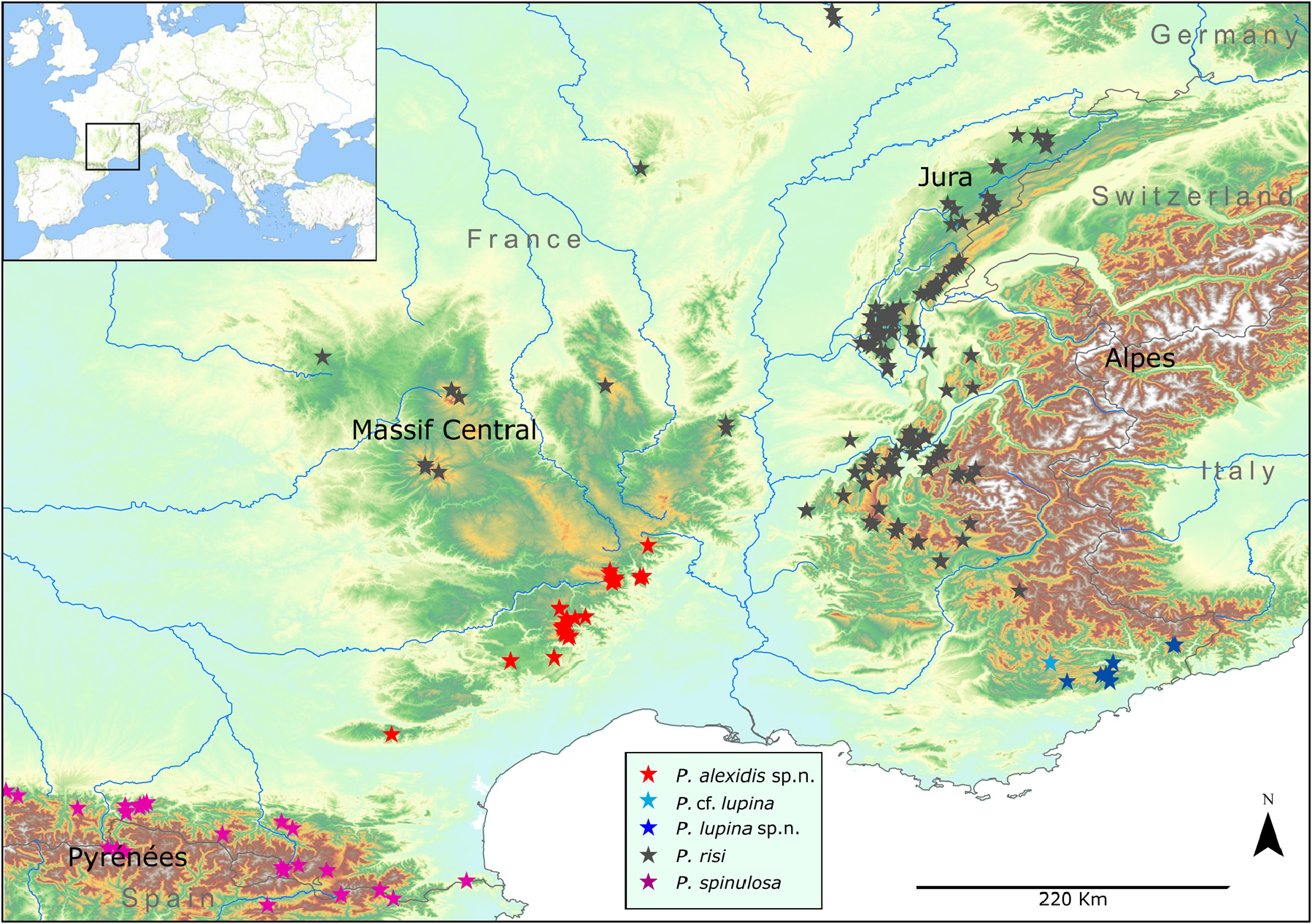
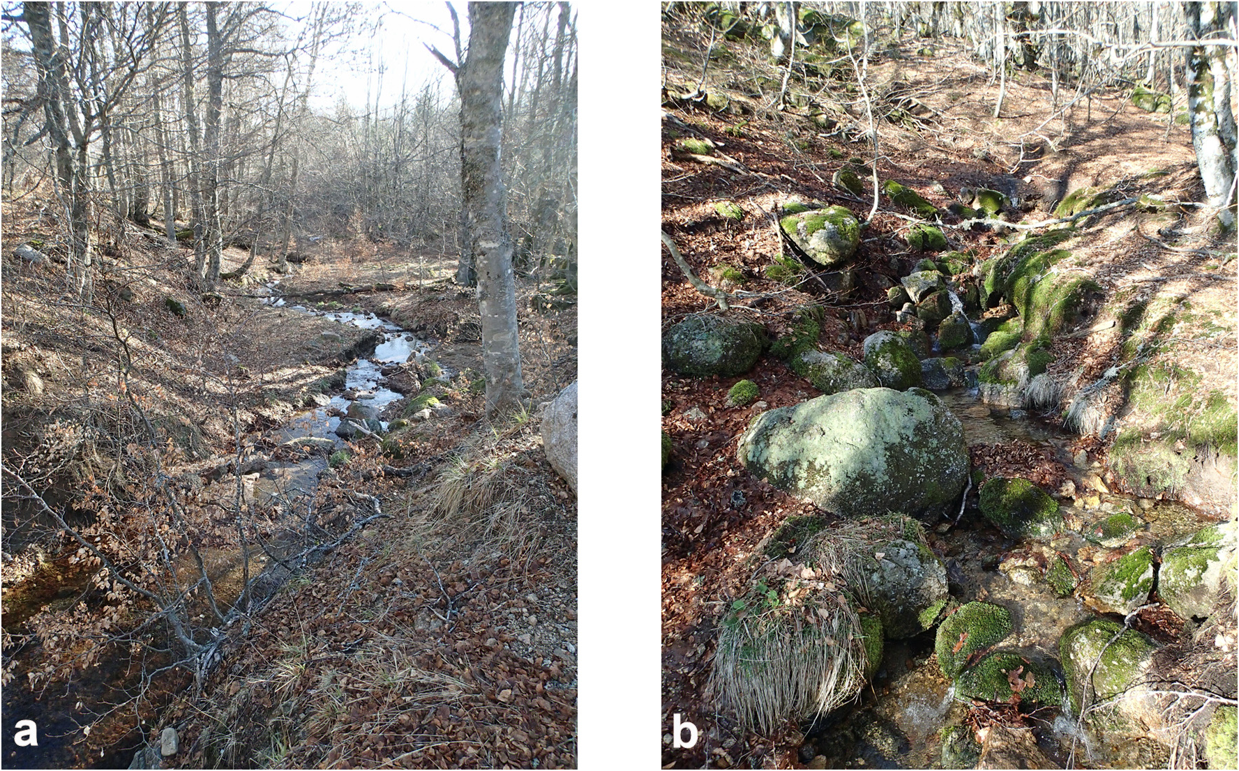
Distribution area and biogeographical notes. Protonemura alexidis sp. n. inhabits the southern flank of the Massif Central: Cévennes, Mont Aigoual, western Larzac, Montagne Noire ( Figs. 41 View Fig , 44). In the northern part of the Massif Central, however, only typical specimens of P. risi were found (unpublished data of the authors; Fig. 41 View Fig ). Protonemura alexidis sp. n. has a wide altitudinal range ( 450–1535 m) and lives in springs and small brooks ( Fig. 44 View FIG ). Adults of both sexes emerge from early spring to autumn (II–X), with a pause during summer.
Derivatio nominis of Protonemura alexidis sp. n. This species is named after Alexis Reding, son of the last author, in recognition of his significant contribution to the molecular genetics of the Plecoptera of the Jura Mountains. The epithet is a noun in the genitive case.
No known copyright restrictions apply. See Agosti, D., Egloff, W., 2009. Taxonomic information exchange and copyright: the Plazi approach. BMC Research Notes 2009, 2:53 for further explanation.
|
Kingdom |
|
|
Phylum |
|
|
Class |
|
|
Order |
|
|
Family |
|
|
Genus |