
Stegana penicillata (Kertész) Pirani & Grimaldi, 2019
|
publication ID |
https://doi.org/ 10.11646/zootaxa.4661.3.2 |
|
publication LSID |
lsid:zoobank.org:pub:A166C53B-EC0F-427B-B6F4-1F39F92A3BD8 |
|
DOI |
https://doi.org/10.5281/zenodo.5929897 |
|
persistent identifier |
https://treatment.plazi.org/id/03B1D930-9109-2252-BB8C-AC2AC111A9C4 |
|
treatment provided by |
Plazi |
|
scientific name |
Stegana penicillata (Kertész) |
| status |
comb. nov. |
Stegana penicillata (Kertész) , New Combination
Pyrgometopa penicillata Kertész, 1901: 419 View in CoL .
Material Examined: HT: Female, Peru: Callanga (examined by D. Grimaldi at the HNHM in 2011, excellent condition) GoogleMaps .
Additional material (2 males, 8 females): BRASIL, SP, Ribeir„o Grande, / P. E. Intervales 24°16’28”S 48°25’20”W / 22.xi.2010 —Malaise/ N.W. Perioto & eq. col. 1 male ( MZUSP) GoogleMaps ; BRASIL, AC, Mancio Lima / Pq Nacional Serra do/ Divisor—Igarapé do Amor / 20.iii–20.iv.2006 — Malaise / Calor, A. R. & Viana, D. cols. 1 female ( MZUSP) GoogleMaps ; BRASIL, AM, Careiro Castanho / BR 319 km-181, Sítio S. Paulo / 4°12’48”S - 60°49’04”W / 10–24, iv.2017, Malaise gde/ J. A. Rafael & F. F. Xavier Fº. 1 female, eggs in one vial, abdomen and terminalia in another vial, right wing double mounted and pinned above specimen and vials ( INPA) GoogleMaps ; Brasil, AM, Manaus. / Res. Km 41 PDBFF / 23–24.VI.2004 / Sub-bosque, R. Querino. 1 female ( INPA) GoogleMaps ; BRASIL, Amazonas, Manaus , ZF-2,/ km-14, 2°35’21”S - 60°06’55”W,/ 25.i–8.ii.2018, Malaise gde solo,/ Poente, J. A. Rafael—Rede BIA. 1 female ( INPA) GoogleMaps ; ( FR-GU) Guyane Française, Mitaraka,/ different sites nr base camp and along/ trails, tropical most forest (different/ sites), 14.iii.2015, SLAM, leg. Julien Touroult & Eddy Poirier (FR-/ GU/Mitaraka/2015)—sample code:/ MITARAKA/191. 2 females ( MNHN) GoogleMaps ; PERU: Cusco, Quincemil / Pte La Cigarra, 13°08’27”S 70°23’14”W, 350m / 01.ix.2012, sweep, JA/ Rafael, RR Cavichioli. 1 female ( INPA) GoogleMaps ; Peru: Cusco, Est Biol Villa Carmen ,/ 12.8911°S, 71.4106°W, 527 m,/ Malaise trap, 12–16.XII.13,/ Norrbom and Sutton. 1 male (genitalia dissected, head mounted and gold coated for SEM; in AMNH), 1 female ( AMNH) GoogleMaps .
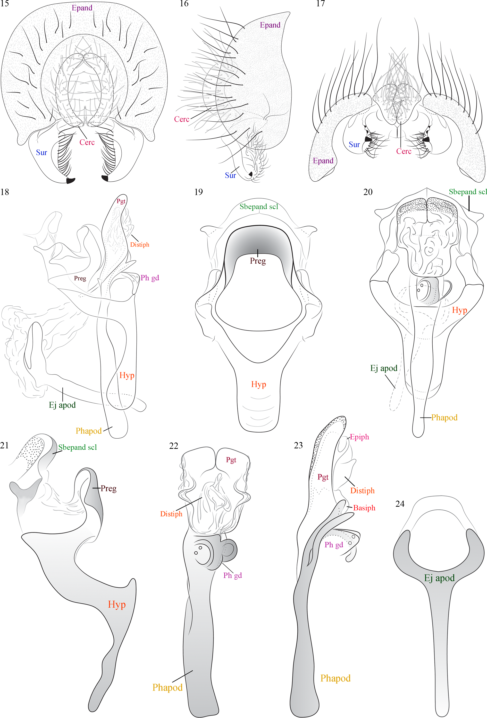
Diagnosis: Well developed ocellar tubercle (both sexes), bearing a tuft of black, spike-like bristles ( Figs. 1–5 View FIGURES 1–6 , 7, 9–10 View FIGURES 7–11 ), many of them with a bifid apex ( Fig. 27 View FIGURES 25–32 ). Postocellars divergent ( Figs. 1, 3 View FIGURES 1–6 ). Acrostichals relatively long and light ( Figs. 3 View FIGURES 1–6 , 8 View FIGURES 7–11 ). Surstylus conical, with a prominent strong spine apically (plus two smaller seen in ventral vista) and a vertical line of regular setae ( Figs. 15–17 View FIGURES 15–24 ); surstyli connected dorsally. Subepandrial sclerite with apical pointed projections, connected to hypandrium by a membrane ( Figs. 19–21 View FIGURES 15–24 ). Hypandrial complex very elaborate, pregonites fused dorsally forming a projected flap ( Figs. 19–21 View FIGURES 15–24 ). Phallic guide present fused to phallapodeme and touching the dorsomedial portion of hypandrium ( Figs. 22–23 View FIGURES 15–24 ).
Description: Body stout, compact ( Figs. 2 View FIGURES 1–6 , 7–8 View FIGURES 7–11 ); arched dorsally, with top surfaces of notum, scutellum, and abdomen in same plane of curvature. Head ( Figs. 1, 3–5 View FIGURES 1–6 , 9–11 View FIGURES 7–11 , 25–30 View FIGURES 25–32 ): attached low on prothorax, just above prosternum, at approximately same level as notopleural suture.
Head ( Figs. 1, 4–5 View FIGURES 1–6 , 9–11 View FIGURES 7–11 , 25–30 View FIGURES 25–32 ): broad, width slightly less than that of thorax, much broader than deep; head depth (without tubercle) 0.42, head width 0.86. Eye without ommatrichia, slightly longer than deep; eye length 0.42, depth 0.34. Frons, face, and clypeus broad; lower frons width 0.37, face width 0.35. Ocellar triangle raised into a tall tubercle ( Figs. 4–5 View FIGURES 1–6 , 10–11 View FIGURES 7–11 , 25–27 View FIGURES 25–32 ), height (0.14) approximately equal to width; ocelli situated on apex; pair of small ocellar setae present, length (0.15) ca. 3.5× ocellus diameter. Center of ocellar tubercle with apical tuft ca. 12 stout, erect, long, spine-like black setae, lengths (0.26 for longest) greater than height of tubercle; longer setae with preapical branch; setae deeply ribbed but not flattened like scales; setal sockets raised. Ocellar tubercle shiny, glabrous, the surrounding frons dull, with dense microtrichia. Postocellar seta positioned on posterior base of ocellar tubercle, approximately same size as ocellar setae, divergent ( Figs. 1, 3 View FIGURES 1–6 ). Proclinate fronto orbital seta at level of middle of frons length; anterior reclinate fronto orbital seta equidistant between other proclinate fronto orbital seta and posterior reclinate fronto orbital seta (absent in some specimens); posterior reclinate fronto orbital seta almost erect and slightly lateroclinate. Inner vertical seta long (0.24), inclinate; outer vertical slightly shorter (0.18), lateroclinate. Face receding ventrally, no carina; gena shallow (0.06); vibrissa fine (length 0.22). Antenna ( Figs. 1, 4 View FIGURES 1–6 , 25, 30 View FIGURES 25–32 ): scape very thin, ring-shaped; pedicel as high as wide, with ca. 10 setulae, 1 erect lateral seta; first flagellomere long, apex extending beyond level of oral margin; arista inserted dorsally on first flagellomere, with ca. 7 dorsal, 3 ventral, 2–3 long inner long branches (plus 6–7 short ones). Palpus paddle-shaped, with 5–6 setulae. Proboscis: short, with an array of fine, whitish scales with apical margins serrate.
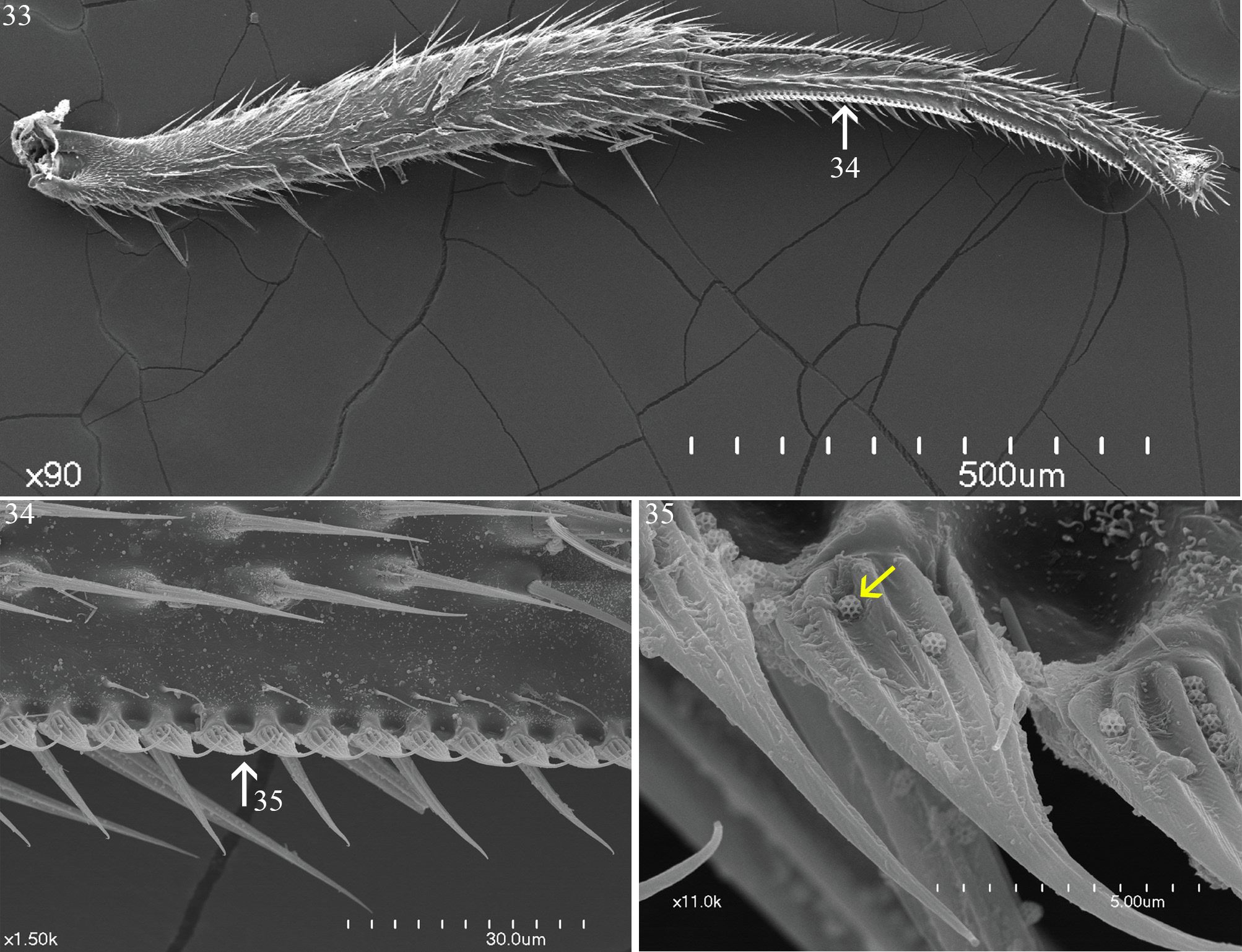
Thorax ( Fig. 3 View FIGURES 1–6 , 8 View FIGURES 7–11 ): Very broad, width of scutum (0.97 mm) almost equal to thorax length (1.17–1.20). Acrostichals relatively long (compared to other drosophilids), fine, light, not arranged in rows. Three pairs prescutellar setae present, lengths 1.5–2.0× the length of acrostichals; anterior dorsocentral slightly longer than acrostichals, posterior dorsocentral length 0.24. One small postpronotal seta; 2 thick and long ventral notopleural setae, one smaller and finer dorsal notopleural seta; 1 large supra-alar and 1 postalar. Scutellum large (length 0.43–0.44), shield-like, flattened and curved; anterior scutellar setae large (lengths 0.31–0.38), parallel, length slightly shorter than length of scutellum; apical scutellar setae small, ca. 0.5× length of anterior one (0.17). Postnotum entirely covered by scutellum. Deep postalar groove in thorax between wing base and anterolateral portion of scutellum. 2 large upright katepisternal setae; length of anterior one 0.28. Legs ( Figs. 33–35 View FIGURES 33–35 ): All coxae short, procoxae inserted just behind head; basitarsus longer (0.27 mm) than combined length of other tarsomeres (0.25 mm). Foreleg: femur slightly longer than tibia (0.48–0.50, vs. 0.37–0.40), lateral surface with ca. 10 upright setae. Tibia with mesal/ventral surface with 4 transverse, comb-like rows of fine, golden-colored setulae. Midleg: femur and tibia approximately equal in length, with preapical dorsal and apical ventral seta (latter one thicker). Tarsomeres 1–4 with 2 longitudinal rows cuneiform, overlapping, scale-like setulae, each having fine, pointed tip. Tarsomere 1 also with longitudinal row of black, spinule-like setulae between cuneiform rows. Hind leg: Tibia with preapical dorsal seta, no apicoventral seta. Tarsomeres 1–4 with one longitudinal row cuneiform, overlapping, scale-like setulae; inner/mesal surface of tarsomeres 1–4 with 2–6 golden-colored setulae in short, oblique, comb-like rows. Wing ( Figs. 6 View FIGURES 1–6 , 31, 32 View FIGURES 25–32 ): length 2.47 mm, width 1.13, apex bluntly pointed; membrane with dense microtrichia, surface irregular and weakly or slightly concave. Sc faint, incomplete; h and Sc break deep; C thickened where it meets R 1; C ending in M 1, with a row of 12 minute thorn-like spinules between R 2+3 and R 4+5 (ventral/adaxial surface of the wing); C spinules gradually decreasing in size apically. CII length (between apices R 1 and R 2+3) 1.22; CIII length (between apices R 2+3 and R 4+5) 0.61. R 2+3 sinuous; R 4+5 slightly curved; distal portion of M 1 faded; apex of M 1 very close to apex of R 4+5 (distance ca. 2.5× thickness of veins). Section III (length of M between crossveins) 0.59, IV (M distal to dm-cu) 0.91. Apical portion of CuA (past dm-cu) abruptly bent. Anal lobe and alula well developed; anal vein present, incomplete; cup, br and bm present.
Abdomen: Short, stout in both sexes; all tergites shiny, with fine, light, scattered setae. Sternites large, well developed and sclerotized. Male terminalia (Figs.: 15–24): Epandrium complex ( Figs 15–17 View FIGURES 15–24 ): Epandrium wider than high with setae and microtrichia covering all its surface. Cercus higher than wide, reniform, with setae over all its surface. Surstylus conical with 3 thick ventral spines (apical spine longer, the only visible on terminal view, Fig. 15 View FIGURES 15–24 ; the other two, shorter, only seen in ventral view, Fig. 17 View FIGURES 15–24 ), plus ca. 15 ordinary setae in a line on the inner surface; both surstyli fused, forming a bridge dorsally. Subepandrial sclerite present, with pointed dorsolateral projections, connected by a membrane to hypandrium. Hypandrial complex ( Figs. 18–21 View FIGURES 15–24 ): Hypandrium Y-shaped, pregonites present and fused to it, extending dorsally and fused to each other, forming a “flap” that projects ventrally. Phallic complex ( Figs. 18, 22 View FIGURES 15–24 ): Phallapodeme well sclerotized, higher than wide, ventral margin longer than ventral margin of hypandrium, not fused to it. Phallic guide present as a ventral extension of the phallapodeme that touches the medioventral portion of hypandrium. Postgonites flanking the phallus, sclerotized, with several little spots covering the anterodorsal surface. Phallus entirely membranous: basiphallus bulbous, positioned between dorsal arms of phallapodeme, epiphallus longer, attached dorsally to basiphallus, distiphallus connected to basiphallus and arising from between the epiphallus and postgonites (phallic divisions better seen on lateral view, Fig. 23 View FIGURES 15–24 ). Ejaculaory apodeme present ( Fig. 24 View FIGURES 15–24 ), racquet-like, connected to the phallic complex by a membrane. Female terminalia ( Figs. 12–14 View FIGURES ): Epiproct round, setose; hypoproct triangular-shaped, bare; cerci long, fused dorsally; one group of medial short setae and another group of longer setae apically. Two spermathecae present, well sclerotized, ovoid, with several spots covering all its surface; spermathecal duct uniting both spermathecae dorsally; smaller sinuous tubes arising from distal ends of each spermathecae. Eggs ( Fig. 14 View FIGURES ): Numerous relatively small eggs per female; oval, spotted, with a small protuberance on one of its apices.
Coloration ( Figs. 1–11 View FIGURES 1–6 View FIGURES 7–11 ): body mainly blackish-brown, with small contrasting light (yellow to orange) areas. Head: frons dark brown, frontal vitta black, dull; anterior half of frons ochre to orange. Eye light red. Face, clypeus, apical half of palp dark brown (proximal half yellowish); labellum whitish. Scape, pedicel, base of flagellomere 1 dark yellow to orange; apical third of flagellomere 1 dark brown. Arista with trunk and bases of branches whitish, apical portion of branches darker. Gena anteriorly yellow (orange), white in the region above the middle of eye and black posteriorly. Occiput dark brown. Scutum, scutellum, pleura dark brown, scutum shiny, scutellum dull (with pile-like microtrichia). Postpronotal lobe lighter, varying from pale yellow to orange. All femora and tibiae dark brown, tarsi lighter. Anterior half of wing dark brown, graded to clear/hyaline posteriorly. Halter light, buff. Tergites and sternites evenly dark brown.
Comments: although the holotype was not dissected, we are confident that the flies we describe here are conspecific. The external morphology of these specimens agrees perfectly with the type. At this point we consider that it is premature to place S. penicillata into one of the currently recognized five subgenera of Stegana , for several reasons: 1. Most of these subgenera were erected and treated many years ago by Enderlein (1922), Hendel (1913), Duda (1923), Okada (1978), Wheeler (1960) and most recently by Sidorenko (2002) and Zhang et al. (2012). The monophyly of subgenera and the phylogenetic relationships between and within them hasn’t been explored satisfactorily (but see Li et al., 2013 and Wang et al., 2017 for considerations about the Stegana of the Oriental region). 2. There are many undescribed species of Stegana , particularly from the Neotropical Region, and it is very likely that S. penicillata may be closely related to any of these undescribed species in a possibly new subgenus. Taxonomic decisions of this nature should better be made in context to a taxonomic revision of the genus or at least a broad study of the Neotropical species.


| HNHM |
Hungarian Natural History Museum (Termeszettudomanyi Muzeum) |
| MZUSP |
Museu de Zoologia da Universidade de Sao Paulo |
| AC |
Amherst College, Beneski Museum of Natural History |
| AM |
Australian Museum |
| INPA |
Instituto Nacional de Pesquisas da Amazonia |
| MNHN |
Museum National d'Histoire Naturelle |
| AMNH |
American Museum of Natural History |
No known copyright restrictions apply. See Agosti, D., Egloff, W., 2009. Taxonomic information exchange and copyright: the Plazi approach. BMC Research Notes 2009, 2:53 for further explanation.
|
Kingdom |
|
|
Phylum |
|
|
Class |
|
|
Order |
|
|
Family |
|
|
Genus |
Stegana penicillata (Kertész)
| Pirani, Gabriela & Grimaldi, David A. 2019 |
Pyrgometopa penicillata Kertész, 1901: 419
| Kertesz 1901: 419 |