
Cercidospora navarroi Y. Joshi, 2022
|
publication ID |
https://doi.org/ 10.11646/phytotaxa.549.2.10 |
|
DOI |
https://doi.org/10.5281/zenodo.6624965 |
|
persistent identifier |
https://treatment.plazi.org/id/03B22465-FF89-FFD5-BCF8-57E87EAFFBCC |
|
treatment provided by |
Plazi |
|
scientific name |
Cercidospora navarroi Y. Joshi |
| status |
sp. nov. |
Cercidospora navarroi Y. Joshi View in CoL , sp. nov. MycoBank no.: MB 843787
( Figs. 1 View FIGURE 1 a-c & 2a-c)
Differs from C. rinodinae in pigmentation of the upper part of the peridium (emerald-green vs brown-black to black with a violaceous hue), smaller asci [(37–)40– 43 –46(–50) µm vs 47–60 µm], smaller ascospores [(8–)8.6– 10.2 –11.8(– 13) × (1.9–)2– 2.3 –2.7(–3) µm vs (12.5‒)14‒17(‒20) × 3.5‒4.5 µm] and habitat preference (corticolous vs saxicolous or humicolous).
Type:— INDIA. Himachal Pradesh, Shimla district, Kufri , towards Chinibunglaw via forest, alt. 2500 m, on thallus and apothecial disc of Rinodina intermedia colonizing Cedrus deodara bark, 22 September 2013, Y. Joshi, S.P. Singh, A.K. Gothwal and party s.n. (holotype RUBL 15 (L)) .
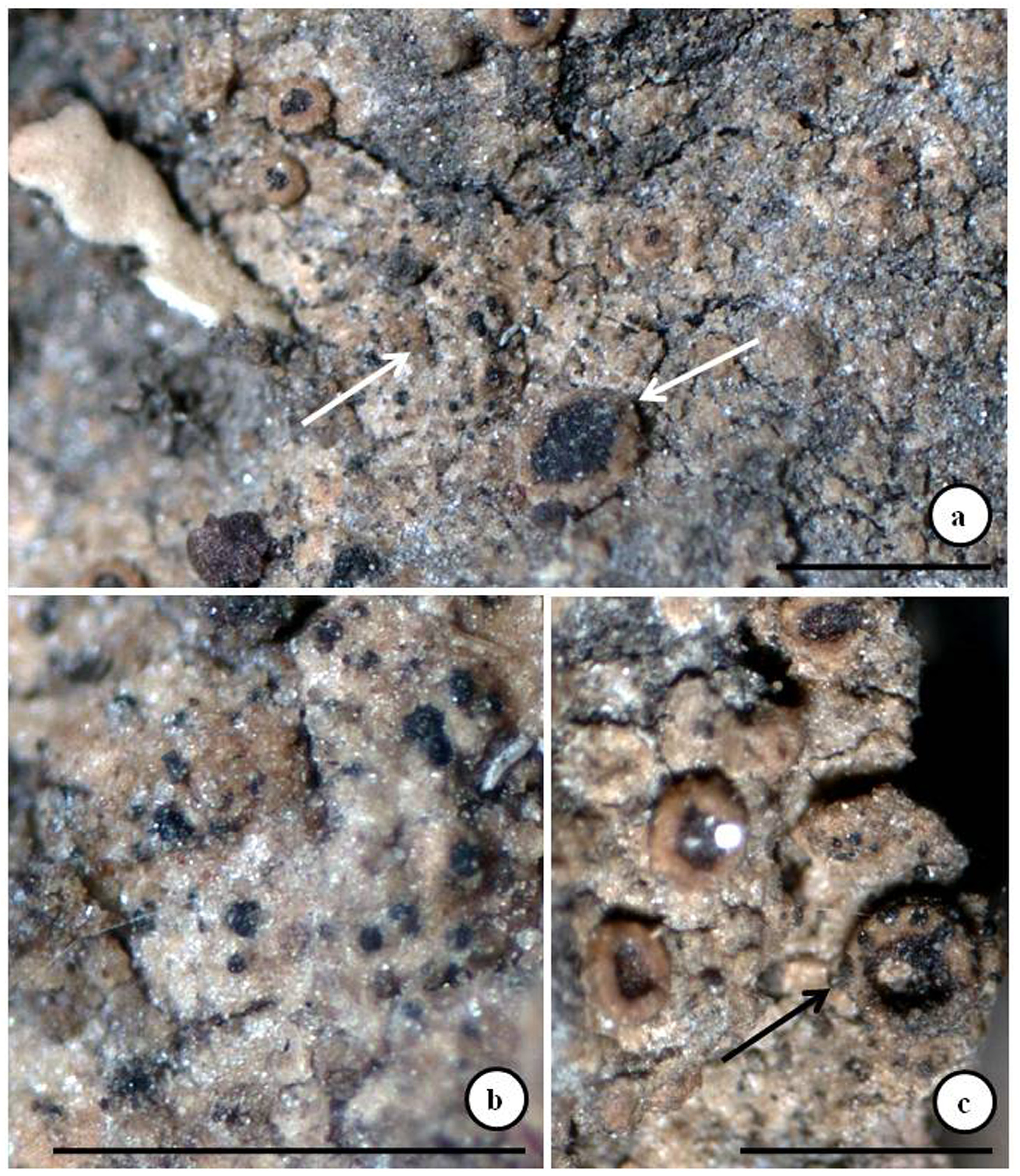
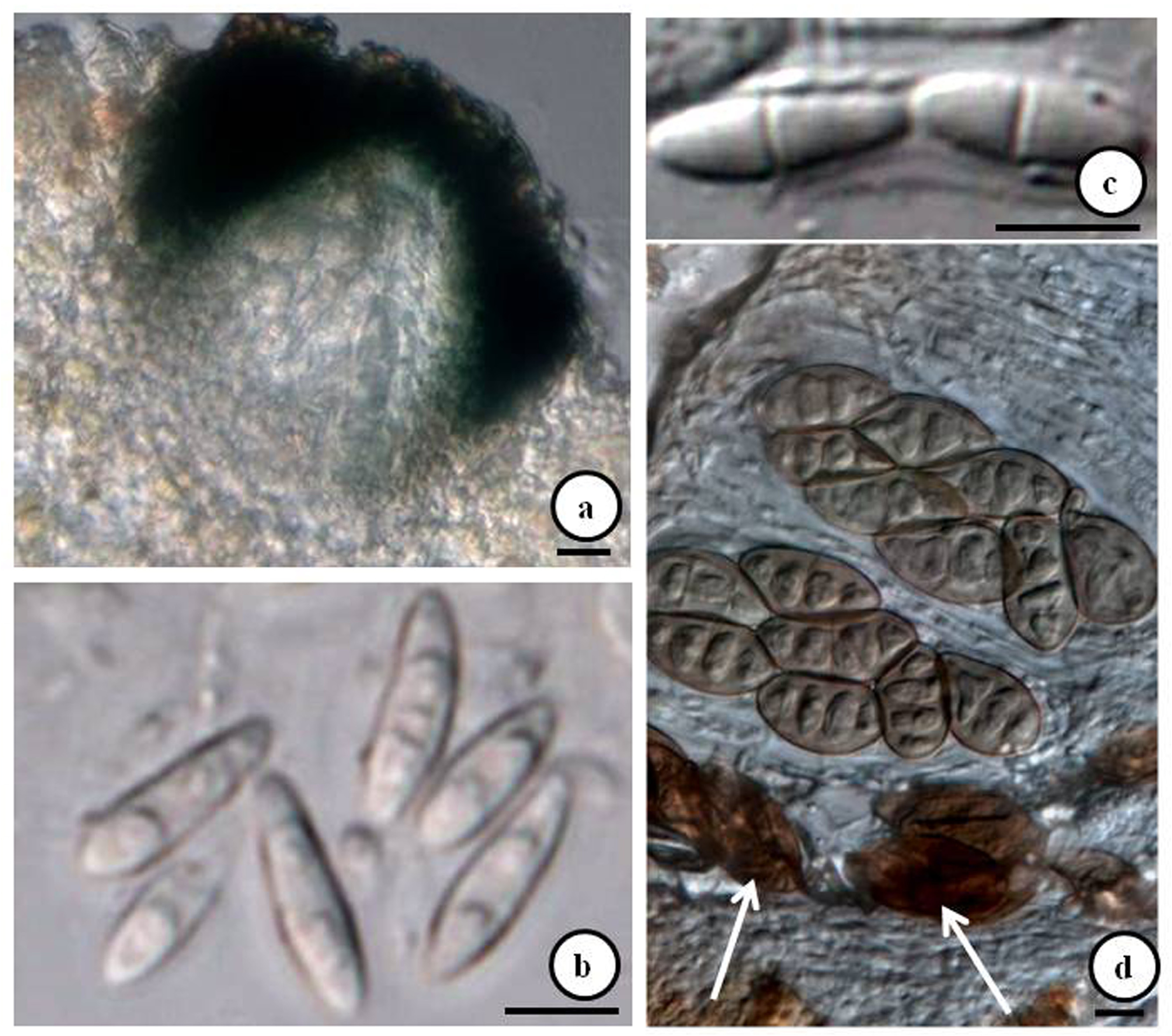
Lichenicolous. Colonizing the thallus and apothecia of Rinodina intermedia ( Fig. 1 View FIGURE 1 a-c). Ascomata perithecioid, black, arising singly or occasionally aggregated, immersed and only protruding at the ostiolar region, breaking through the cortex of the host, (70–)80– 85 –90(–95) µm diam. (n = 25), orbicular; ostiole c. 15 µm diam. Peridium in the upper part emerald green, c. 10–20 µm thick, in the lower part hyaline and then indistinctly delimited from the host ( Fig. 2a View FIGURE 2 ), c. 10–15 µm thick, wall composed of several rows of cells and in surface view recalling textura angularis or textura epidermoidea, K+ blue-black, N+ dull purple-violet; hymenial gel I-, K/I-. Hamathecium composed of abundant interascal filaments, c. 1 µm thick, simple to branched, septate but individual cells not easily distinguishable, apically not swollen. Asci bitunicate, fissitunicate, subcylindrical to elongate-clavate, with flattened apex, short-stalked, ascoplasm K/I+ orange (dextrinoid reaction), 4-spored, uniseriate, with a small ocular chamber, (37–)40– 43 –46(–50) × (4.5–)5– 6 –7(–8) µm (n = 25). Ascospores 1-septate, rarely simple, hyaline, smooth-walled, ellipsoid, with pointed end cells, ±heteropolar, with one cell normally more slender, not constricted at the septum which is very thin, with one large oil-droplet per cell (prominent after treatment with K), (8–)8.6– 10.2 –11.8(–13) × (1.9–)2– 2.3 –2.7(–3) µm, l/b = (2.3–)3.7– 4.6 –5.5(–6.5) without perispore (n = 50) ( Fig. 2b&c View FIGURE 2 ). Conidiomata and vegetative hyphae were not observed.
Etymology: The species is named in honor of Dr. Pere Navarro-Rosinés, a Spanish lichenologist, for his immense contribution to the lichenicolous genus Cercidospora .
Notes: Of the 40 binomials of Cercidospora known so far ( Diederich et al. 2018), the lichen genus Rinodina hosts only two species – C. exiguella (Nyl.) Arnold , and C. rinodinae Etayo & van den Boom. The color reaction of the upper part of the peridium (K+ blue-black and HNO 3 + purple-violet) and the ellipsoid, 1-septate ascospores without halo brings Cercidospora navarroi closer to C. rinodinae ( Etayo & van den Boom 2005) . However, the new taxon differs from C. rinodinae in pigmentation of the upper part of the peridium (emerald-green vs brown-black to black with a violaceous hue), shorter asci [(37–)40– 43 –46(–50) µm vs 47–60 µm], shorter and narrower ascospores [(8–)8.6– 10.2 –11.8(–13) × (1.9–)2– 2.3 –2.7(–3) µm vs (12.5‒)14‒17(‒20) × 3.5‒4.5 µm], and habitat preference (corticolous vs saxicolous or humicolous) (Table).
Cercidospora exiguella , colonizing saxicolous species of Rinodina , differs from the new taxon in having a blue ( Etayo & van den Boom 2005) or brown to brown-black ( Ihlen & Wedin 2008) peridium, longer and wider asci [47–52 × 9–10 µm vs (37–)40– 43 –46(–50) × (4.5–)5– 6 –7(–8) µm] and very long and wide ascospores [21‒27 × 6‒8 µm ( Vouaux 1913) or 13.5‒27 × 4.5‒6.5 µm ( Zhurbenko 2002; Zhurbenko & Triebel 2003) vs (8–)8.6– 10.2 –11.8(–13) × (1.9–)2– 2.3 –2.7(–3) µm].
Zhurbenko (2017) while examining a specimen of Rinodina roscida (Sommerf.) Arnold (1887: 133) collected from Northwest Caucasus found it to be infected with Cercidospora and observed that the examined material differs from C. rinodinae in having a brown to olive-gray pigmented upper part of the exciple which is often green or bluish-green around the ostiole and is K-, N+ moderate red; wider interascal filaments and slightly wider ascospores occasionally with a halo up to 1.5 µm thick and treated it as C. cf. rinodinae . This material also differs from the new taxon in several characters (Table).
Distribution and ecology: To date, the taxon is known only from the type locality in the temperate region of Himachal Pradesh ( India), where it was growing as a weak parasite on the thallus, apothecial disc, and margin of Rinodina intermedia colonizing the bark of Cedrus deodara (Roxb.) G. Don. The asci of the host lichen contain some damaged along with healthy ascospores; therefore we suggest that this lichenicolous fungus might be considered a weak parasite ( Fig. 2d View FIGURE 2 ).
No known copyright restrictions apply. See Agosti, D., Egloff, W., 2009. Taxonomic information exchange and copyright: the Plazi approach. BMC Research Notes 2009, 2:53 for further explanation.