
Carlogonus gayathri, Sankaran & Sebastian, 2020
|
publication ID |
https://doi.org/ 10.11646/zootaxa.4868.1.2 |
|
publication LSID |
lsid:zoobank.org:pub:B869EB5E-073D-471D-A9F3-D390D57B25E6 |
|
DOI |
https://doi.org/10.5281/zenodo.4427141 |
|
persistent identifier |
https://treatment.plazi.org/id/03B27A7E-FFD3-0061-FF44-5FF1FEB4FE57 |
|
treatment provided by |
Plazi |
|
scientific name |
Carlogonus gayathri |
| status |
sp. nov. |
Carlogonus gayathri View in CoL sp. nov.
Figs 1–7 View FIGURE 1 View FIGURE 2 View FIGURE 3 View FIGURE 4 View FIGURE 5 View FIGURE 6 View FIGURE 7 , 9 View FIGURE 9
Type material. Holotype: Male (MILLI-ADSH0012), INDIA, Kerala: Palakkad, Thrippalur , Pullodu , 10°38’16.58’’N, 76°33’52.87’’E, 70 m alt., 23 October 2017, M.S. Pradeep leg., from ground, by hand GoogleMaps . Paratypes 3 males, 9 females (MILLI-ADSH0013), same data as holotype except for the collecting date of GoogleMaps 3 paratype females (11 November 2019).
Etymology. The specific epithet refers to the name of the Gayathripuzha River, one of the main tributaries of Bharathapuzha River, flowing through the Palakkad district on the bank of which the type locality (Thrippalur) of the new species lies.
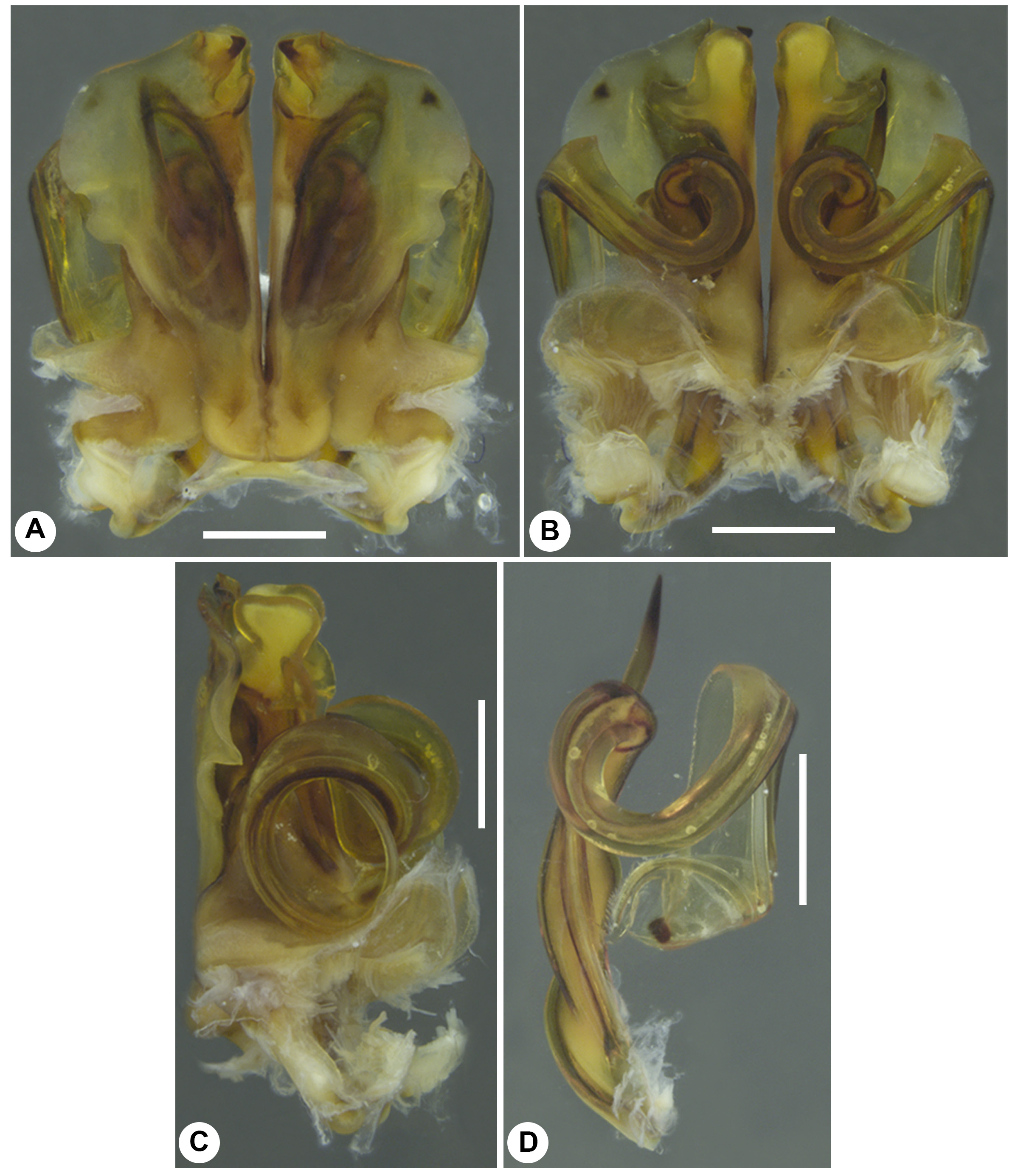
Diagnosis. Within the exaratus -group, C. gayathri sp. nov. is most similar to C. subvalidus as both have an antero-mesal hook-like process of the anterior coxal fold, a sharply curved tibial spine and a distally twisted telopodite, but both can be separated from one another by the following combination of characters: lateral margin of anterior coxal fold without a spine-like outgrowth (lateral margin of anterior coxal fold of C. subvalidus with a spine-like outgrowth), narrow lateral flat process of the posterior coxal fold ( C. subvalidus with a wide lateral flat process of the posterior coxal fold) and a femoral spine without basal twists (femoral spine of C. subvalidus has basal twists) (compare Figs 5A, B, D View FIGURE 5 , 7A, B View FIGURE 7 , F–H with Carl 1941: figs 144–147 and herein Fig. 8G View FIGURE 8 ).
Description. Measurements: male with 65 body rings (64 podous and 1 apodous (telson)), circa 127 mm long, 6.4 mm wide. Female with 66 body rings (65 podous, 1 apodous), circa 133 mm long, 7.9 mm wide.
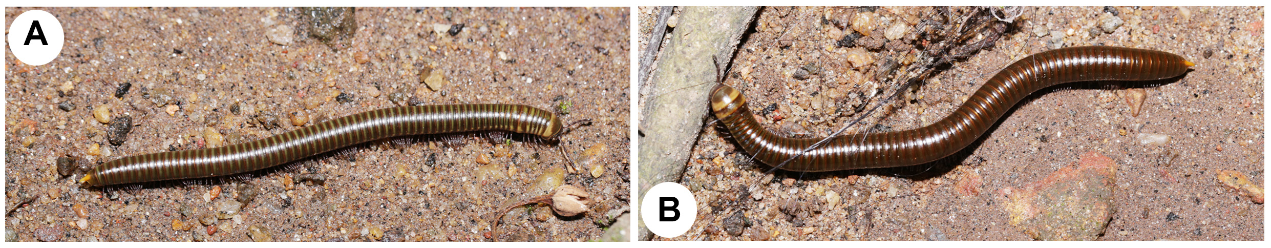
Colour. Head, collum yellowish with broad coffee-brown patches on both. Antennae, anal valves, legs coffeebrown. Clypeal margin with a coffee-brown, serration-like pattern ( Fig. 3F View FIGURE 3 ). Body rings coffee-brown, baso-laterally with yellowish patch. Posterior margin of metazonae light brown. Metazonae up to ring 64 dorso-laterally with olive-green transverse patch. Preanal process (anal spine) yellowish. Colouration of preserved material: overall dull brown with greyish patches and stripes; parts with yellowish colour faded to straw-colour. Metazonae lose dark olive-green colouration.
Head. Head smooth. Each eye patch with circa 55–59 ommatidia arranged in 7 or 8 horizontal rows. Axial sulcus prominent, reaching up to frons. Labrum with three smoothly rounded teeth and a single row of 6 or 8 short labral setae ( Fig. 3F View FIGURE 3 ). Clypeus with six setiferous foveolae, three on each side ( Fig. 3F View FIGURE 3 , arrows 3–8). Antennal cavity present, nearly circular in outline. Antennae moderately long ( Fig. 3G View FIGURE 3 ), protruding back to ring 4. Relative length of antennomeres: 1<2>3>4<5>6. Terminal antennomere (disc) with four large sensory cones located together inside a membranous area ( Fig. 4A View FIGURE 4 ). Antennomere 6 apico-laterally with field of 9 or 10 rows of narrow, long sensilla basiconica, disc laterally with 1 or 3 rows of irregularly clustered tiny sensilla basiconica ( Figs 4 View FIGURE 4 A–C, arrows 1 and 2 respectively).
Gnathochilarium. Usual for spirostreptideans ( Fig. 3D View FIGURE 3 ). Mentum smooth with more or less straight posterior margin with less developed prebasilare ( Fig. 3D View FIGURE 3 , arrow 1). Hypostoma flat, medially smooth, laterally with oblique striations. Lamellae linguales each with five spine-like setae, two proximally, three distally. Stipites with a slightly wavy lateral margin, with disto-ventral bulging, medially with oblique excavation, each with a short, thick, thornlike disto-ventral seta placed on a small conical mount, each with four stout apico-lateral setae ( Fig. 3D View FIGURE 3 ), with 11 or 12 short basal spine-like setae. Only 1st pair of palpi with numerous sensilla ( Fig. 4E View FIGURE 4 ). Hypopharyngeal crest with weakly developed field of spine-like structures, distally with a membranous lamina bearing a series of short marginal setae ( Fig. 4E View FIGURE 4 ). Central pads of endochilarium divided to a short distance by a groove and a ridge into two separate regions ( Fig. 4E View FIGURE 4 ; CP, Endo).
Mandible. Stout and short. External tooth simple, elongately triangular ( Fig. 4F View FIGURE 4 ; ET); inner tooth with four cusps ( Fig. 4F; 4 View FIGURE 4 IT). Nine or 11 rows of pectinate lamellae ( Fig. 4F View FIGURE 4 ; PL). Medio-basal margin of pectinate area smooth ( Fig. 4F View FIGURE 4 ); basal margin with 3 or 4 transverse rows of small spines. Molar plate long with a distal excavation ( Fig. 4F View FIGURE 4 , arrow 3; MP).
Collum. Lateral margin rounded, extending beyond the tips of ring 2, surface smooth ( Fig. 3A, E, M View FIGURE 3 ).
Body rings. Divided by sutures in two transverse zones, pro- and metazonae. Pro- and metazonae dorsally micropunctuated, ventrally with numerous weak longitudinal striations. Ozopore located on metazona, starting with ring 6, located close to, but not touching the suture between pro- and metazonae. Preanal ring/epiproct sharp-edged, extending beyond the anal valves/paraprocts as a long, smooth claw-like preanal process slightly curved downwards ( Fig. 3C, N View FIGURE 3 ). Anal valves with well developed lips, but with neither micropunctuations, grooves, nor setae. Subanal scale/hypoproct, small, widely triangular.
Legs. Coxae 1 short, subcylindrical, coxae 2 rectangular in outline, fused together ( Fig. 3 View FIGURE 3 H–I, O–P); coxae 3 and 4 elongately triangular, fused ( Fig. 3J, Q View FIGURE 3 ); others stout, broadly triangular, unfused. Prefemur 1 and 2 moder- ately long, subcylindrical, unfused ( Fig. 3 View FIGURE 3 H–I, O–P). Leg-pair 2 longer than 1 ( Fig. 3I View FIGURE 3 ); each podomere with apical/ventral/dorsal/mesal/lateral tiny, stout and slender setae; postfemur and tibia from leg-pair 3 onwards bear well developed, complete pads; in anterior leg-pairs, pads with tip curved down ( Fig. 3K View FIGURE 3 ). Length of mid-body legs circa 5.7 mm in males, circa 5.1 mm in females. Claw large, slightly curved ( Fig. 3K View FIGURE 3 ).
Male sexual characters. Podomeres lack modifications ( Fig. 3 View FIGURE 3 H–J). Only body ring 7 conspicuously enlarged ( Fig. 3A View FIGURE 3 ). Tips of gonopods visible in lateral view ( Fig. 3A View FIGURE 3 ).
Penis. Short, bluntly triangular with thin, flat apical part ( Fig. 3I, L View FIGURE 3 , arrow 2).
Gonopods ( Figs 5 View FIGURE 5 A–D, 7A–I). Anterior coxal fold ( Figs 5A View FIGURE 5 , 7A View FIGURE 7 ; AC): lateral margin less modified, with a weak indentation ( Figs 5A View FIGURE 5 , 7A View FIGURE 7 , arrow 1); antero-mesally with a thumb-like and a hook-like processes, antero-laterally with a short process ( Fig. 7A View FIGURE 7 ; amtAC, amhpAC, alpAC). Posterior coxal fold ( Fig. 7C View FIGURE 7 ; PC): basally with moderately high lateral paracoxites, distally flat to accommodate telopodite ( Fig. 7 View FIGURE 7 A–B, D; PX); internal mesal fold with antero-mesal wide process ( Fig. 7E View FIGURE 7 ; imPC); baso-laterally with a mount-like process lying near to the basal part of telopodite, distally with an anterior roughly globular and a lateral flat processes ( Fig. 7 View FIGURE 7 B–C; blpPC, apPC, lpPC). Telopodite ( Figs 5D View FIGURE 5 , 7B View FIGURE 7 , F–I; T): flat resting on paracoxite, with ventral and lateral twists ( Figs 5D View FIGURE 5 , 7B View FIGURE 7 ); femur with a single strong femoral spine having abrupt basal and smooth distal curvatures ( Figs 5D View FIGURE 5 , 7B View FIGURE 7 , F–G; FS); tibial spine long, slender, smoothly curved with a basal apically bifurcated seta-like side branch ( Fig. 7B, D View FIGURE 7 , G–H, arrow 2; TS); palette wide, flat, narrowing towards tip, dorsally with a longitudinal crest, with a short lateral thorn-like process, with a single dorsal row of more than 10 pale, brownish apically bifurcated blepharochaetae ( Fig. 7 View FIGURE 7 H–I; P, C, dtpP, B).
Female copulatory organ (vulva) ( Fig. 6 View FIGURE 6 A–G). Located in membranous pouches attached to coxae 2 and 3 and to the inner lateral margin of ring 1 ( Fig. 6A View FIGURE 6 , arrows 1 and 2). Simple, looking like a rose flower bud ( Fig. 6 View FIGURE 6 B–D), consisting of two simple, subequally-sized, moderately sclerotised valves: a large aboral valve covering the oral valve, with lateral longitudinal invagination, with an antero-lateral membranous extension to attach with the inner margin of ring 1, with ventro-lateral ridges internally ( Fig. 6E, G View FIGURE 6 , arrow 5). Oral valve short, being covered by meso-lateral part of aboral valve, externally with tiny thorn-like processes and ridges ( Fig. 6 View FIGURE 6 E–F, arrow 4). No valval setae.
Natural history. Carlogonus gayathri sp. nov. is living in open grounds covered with dried leaves and debris. It can be seen during the onset of the rainy season. Rarely it could be observed crawling inside buildings (Sankaran, pers. obs.).
Barcode. Partial barcode, positions 1490–2198 of the standard barcode: CAAAAAATCAGAATAAGTGTTGGTATAAAATAGGGTCTCCTCCACCAGCAGGGTCAAAGAAAGAATAT- TAAAATTTCGGTCAGTAAGTAATATTGTGATAGCTCCGGCTAGCACTGGCAAAGATAGTAAAGGAGA- ATGGCTGTAATTTTTACAGCTCATACGAAAAGGGGTATTTGTTCGAATAATATACCTGCTGTTCG- TATATTAATGATGGTTGTAATGAGTTGATTGCACCTAGGATTGATGAAGCACCTGCTAAATGTAAAGAAAAAATAGC- TATATCTACTGATGGGCCGGTATGTGCTAAGGTGGAAGCTAAAGGAGGGTAAACGGTTCAACCT- G TA C C A G C T C C T T T T T C TA C G G C T G A A G A G G C TA G TA ATA G G A ATA A G G C A G G G G G G A G - CAATCAAAAGCTTATATTATTTATTCGAGGGAAGGCTATATCTGGGGCACCTAATATAAGGGGGACTAGT- CAATTTCCGAAACCACCAATTATAATTGGTATTACTATAAAGAAAATTATTACAAAAGCGTGGGCTGT- TACGATTACATTATAAATTTGATCGTCTCCAATTAAGCTGCCAGGTTGGCTTAATTCTAAGCGGATTA- ATATACTTAAAGAGGATCCAACTAATGCTGCTCAAGCACCAAAAATTAAATATATAGTTCCAATATCTTTAT
GenBank accession number. The GenBank accession number of the ORF region (positions 292–462) of the new sequence is MN887639 View Materials .
Phylogeny. Phylogenetic analysis shows that Carlogonus is the sister-taxon to Thyropygus , with Anurostreptus in a basal position ( Fig. 1 View FIGURE 1 ).
Note. A broken femoral spine was found in the left vulva of one of the female specimens of C. gayathri sp. nov. ( Fig. 6B View FIGURE 6 , arrow 3). One male with a broken-off femoral spine on the left telopodite was also found. The occurrence of a broken femoral spine in the vulva may indicate its use as an accessory copulatory structure. It can be hypothesised that during copulation, the male C. gayathri sp. nov. (and possibly the males of other known Carlogonus species) may use the femoral spine to keep the valves of the vulva open in order to support the insertion of the telopodite.


No known copyright restrictions apply. See Agosti, D., Egloff, W., 2009. Taxonomic information exchange and copyright: the Plazi approach. BMC Research Notes 2009, 2:53 for further explanation.
|
Kingdom |
|
|
Phylum |
|
|
Class |
|
|
Order |
|
|
Family |
|
|
Genus |