
Zoica puellula ( Simon, 1898 )
|
publication ID |
https://doi.org/ 10.11646/zootaxa.4276.1.11 |
|
publication LSID |
lsid:zoobank.org:pub:766C548B-9F57-4651-94E3-BD4B26414576 |
|
DOI |
https://doi.org/10.5281/zenodo.6016924 |
|
persistent identifier |
https://treatment.plazi.org/id/03B287A5-0B2F-FFDF-CBD1-FF08FBD1FC98 |
|
treatment provided by |
Plazi |
|
scientific name |
Zoica puellula ( Simon, 1898 ) |
| status |
|
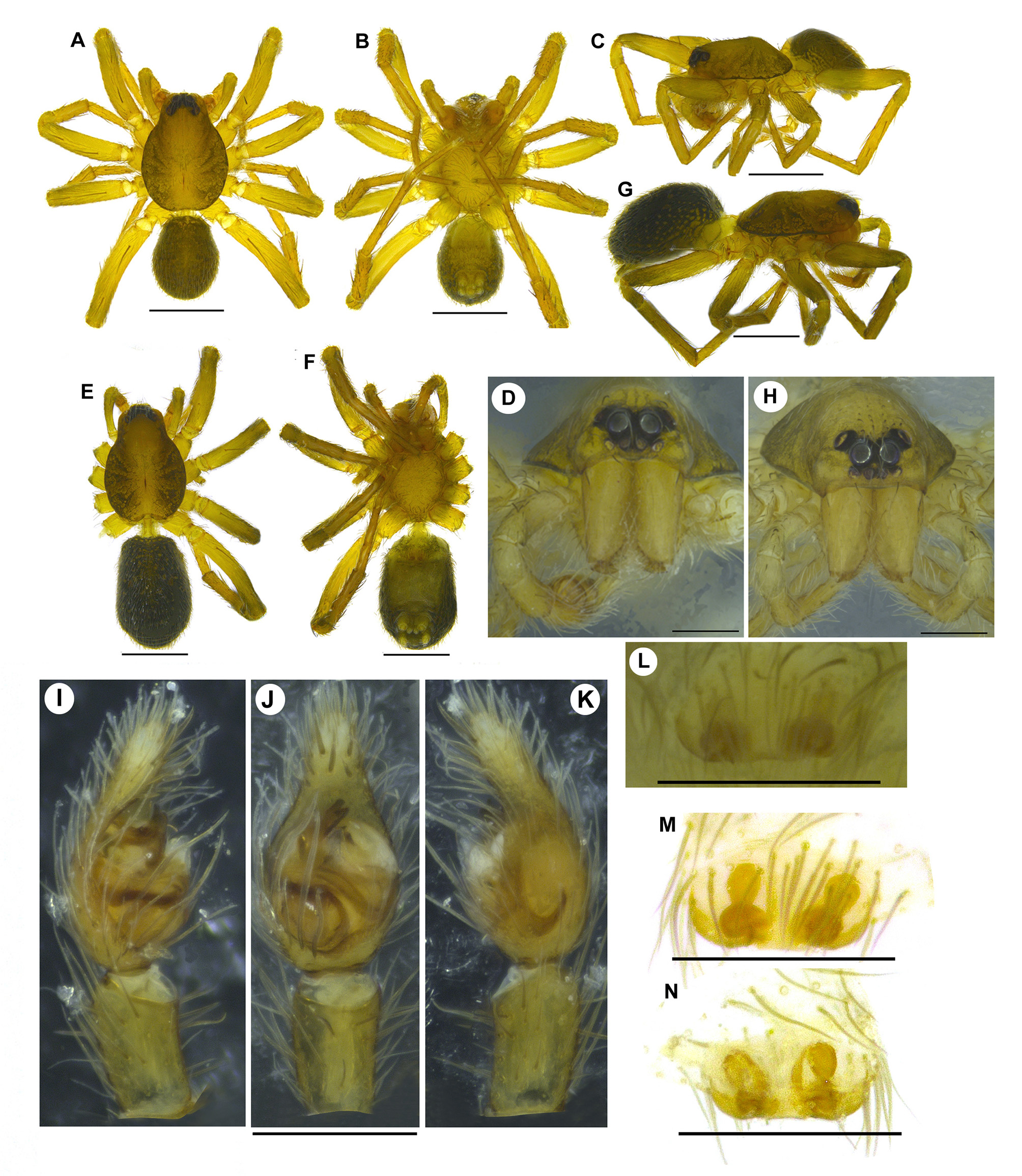
Zoica puellula ( Simon, 1898) View in CoL ( Figs 1–4 View FIGURE 1 View FIGURE 2 View FIGURE 3 View FIGURE 4 )
Flanona puellula Simon, 1898: 349 . Gravely 1924: 588; Roewer 1960: 839, fig. 465; Tikader & Malhotra 1980: 367, figs 236–237. Zoica puellula View in CoL — Lehtinen & Hippa 1979: 16, figs 48, 57.
Type material. Lectotype female (here designated) of Flanona puellula Simon, 1898 from SRI LANKA: Taprobane (5o58'03.94''N, 80o25'32.65''E, 0 m alt.) or Galle (6o03'12.67''N, 80o13'15.52''E, 13 m alt.); collector unknown, possibly E. Simon leg.; no date ( MNHN 16196 View Materials ). Paralectotype: penultimate female, data as lectotype (all examined based on photographs). GoogleMaps
Other material examined. INDIA, Kerala: Malappuram, Akambadam (11o18'37.42''N, 76o12'31.68''E), 41 m alt., 23 August 2013, M.S. Pradeep leg., from forest litter, by hand, 1 male, 5 females ( ADSH 5101119 View Materials ), 1 female ( ADSH 5101120 View Materials ) (all females with egg sac). GoogleMaps
Diagnosis. Zoica puellula resembles Zoica carolinensis Framenau, Berry & Beatty, 2009 , but can be separated by the following combination of characters: Males. Bilobed lateral apophysis ( Z. carolinensis with unilobed lateral apophysis), tegulum without deep retrolateral excavation near median tegular lobe (tegulum in Z. carolinensis with deep retrolateral excavation near median tegular lobe) and tegulum with median tegular lobe and paired additional ventral lobes (tegulum in Z. carolinensis with only median tegular lobe). Females. Large median plate ( Z. carolinensis with small median plate), spermathecal stalk with distal twist (spermathecal stalk in Z. carolinensis without twist), large, oval spermathecal head ( Z. carolinensis with small, circular spermathecal head) and long, longitudinally oriented fertilization duct (fertilization duct in Z. carolinensis short, transversely oriented) (compare Figs 3 View FIGURE 3 B–G with Framenau et al. 2009: figs 5–8).
Redescription. Male (based on ADSH5101119) ( Figs 2 View FIGURE 2 A–D). Prosoma brownish, postero-laterally mottled with fine black circular patches. Eye field black; AER very slightly recurved, PER moderately recurved ( Fig. 2 View FIGURE 2 D). Clypeus, chelicerae, sternum, endites, labium, and legs yellowish-brown. Cheliceral promargin with three and retromargin with two teeth; second promarginal tooth the largest. Opisthosoma squarish, hirsute; dorsum and laterals black with numerous small, irregularly scattered white spots; venter greyish. Spinnerets yellowish-brown with black patches. All metatarsi and tarsi without scopulae. Body length 1.93. Prosoma length 1.11, width 0.80. Opisthosoma length 0.82, width 0.63. Eye diameters: ALE 0.02. AME 0.02. PLE 0.05. PME 0.06. Eye interdistances: AME–ALE 0.01. AME–AME 0.01. AME– PME 0.04. PLE–PLE 0.21. PME–PLE 0.05. PME–PME 0.04. Clypeus height at ALE 0.02, at AME 0.02. Chelicerae length 0.39. Measurements of pedipalp and legs. Pedipalp 1.23 [0.41, 0.20, 0.23, 0.39], I 3.00 [0.83, 0.35, 0.68, 0.63, 0.51], II 2.75 [0.81, 0.36, 0.56, 0.61, 0.41], III 2.45 [0.67, 0.31, 0.51, 0.58, 0.38], IV 3.75 [1.02, 0.37, 0.88, 0.95, 0.53]. Leg formula: 4123. Spination of pedipalp: femur 1p, 2d; patella 1d; tibia 1d; cymbium/tarsus 1-2- 1v. Spination of legs: leg I femur 1p, 2d; patella 2d, tibia 1p, 1r, 2-2- 1v; metatarsus 1p, 2-2- 1v. Leg II femur 1p, 2d, 1r; patella 2d, tibia 2p, 1d, 1-1- 1v; metatarsus 1p, 1- 2v. Leg III femur 1p, 2d, 1r; patella 2d, tibia 2p, 1d, 2r, 1- 2v; metatarsus 3p, 3r, 2- 1v. Leg IV femur 1p, 2d, 1r; patella 2d, tibia 2p, 1d, 2r, 1-2- 2v; metatarsus 3p, 3r, 2- 1v. Pedipalp ( Figs 2 View FIGURE 2 I–K, 3A–E). Pedipalp segments yellowish-brown; cymbium disto-ventrally with four spines, baso-prolaterally with three very long setae ( Figs 2 View FIGURE 2 I, 3A, arrows). Tegulum with a disto-median tegular lobe, with short, paired ventral lobes, a prolateral one narrow and a retrolateral broad ( Fig. 3 View FIGURE 3 E). Lateral apophysis bilobed; anterior lobe slightly longer in prolateral view, with a tiny apicoprolateral projection ( Fig. 3 View FIGURE 3 E). Terminal apophysis short, originating near the basal part of lateral apophysis, lying behind median tegular lobe, its tip with a slight prolateral bend ( Fig. 3 View FIGURE 3 C, E). Conductor broad ( Fig. 3 View FIGURE 3 E). Embolus short, narrow, originating distally from the bulb, concealed entirely by terminal apophysis and tegular lobe ventrally, with a smooth median U-shaped curve ( Fig. 3 View FIGURE 3 E).
Female (based on ADSH5101120) ( Figs 2 View FIGURE 2 E–H, 3I). Like male except the following. Body length 2.53. Prosoma length 1.29, width 0.90. Opisthosoma length 1.24, width 0.75. Eye diameters: ALE 0.03. AME 0.03. PLE 0.06. PME 0.07. Eye interdistances: AME–ALE 0.02. AME–AME 0.02. AME–PME 0.03. PLE–PLE 0.21. PME–PLE 0.05. PME– PME 0.05. Clypeus height at ALE 0.04, at AME 0.04. Chelicerae length 0.41. Measurements of palp and legs. Palp 1.28 [0.43, 0.21, 0.25, 0.39], I 3.01 [0.85, 0.39, 0.62, 0.66, 0.49], II 2.86 [0.81, 0.34, 0.62, 0.62, 0.47], III 2.75 [0.79, 0.35, 0.56, 0.63, 0.42], IV 4.23 [1.15, 0.47, 1.01, 1.05, 0.55]. Spination of palp: femur 2d; patella 2d; tibia 1p, 1d; tarsus 1p, 1v. Spination of legs: leg I femur 1p, 2d; patella 2d; tibia 1p, 1r, 2- 2v; metatarsus 2p, 2- 1v. Leg II femur 1p, 2d; patella 2d, tibia 2p, 1d, 1r, 1- 1v; metatarsus 1p, 1r, 1- 1v. Leg III femur 1p, 2d; patella 2d; tibia 2p, 1d, 2r, 1- 1v; metatarsus 3p, 3r, 2- 1v. Leg IV femur 1p, 2d; patella 2d; tibia 2p, 1d, 2r, 1-1-1- 2v; metatarsus 3p, 3r, 2- 1v. Epigynum ( Figs 2 View FIGURE 2 L–N, 3F–H). Median plate hirsute with a straight posterior border ( Figs 2 View FIGURE 2 L–M, 3F). Copulatory openings lie antero-laterally to the median plate. Copulatory duct short, with distal single counterclockwise twist ( Fig. 3 View FIGURE 3 G–H). Spermathecal head oval, lying nearly parallel to each other ( Fig. 3 View FIGURE 3 G). Fertilization ducts very long, lying parallel to spermathecal head, semicircular with a slight median bulge ( Figs 2 View FIGURE 2 N, 3G).
Remarks. In his original description, Simon (1898) mentioned the type locality of Z. puellula as Taprobane, but the label contained in the vial of the type specimens shows Galle, which is a city close to Taprobane ( Fig. 1 View FIGURE 1 D, see also Lehtinen & Hippa 1979). According to Roewer (1960) and Lehtinen & Hippa (1979), the Z. puellula type series in the MNHN collection contained a female “holotype” together with a male “alloparatype” and several other “paratypes”. However, the MNHN collection currently has only a single tube containing two females (one adult and one penultimate; voucher number 16196, Elise-Anne Leguin, pers. comm.). Although Lehtinen & Hippa (1976) used the word “holotype” to refer to a specific female specimen from the type series, this does not meet the requirements of articles 74.5 and 74.6 of the ICZN (1999) for a lectotype designation. Thus, we herein designate the only adult female found at the MNHN as the lectotype.
The tegular lobes of the male pedipalp were misidentified by Lehtinen & Hippa (1979) as lobes of the conductor, although they are clearly connected to the tegulum, and independent of the conductor ( Fig. 3 View FIGURE 3 E).
Distribution. India and Sri Lanka ( Fig. 4 View FIGURE 4 ).
Natural history. At Malappuram, Z. puellula were collected from the forest floor among litter. All females were collected along with a tiny white coloured egg sac of irregular shape, containing 6– 9 eggs.


No known copyright restrictions apply. See Agosti, D., Egloff, W., 2009. Taxonomic information exchange and copyright: the Plazi approach. BMC Research Notes 2009, 2:53 for further explanation.