
Myxinidocotyle eptatreti, Vaughan, David B. & Christison, Kevin W., 2010
|
publication ID |
https://doi.org/ 10.5281/zenodo.198807 |
|
DOI |
https://doi.org/10.5281/zenodo.6211789 |
|
persistent identifier |
https://treatment.plazi.org/id/03B287F3-FF8D-D24D-FF67-FC0BFE9F300F |
|
treatment provided by |
Plazi |
|
scientific name |
Myxinidocotyle eptatreti |
| status |
sp. nov. |
Myxinidocotyle eptatreti View in CoL n. sp.
( Figs 1–4 View FIGURE 1 View FIGURE 2 View FIGURE 3 View FIGURE 4 )
Type-host: Eptatretus hexatrema (Müller) .
Type-locality: Originally collected from Kommetjie (34°8’36.03”S, 18°19’14.24”E), South Africa in October 2009 and held at Two Oceans Aquarium, Dock Road, Victoria and Alfred Waterfront, Cape Town, 8002, South Africa.
Additional locality: Jacobsbaai, West coast, South Africa. (32°57’41.65”S, 17°53’7.83”E)
Location on host: Skin.
Material examined: SAMCTA 29490 (holotype), SAMCTA 29491 (15 whole-mounted paratypes), SAMCTA 29492 (13 whole mounted vouchers), SAMA AHC 29932–29941 (10 whole-mounted paratypes), SAMA AHC 29942 (10 whole-mounted vouchers), SMNH Type-7845–7849 (5 whole-mounted paratypes), SMNH 107810–107814 (5 vouchers).
Etymology: This species is named after the host genus.
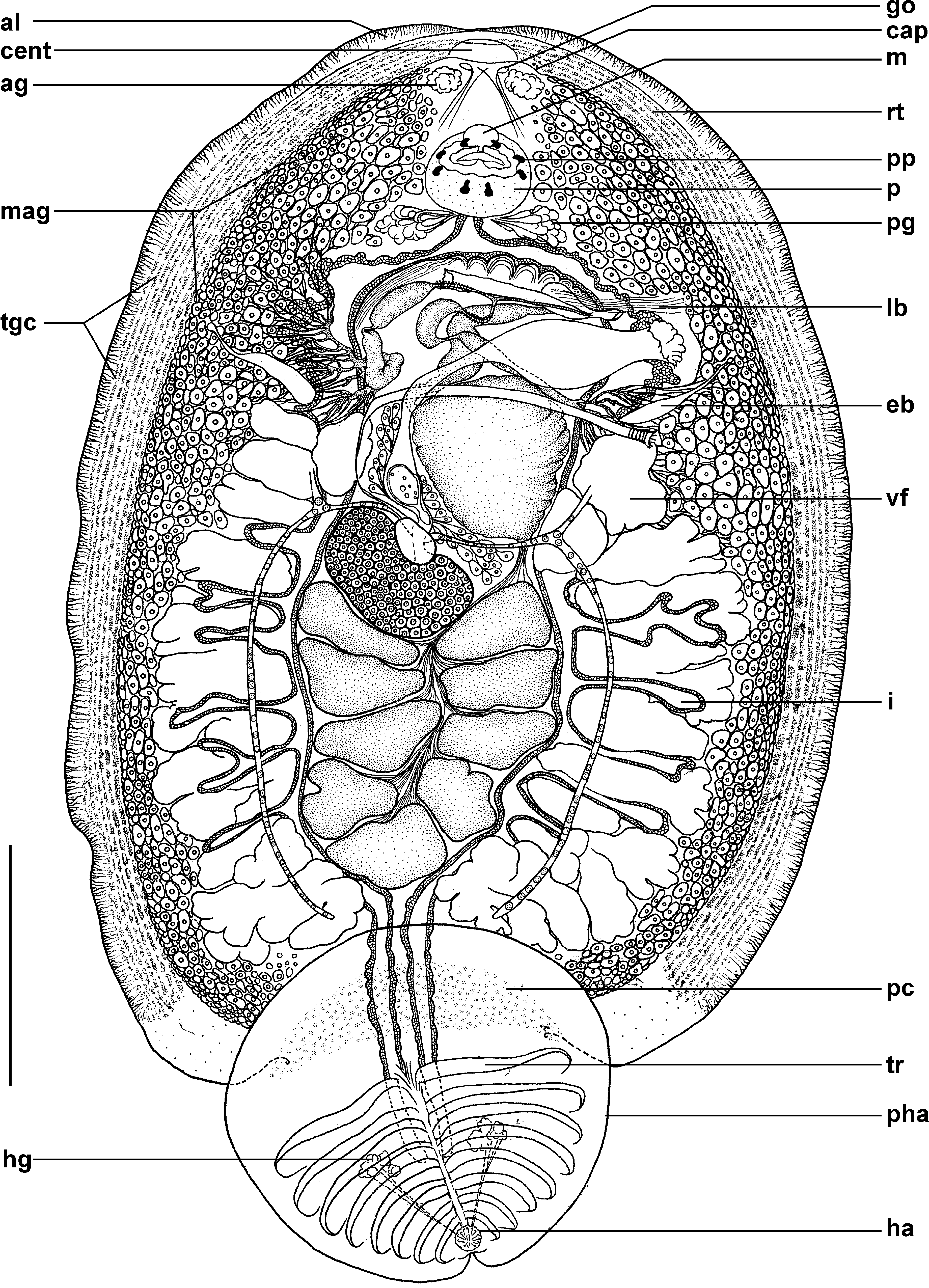
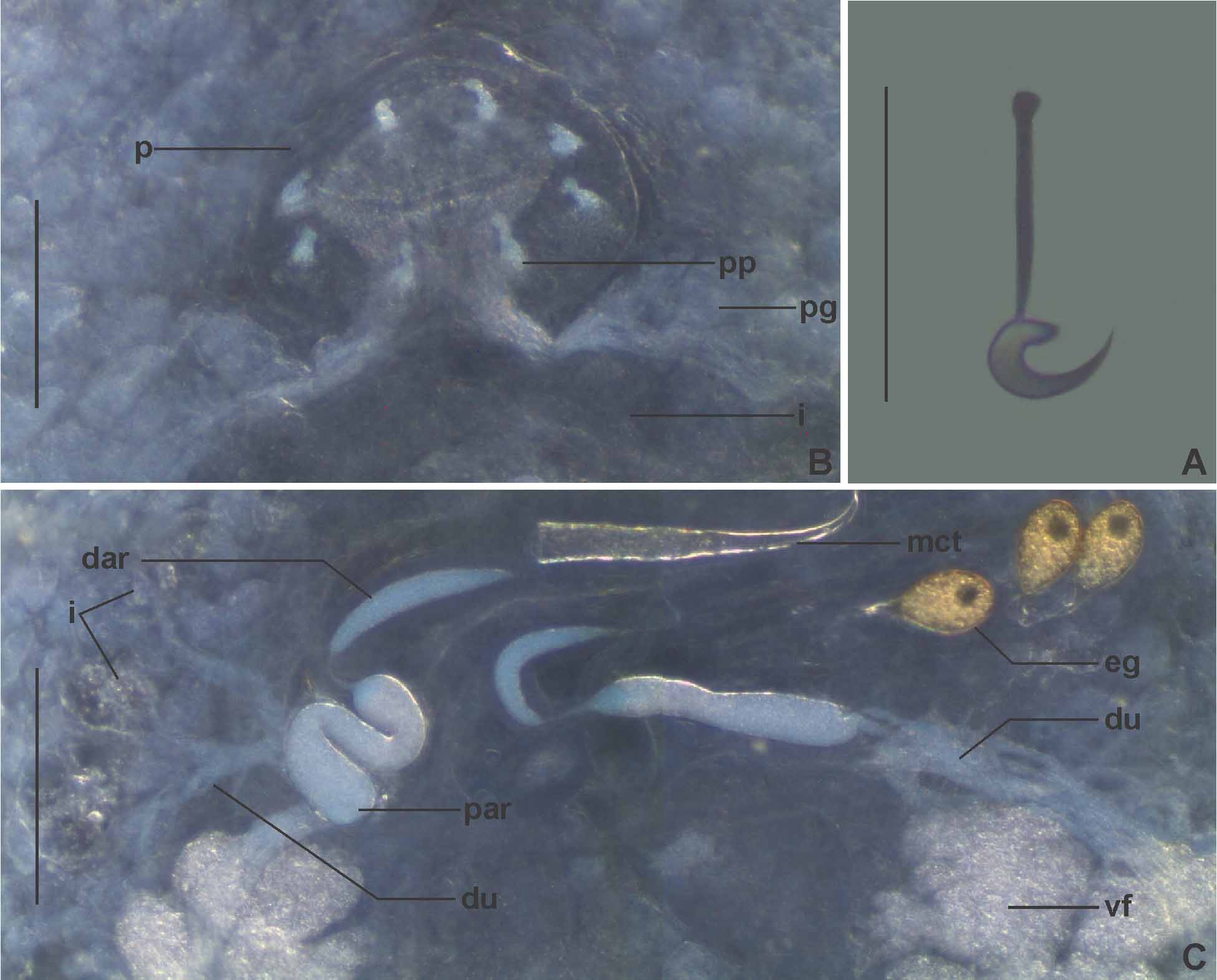
Description: Based on the mounted holotype and 26 paratypes, 4 adult specimens temporarily mounted in ammonium picrate solution, and observations made from 26 live adult specimens in vitro left unhindered or mounted in seawater under coverslip pressure. Total body ( Fig. 1 View FIGURE 1 ) including pseudohaptor 2053 + 517.2(1074–2967, n = 27) long, 1275 + 302.2(689–1801, n = 27) wide at widest part. Ventral furrows not observed. Pseudohaptor transversely ovoid 581 + 102.0(384–782, n = 27) long, 643 + 108.4(436–918, n = 27) wide with 8 transversely paired ridges decreasing in length from anterior to posterior. Pseudohaptor contains paired haptoral glands located roughly dorsal to third or fourth ridge either side of mid line with ducts leading to posterior part of haptor ( Fig. 1 View FIGURE 1 ). Haptoral glands produce adhesive secretion. Posterior portion of pseudohaptor turning inward near base of haptor. Anterior portion of pseudohaptor contains field of pigmented cells of unknown function with granular appearance ( Fig. 1 View FIGURE 1 ). Haptor circular, 47 + 3.9(41–54, n = 18) in diameter, ventral and posterior to pseudohaptor ( Fig. 1 View FIGURE 1 ), containing 16 marginal hooks; 14 peripheral and 2 central 16 + 0.5(15–17, n = 17) long ( Fig. 2 View FIGURE 2 A). Extensive field of gland cells arranged in longitudinal rows with fine ducts extends along the tegument from anterior-most portion of body to just short of posteriormost portion ( Fig. 1 View FIGURE 1 ). Mucous-like secretions associated with this field present in live specimens. Anterior end with two lobes and raised portion of tegument ( Fig. 1 View FIGURE 1 ). Confluent, adhesive protuberances with glands and gland openings situated anterior to pharynx, posterior to single, rounded anterior protuberance with groove ( Fig. 1 View FIGURE 1 ). Mouth ventral, anterior to pharynx ( Fig. 1 View FIGURE 1 ). Muscular pharynx 134 + 35.2(74–222, n = 25) long, 189 + 47.9(107–284, n = 25) wide with 8 papillae, symmetrically arranged in pairs; 1 pair located at the anterior portion of the pharynx, 2 lateral pairs, and 1 posterior pair ( Figs 1 View FIGURE 1 , 2 View FIGURE 2 B). Paired cluster of pharyngeal glands present on either side of posterior portion of pharynx ( Figs 1 View FIGURE 1 , 2 View FIGURE 2 B). Intestinal caeca bifurcate laterally posterior to pharynx before travelling posteriorly without undulating curves between field of testes and vitellarium. Lateral diverticula extending between vitelline follicles ( Fig. 1 View FIGURE 1 ). Intestinal caeca enter pseudohaptor, ending blindly near centre ( Fig. 1 View FIGURE 1 ). Excretory bladders present either side of body anterior to first vitelline follicle ( Fig. 1 View FIGURE 1 ).
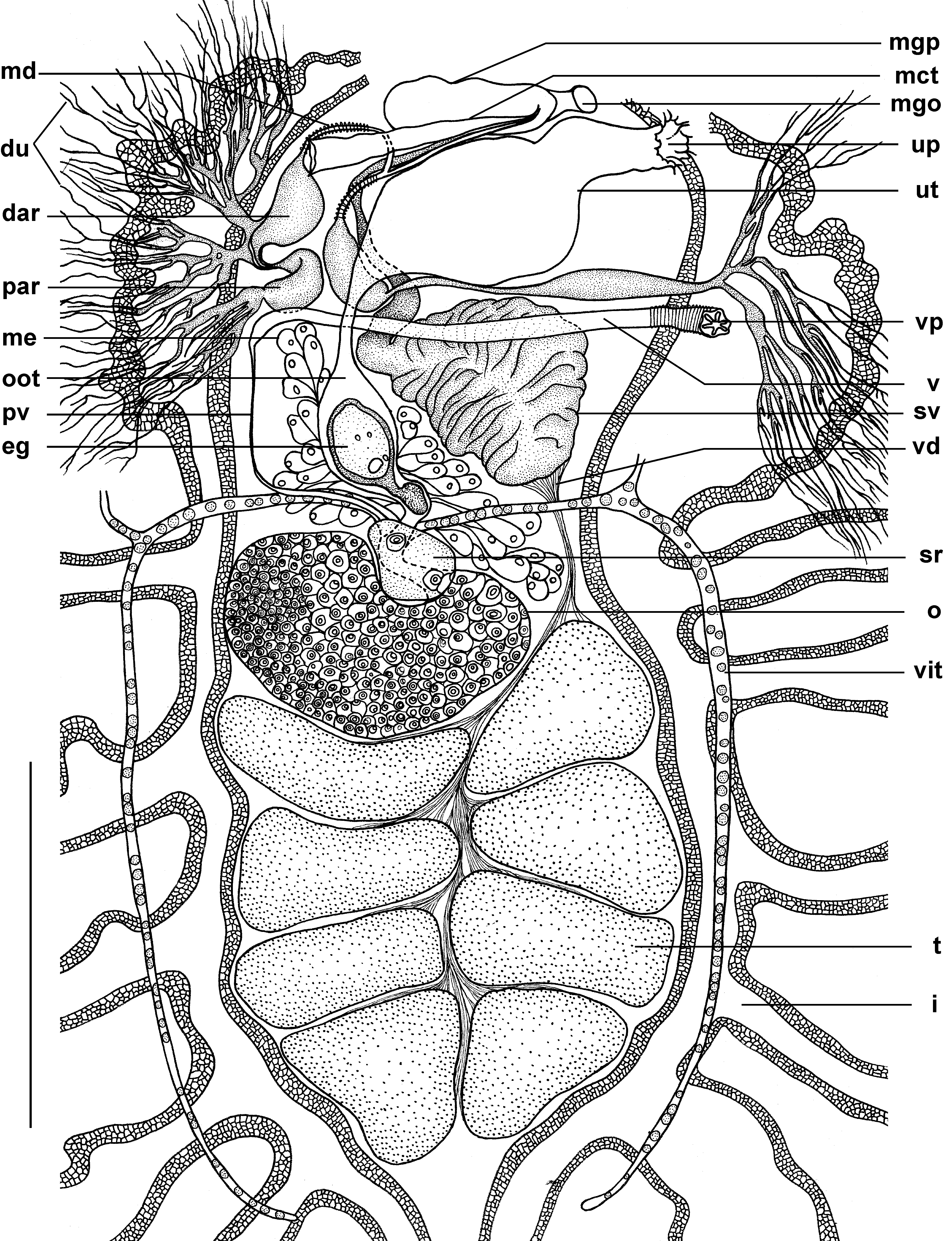
Eight or 9 testes, each 133 + 37.2(65–205, n = 27) long, 202 + 51.2(105–286, n = 27) wide situated in posterior half of body with anterior-most testis situated left or left and slightly posterior to ovary ( Figs 1 View FIGURE 1 , 3 View FIGURE 3 .) Vasa efferentia from each testis form a thin vas deferens travelling anteriorly, left of ovary to base of large expanded seminal vesicle ( Figs 1 View FIGURE 1 , 3 View FIGURE 3 ). Anterior region of seminal vesicle narrows abruptly to form tubular proximal portion of vas deferens travelling left, running ventral to male accessory gland reservoir junction, following length of male copulatory tube and terminating within male genital pocket ( Figs 1 View FIGURE 1 , 3 View FIGURE 3 ). Male accessory gland reservoirs located in anterior portion of body, either side of seminal vesicle; separated into distal and proximal parts ( Figs 1 View FIGURE 1 , 2 View FIGURE 2 C, 3). Proximal part termini of male accessory gland reservoirs associated with vast intricate network of ducts ( Figs 1 View FIGURE 1 , 2 View FIGURE 2 C, 3) leading ventrally over respective caecum from extensive field of male accessory gland cells extending the length of body proper ( Fig. 1 View FIGURE 1 ). Proximal part extends anteriorly, narrowing abruptly to form thin connection to swollen muscular distal part ( Figs 1 View FIGURE 1 , 2 View FIGURE 2 C, 3). Ducts leading from narrowing distal parts join dorsally and slightly ventral to, or in line with male copulatory tube forming common duct ( Figs 1 View FIGURE 1 , 3 View FIGURE 3 ). Common duct leads along male copulatory tube, terminating at its distal portion within male genital pocket ( Figs 1 View FIGURE 1 , 3 View FIGURE 3 ). Single hollow-tubular, sclerotised male copulatory tube 269 + 64.0(143–369, n = 27) long, 39 + 9.6(22–53, n = 27) wide at its base curved distally, tapering to point, positioned posterolateral to pharynx ( Figs 1 View FIGURE 1 , 2 View FIGURE 2 C, 3, 4A). Male genital pocket 163 + 37.4(97–256, n = 17) long, 73 + 13.7(46–105, n = 17) wide, with ventral opening anterior to uterine pore ( Figs 1 View FIGURE 1 , 3 View FIGURE 3 ). Male genital pocket associated with short lateral band of muscle fibres ( Fig. 1 View FIGURE 1 ).
Ovary roughly kidney-shaped, 152 + 40.6(56–234, n = 27) long, 239 + 53.7(124–311, n = 27) wide, positioned right of body mid-line ( Figs 1 View FIGURE 1 , 3 View FIGURE 3 ). Small ovoid seminal receptacle 51 + 11.2(32–75, n = 24) long, 75 + 13.2(49–93, n = 24) wide, cushioned within the anterior concave portion of ovary, ventral to short common vitelline duct ( Figs 1 View FIGURE 1 , 3 View FIGURE 3 ). Short ovovitelline duct exits left side of common vitelline duct, posterior to transverse vitelline duct, entering base of oötype. Oötype crossing ventrally under transverse vitelline duct, travelling anteriorly, right of proximal portion of seminal vesicle ( Figs 1 View FIGURE 1 , 3 View FIGURE 3 ). Extensive network of Mehlis’ glands present ( Figs 1 View FIGURE 1 , 3 View FIGURE 3 ). Oötype narrows to form uterus crossing ventrally, left under distal portion of seminal vesicle, opening at uterine pore left and posterior to male genital pore ( Figs 1 View FIGURE 1 , 3 View FIGURE 3 ). Eggs (in utero) 126 + 11.4(102–151, n = 21) long, 54 + 4.3(44–63, n = 21) wide with short polar filament with basal disc ( Figs 1 View FIGURE 1 , 2 View FIGURE 2 C). Eggs stored in distal portion of uterus ( Fig. 2 View FIGURE 2 C). Vaginal pore inconspicuous, muscular, left of body, anterior to first vitelline follicle ( Figs 1 View FIGURE 1 , 3 View FIGURE 3 ). Vagina 759 + 171.9(390–1043, n = 17) long, travels laterally across body, ventral to anterior region of proximal portion of seminal vesicle, dorsal to oötype ( Figs 1 View FIGURE 1 , 3 View FIGURE 3 ). After passing behind oötype, vagina widens slightly, with sharp posterior turn, ventral to intestinal caecum, narrowing abruptly as it travels ventral to vitelline follicles before widening, turning left and following transverse vitelline duct ventrally before entering seminal receptacle ( Figs 1 View FIGURE 1 , 3 View FIGURE 3 ). Field of lobed vitelline follicles arranged laterally extends along either side of body beginning at level of oötype, ending short of pseudohaptor ( Fig. 1 View FIGURE 1 ).
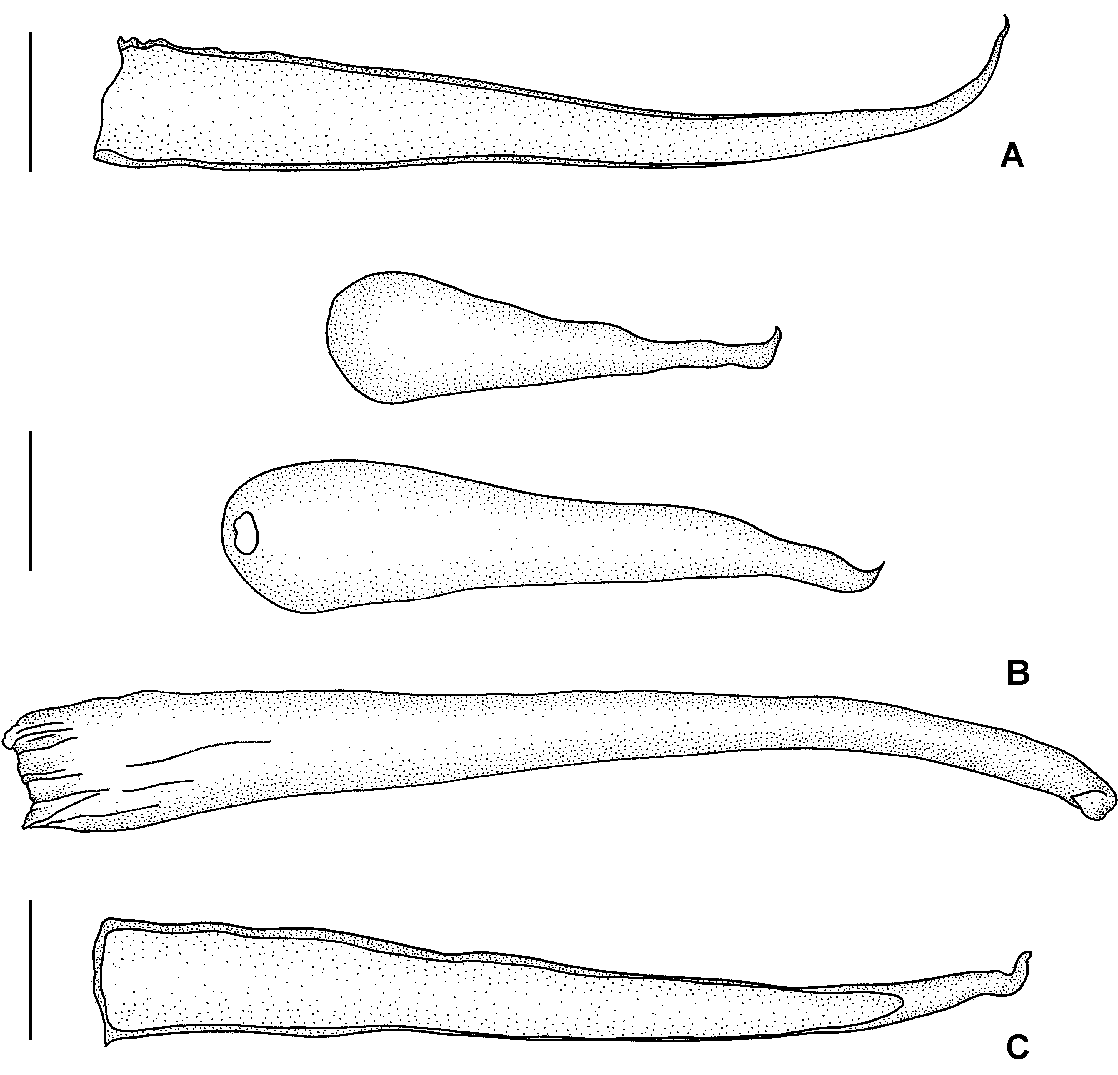
Remarks: Myxinidocotyle eptatreti is distinguished from M. californica and M. japonica primarily by the shape of the single sclerotised male copulatory tube which is curved distally and tapers to a point ( Fig. 4 View FIGURE 4 A). Although the male copulatory tubes of M. japonica also taper to a point, this species has 2 or 3 tubes arranged above one another and the curve of the tip of each tube is more acute ( Fig. 4 View FIGURE 4 B). The single sclerotised male copulatory tube of M. californica ends in a hook-like prong ( Fig. 4 View FIGURE 4 C). Myxinidocotyle eptatreti is further distinguished from other species as it is currently the only species with an intestinal caecum with lateral diverticula.
No known copyright restrictions apply. See Agosti, D., Egloff, W., 2009. Taxonomic information exchange and copyright: the Plazi approach. BMC Research Notes 2009, 2:53 for further explanation.