
Agnetaria Bruce, 1953
|
publication ID |
https://doi.org/ 10.37520/aemnp.2022.006 |
|
publication LSID |
lsid:zoobank.org:pub:42A5070B-F287-4B14-84A1-A57F7E274CE6 |
|
persistent identifier |
https://treatment.plazi.org/id/03B2F470-DB79-5579-D282-FF74A7B2F6B4 |
|
treatment provided by |
Felipe |
|
scientific name |
Agnetaria Bruce, 1953 |
| status |
|
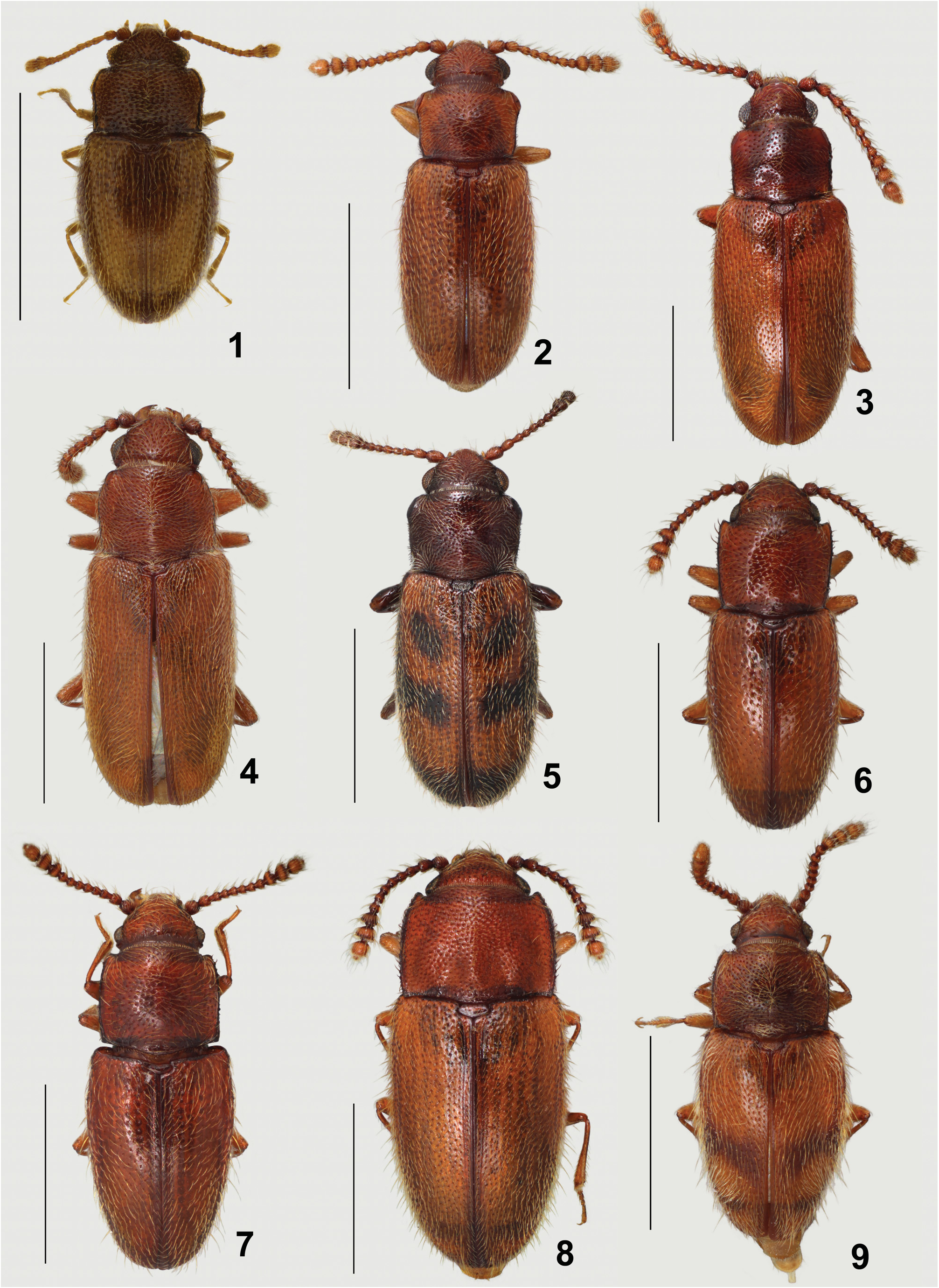
( Fig. 1 View Figs 1–9 )
Agnetaria Bruce, 1953: 790 . Type species: Agnetaria cryptophagoides Bruce, 1953 , by monotypy.
Type material examined. Label data for the holotype / lectotype are as follows: “Type [red-bordered circle] // Typus [red label] // Port Lincoln, / S. Australia. / (Blackburn) // Port Lincoln / Blackburn [handwritten] // Sharp Coll / 1905-313.// Agnetaria cryp- / tophagoides n.sp. [handwritten] / N. BRUCE det. // HOLOTYPE / Agnetaria cryptophagoides / Bruce / det.Gimmel & Leschen [red label] // LECTOTYPE / Mycetaea pilosella / Blackburn / des.Gimmel & Leschen [red label]” (deposited in NHMUK).
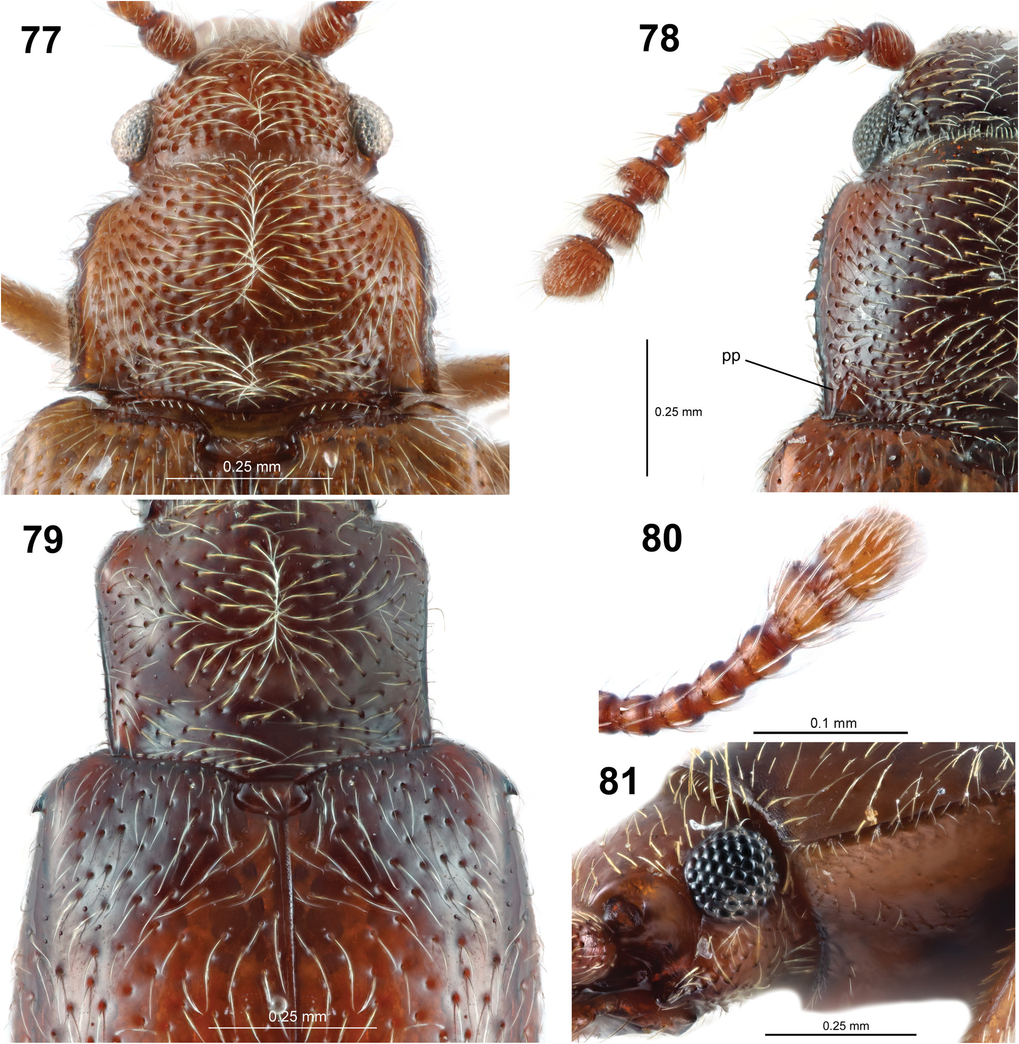
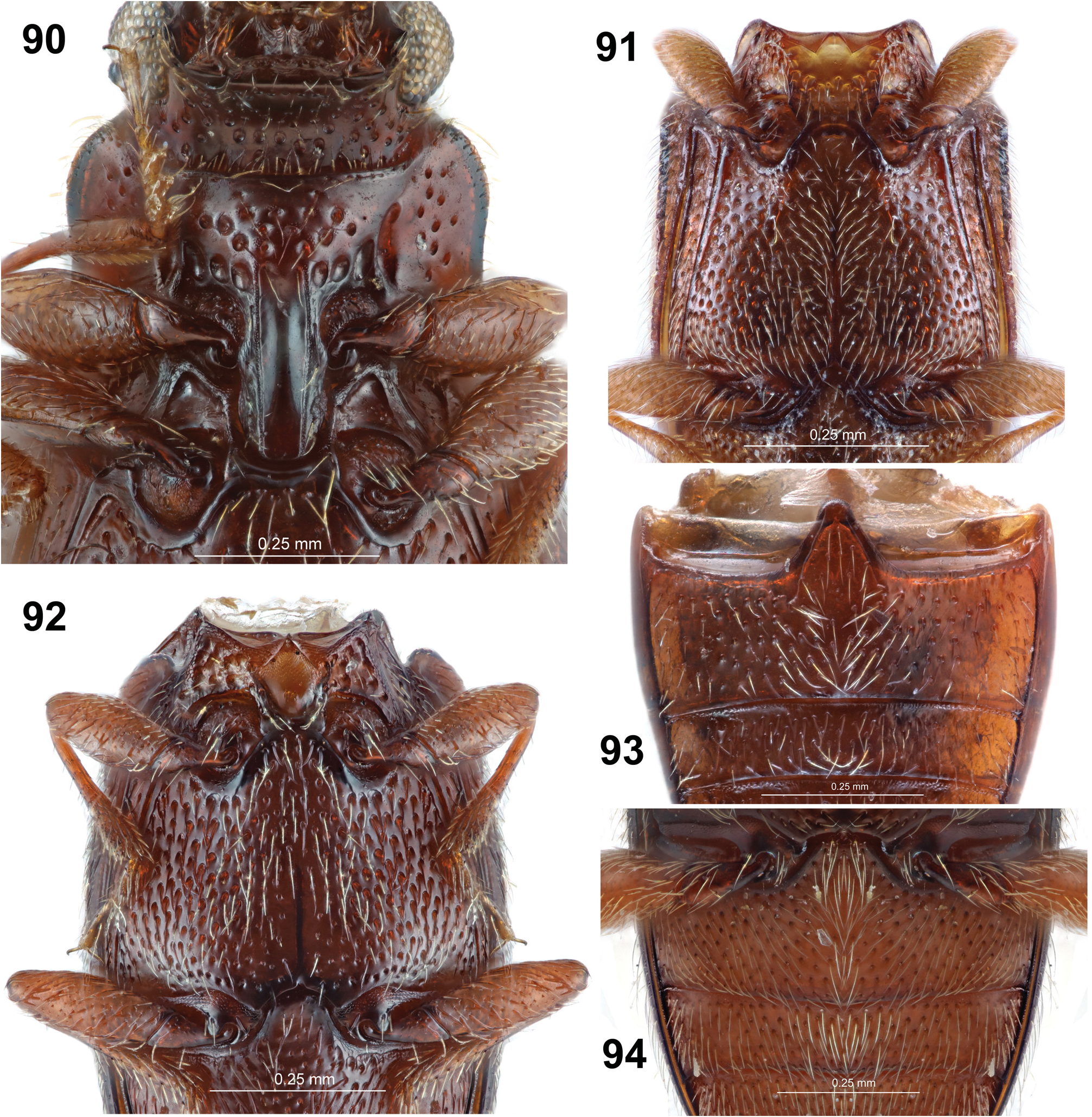
Diagnosis. This genus may be recognized among Picrotini by the free abdominal ventrites, lack of pronotal platforms or tumidities (although the pronotum is slightly expanded in the anterior 1/3), smooth lateral pronotal carina, densely setose dorsal surface and antennal club clearly consisting of only two antennomeres. The only other genera of Picrotini with such an antennal club are Paragnetaria ( Fig. 80 View Figs 77–81 ) and Picrotus , but in these either abdominal ventrites 2–3 are connate or the body surface is mostly glabrous (with few or no setae), or both. Additional diagnostic characters for Agnetaria include the short discrimen, less than half the length of metaventrite, and lack of postcoxal lines on abdominal ventrite 1 (as in Fig. 94 View Figs 90–94 ).
Redescription. Length 1.20–1.39 mm. Body form ( Fig. 1 View Figs 1–9 ) stout, somewhat shining dorsally, with dense decumbent setae and long, sparse, erect setae on head and elytra; cuticle unicolorous. Head with tempora inconspicuous, length less than 1/4 length of eye; vertex with very weak temporal depression immediately anterior to ridge; band of reticulate sculpture absent. Frontoclypeus not projecting laterally; raised portion of frons between antennal insertions constricted, about as narrow as width of antennal club. Transverse ridge above antennal insertions absent. Eye medium-sized, not reduced, conical, contacting antennal cavity; interfacetal setae present. Antennal club consisting of 2 antennomeres; antenna inserted into small cavity; antennomere 9 subequal in width to antennomere 8. Mandible with apex bifid, subapical serrations present. Maxillary palpomere 4 distinctly longer than 3; palpomere 4 not subulate. Gena with weakly indicated antennal groove ventral to eye; genal spines acute. Gular sutures incomplete, not reaching occipital foramen. Pronotum moderately explanate, narrower than elytra and not constricted at base, and widest anteriorly; anterior angles obtuse, slightly projecting anteriorly (extending anterior to cervical foramen of prothorax), without a distinct flat surface, platform or tumidity, but pronotal edges slightly projecting laterally about 1/3 from apex; lateral carina complete, smooth, lacking teeth, crenulations, or setigerous tubercles, with lateral glabrous space narrow, width of lateral bead wider than discal puncture and narrower than antennal funicle; disc with transverse basal impression; paramedial carinae and paralateral plicae absent; posterolateral angles about right angled. Prothoracic hypomeron fused to prosternum. Prosternum with anterior margin on same plane as disc; prosternal process with lateral marginal beads parallel, with central longitudinal carina, slightly expanded apically, broadly rounded and crenulate with minute setae; procoxal cavity with anterolateral notch. Scutellar shield clearly visible, transverse, truncate apically. Elytron without humeral tooth; subbasal and subapical impressions absent; subapical gape present; punctation confused and well impressed; vestiture dual with long, sparse, erect setae present, decumbent setae uniformly directed posteriorly. Hind wing well developed. Mesoventrite with mesoventrital cavity bowl-like and flanked by sharp carinae. Mesanepisternal pit present and lined with setae. Metaventrite with indistinct, scalloped postcoxal lines; discrimen less than 1/2 length of metaventrite, posterior notch of metaventrite absent. Metendosternite with anterior tendons approximate. Tarsi 5-5- 5 in female, male condition unknown; tarsi moderately slender, tarsomere 5 as wide as preceding tarsomeres in lateral view; pro- and mesotarsomere 4 without ventral setae; mesotarsomere 3 unlobed; mesotarsomeres 1–4 of equal lengths, mesotarsomere 5 about as long as mesotarsomeres 3 and 4 combined. Abdominal ventrites free and lacking calli, intersegmental crenulations absent; ventrite 1 with intercoxal process broadly rounded, lacking postcoxal lines; medio-basal thickenings of ventrites 3–5 absent; apex of ventrite 5 lacking crenulations. Abdominal spiracles with opening on segment VII present and larger in diameter than spiracle VI, texture granulate and atrium rounded and saclike. Aedeagus not examined.
Remarks. Regrettably, LൾඌർHൾඇ & Gංආආൾඅ (2012) overlooked a BඅൺർKൻඎඋඇ (1891: 122) species name which has priority over the Bඋඎർൾ (1953: 790) species name. According to MൺඍඍHൾඐඌ (1992: 11), Nils Bruce unknowingly redescribed the unlabeled syntype series of Mycetaea pilosella Blackburn, 1891 (originally placed in Mycetophagidae ) as Agnetaria cryptophagoides Bruce, 1953 . We here designate the lectotype of M. pilosella as the holotype of A. cryptophagoides , thus formalizing the objective synonymy: Agnetaria pilosella ( Blackburn, 1891) = Agnetaria cryptophagoides Bruce, 1953 , syn. nov. Male specimens of this species were not available for study. A habitus line drawing of A. pilosella was provided in MൺඍඍHൾඐඌ (1992: fig. 44).
Biology. The three specimens we examined were collected by Berlese funnel of mallee litter. The gut of one dissected specimen was packed with unidentifiable material, suggesting saprophagy.
Distribution. Australia.
Included species (1). Agnetaria pilosella ( Blackburn, 1891) .
| NHMUK |
Natural History Museum, London |
No known copyright restrictions apply. See Agosti, D., Egloff, W., 2009. Taxonomic information exchange and copyright: the Plazi approach. BMC Research Notes 2009, 2:53 for further explanation.
|
Kingdom |
|
|
Phylum |
|
|
Class |
|
|
Order |
|
|
Family |
Agnetaria Bruce, 1953
| Gimmel, Matthew L. & Leschen, Richard A. B. 2022 |
Agnetaria
| BRUCE N. 1953: 790 |