
Euthygomphus schorri, Kosterin, Oleg E., 2016
|
publication ID |
https://doi.org/ 10.11646/zootaxa.4171.1.2 |
|
publication LSID |
lsid:zoobank.org:pub:28AA1836-9D39-4CD9-990B-C1E900863FE5 |
|
DOI |
https://doi.org/10.5281/zenodo.6070310 |
|
persistent identifier |
https://treatment.plazi.org/id/03B30362-2362-1E25-EBFB-FE2C9DCBFDB7 |
|
treatment provided by |
Plazi |
|
scientific name |
Euthygomphus schorri |
| status |
sp. nov. |
Euthygomphus schorri View in CoL sp. nov.
Fig. 7 View FIGURE 7 c–d, 8–11
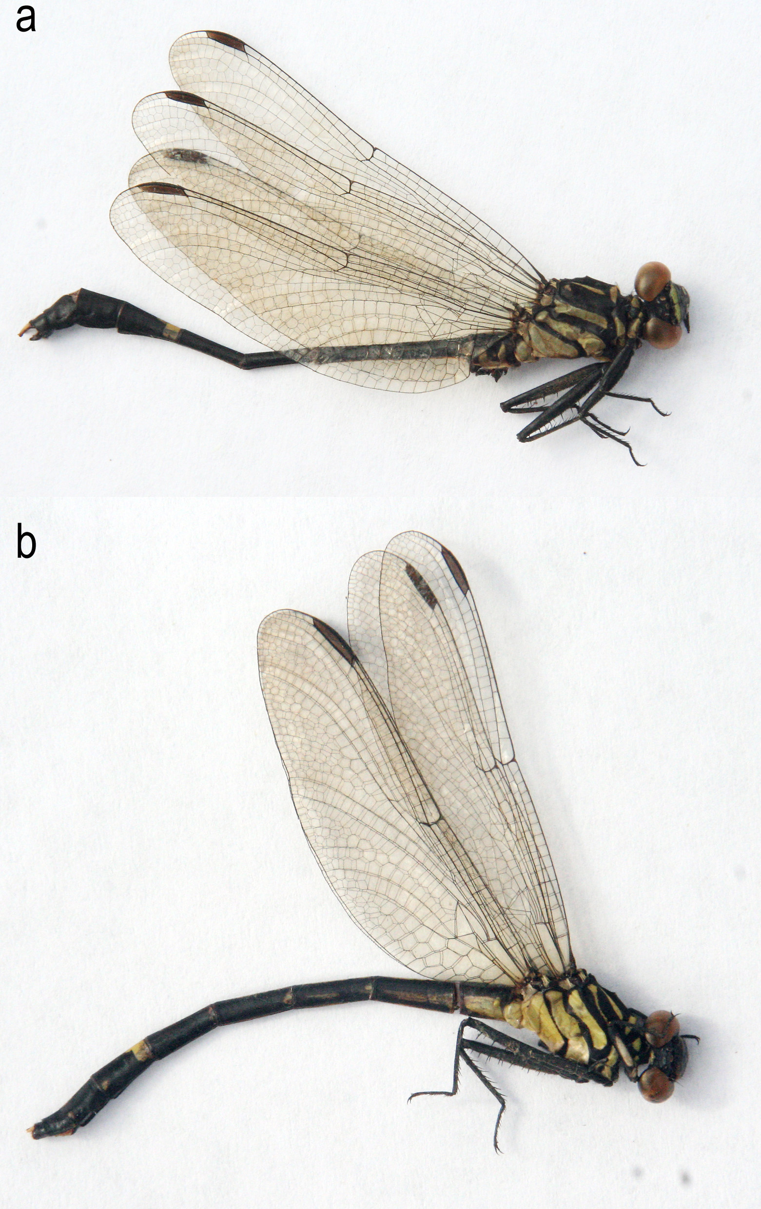
Type material. Holotype: ♂ (dry in envelope) ( Fig. 9 View FIGURE 9 a, 10a–d, 11a–j), Cambodia, Mondulkiri Province, 3.5–3.8 km ESE of Sen Monorom, ‘ Culminicola Rivulet’ , 12°26'43–53'' N 107°13' 00–20'' E, 689–691 m a.s.l., 8 vi 2014, O. Kosterin leg., deposited in RMNH . Paratypes: 1♀ (female 1, dry in envelope) ( Fig. 10 View FIGURE 10 j), the same locality and date (in RMNH) ; 1♀ (female 2, dry in envelope) ( Fig. 9 View FIGURE 9 b, 10e–i, 11k), Cambodia, Mondulkiri Province, downstream of Buu Sraa Waterfalls, ‘ Loringae brook’ just downstream from its own waterfall, 12°34’01'' N, 107°24'50'' E, 490 m a.s.l., 15 vi 2014 O. Kosterin leg (in the author’s collection). GoogleMaps
Etymology. The new species is dedicated to Martin Schorr, a passionate odonatological enthusiast who for a long time has edited and managed the International Dragonfly Fund, which supports so much important odonatological research worldwide (including this Cambodian survey) and publishes an excellent, rapidly issued, richly illustrated, flexible in format, well-known and respected odonatological journal IDF-Report.
Short diagnosis. A Euthygomphus with unique male cerci and female vulvar scale. Male cerci simple, yellow, arched in lateral view and diverging by curving outside each other in dorsal view, apically furnished with a yellow tooth directed up and behind, two small black terminal tubercles and two small ventral knobs. Vulvar scale triangular, with its lobes touching each other for quite a distance and pointed apically, with straight outer and rounded inner margins. Males without and female with expressed antehumeral yellow stripes; occiput black in males, with a broad yellow spot behind ridge and a pair of very closely set, long spines in females.
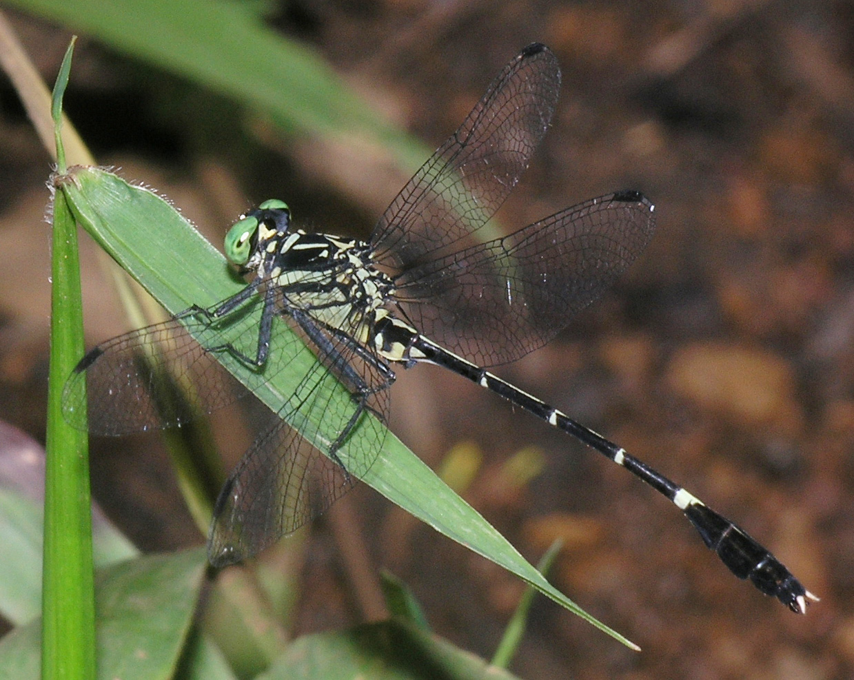
Holotype male. Body black with yellow markings ( Fig. 8 View FIGURE 8. A , 9 View FIGURE 9 a, 10a–c).
Head. Eyes green in life ( Fig. 8 View FIGURE 8. A ). Face black with the following yellow pattern ( Fig. 10 View FIGURE 10 c). Labium with blackish brown prementum and postmentum contrasting with yellowish sides of postmentum and lateral lobes ( Fig. 10 View FIGURE 10 a). Mandible bases entirely yellow. Labrum with a pair of large lateral yellow spots shifted to its dorsal margin, with black space between them as broad as 2/3 of their length. A broad, straight, yellow stripe across frons dorsal surface, its hind border notched at middle, its sides below bases of antennae narrower, ends rounded. Antennal segments 1 and 2 black with light brownish grey apices, rest of antenna brown. Top of head black, a pair of gentle roundish ridges behind lateral ocelli, behind them vertex flat, occiput perfectly straight ( Fig. 10 View FIGURE 10 d).
Thorax black with yellow pattern ( Figs. 9 View FIGURE 9 a, 10a–b).
Prothorax with a pair of moderately large yellow spots at lateral sclerites; median lobe with large lateral spots and a pair of small, oval spots close to each other; anterior lobe yellow; posterior lobe black, hairy. Synthorax with following pale pattern. Collar stripes well separated dorsally from each other. Straight, broad dorsal stripes, pointed anteriorly and rounded posteriorly, separated from collar stripes for a distance equal to the width of the latter ( Fig. 10 View FIGURE 10 b). Relatively large, mushroom-shaped antealar spots. Large yellow spot occupying ventral and most of central parts of inframesepisternum. Sides ( Fig. 10 View FIGURE 10 a) yellow with two broad, continuous black stripes along sutures, 1st one bent behind and 2nd one somewhat expanding forward in their upper parts, forming a 'neck' of 2nd yellow space. A short and narrow black streak at upper part of metepimeron hind margin.
Legs black but profemora outer sides yellowish; coxae with broad yellow stripes at outer surface. All femora with two ventral rows of numerous small short spines, metafemora also with some similar spines between them; femora sides with sparse smaller pointed knobs, on mesofemora indistinctly and metafemora distinctly arranged into a medial row; metafemora in distal 2/3 also with six strong and very long ventral spines. Metafemora extend to midway between posterior hamuli and vesicle. Tibiae with four sharp ridges, of which two inner rows with numerous, dense small spinules and two adaxial rows set with rather sparse strong spines as follows: mesotibia: 9– 10 in outer row, 12 in inner row; metatibia: 10 in inner and outer rows; protibia: 8–9 strong spines followed in apical part by some 7–8 very dense and shorter brownish spines, and numerous further smaller spines appear between outer rows.
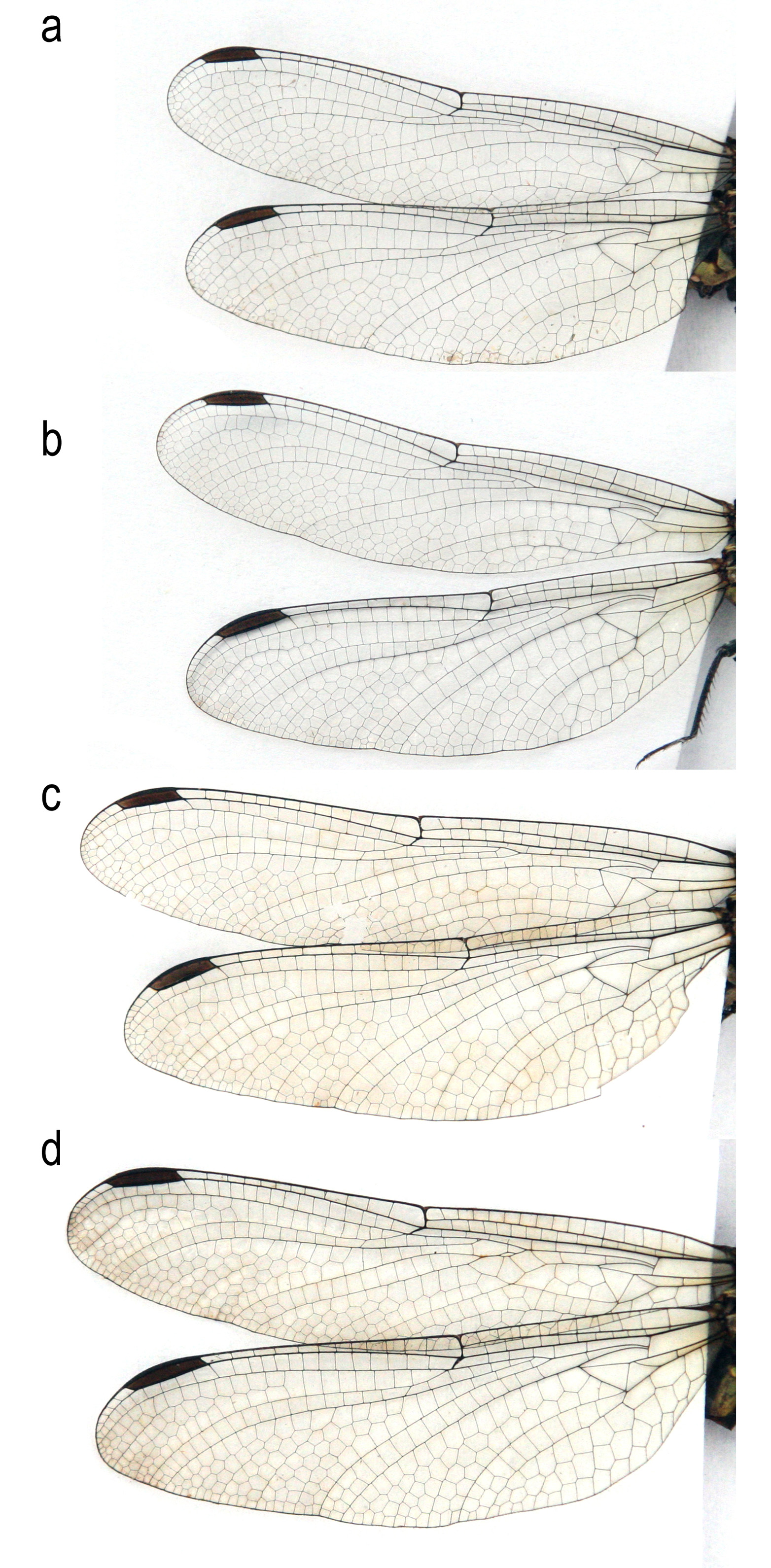
Wings ( Fig. 7 View FIGURE 7 c) hyaline, very slightly tinted brownish; venation blackish brown. Arculus opposite 2nd antenodal. Antenodals (1st and 5th primary) 14 on forewings, 10 (right) – 11 (left) on hindwings; in addition, forewings with an incomplete basal antenodal. Postnodals 10 (right) – 12 (left) on forewings, 9 (left) – 11 (right) on hindwings. One cubito-anal vein on all wings. Triangles not crossed. Three crossveins between Arc and R1-R4 junction on forewings and one on hindwings, both above and below Rs. Anal loop not defined. Anal triangle well formed, 3-celled, middle cell very large, wing margin straight here. Tornus gently rounded. Membranule extremely narrow, almost absent, brownish. Pterostigmata brown, with black bordering veins, of which lengthwise ones swollen, below covering 4 cells.
Abdomen narrow at S3–6, slightly expanding at S7–9, black with yellow markings as follows ( Fig. 8 View FIGURE 8. A , 9 View FIGURE 9 a): S1 with a dorsal triangle directed anteriorly, sides entirely yellow. S2 with a dorsal yellow spot of a complicated shape, consisting of a central elongate hexagonal ‘shield’, a distal ‘neck’ and a fine proximal streak; ventral sides yellow with that colour expanding behind auricles, as a rectangular projection; auricles also yellow. S3–4 with a fine yellow dorsal streak and anterolateral yellow spots (that on S3 larger and triangular); ventral margins narrowly yellow (but this colour fades at S4 distal part). S5–6 only with small anterolateral spots. S7 with an anterodorsal yellow rings with rectangular ventral sides, occupying ¼ of segment length, which a central proximal extension shaped as candle flame and extending to 40% of segment length. S8–10 entirely black.
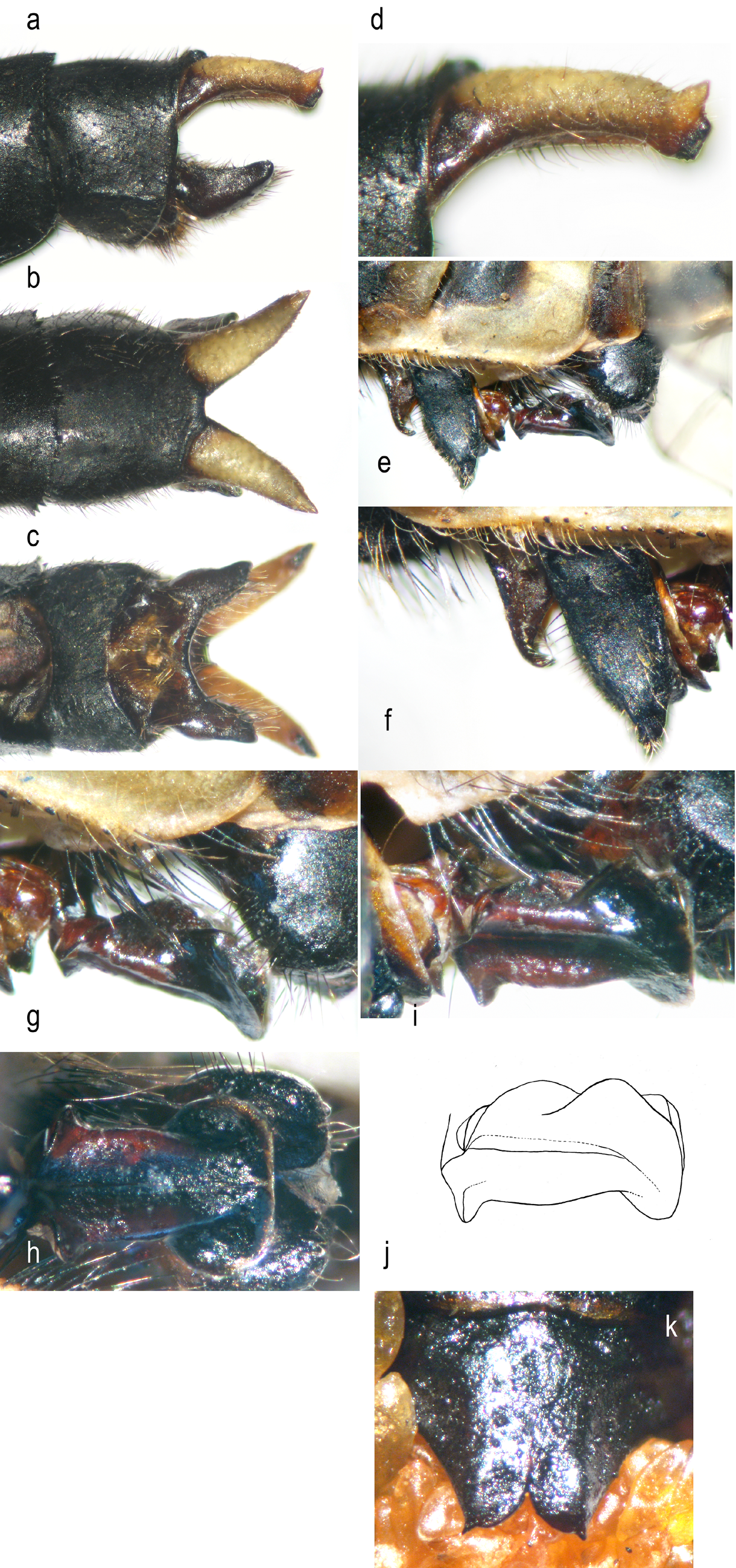
Cerci ( Fig. 11 View FIGURE 11 a–d) yellow, brownish at very base and inner margin; epiproct brownish black. Cerci simple, in lateral view ( Fig. 11 View FIGURE 11 a,d) slightly arched and ending with a yellow, dorsoposteriorly directed apical tooth, two blunt black terminal knobs and two small black subapical ventral teeth on an elongate black spot ( Fig. 11 View FIGURE 11 c). Epiproct branches ca 3/4 as long as cerci, in lateral view ( Fig. 11 View FIGURE 11 a) broadly sickle-shaped, with evenly slightly concave upper margin and evenly strongly convex lower margin, rounded apically. In dorsal view ( Fig. 11 View FIGURE 11 b), cerci diverging by evenly curving out from each other, pointed; diverging epiproct branches slightly extending beyond cerci. In ventral view ( Fig. 11 View FIGURE 11 c), epiproct branches slightly diverging, rounded, with a large evenly roundish incision between them.
Accessory genitalia ( Fig. 11 View FIGURE 11 e–j): Anterior lobe moderately prominent, black, with dense, long, light hairs. Tergite margins with two series of light hairs, short ones behind hamuli and long ones on a ledge between hamuli and vesicle. Anterior hamulus broad at base, apically tapering, elegantly curving and hooked behind ( Fig. 11 View FIGURE 11 e–f). Posterior hamulus in lateral view ( Fig. 11 View FIGURE 11 e–f) leaf-shaped, tapering and slightly and bluntly hooked ahead at a pointed apex, with inner blunt projections seen from sides (those of both hamuli touch each other), with dense short light hairs at a coarse anterior margin and sparse hairs at lateral sides. Anterior hamulus dark brown, posterior hamulus black ( Fig. 11 View FIGURE 11 e–f). Vesicle of penis (V1) in lateral view rounded triangular, with sparse moderately long light hairs ( Fig. 11 View FIGURE 11 e). Penis distal segment (V4) generally of a complicated saddle-like shape ( Fig. 11 View FIGURE 11 g–j), in ventral view rather mushroom-shaped ( Fig. 11 View FIGURE 11 h). It has a broad base and a very broadly expanding apex. Its ventral surface has a central lengthwise groove. In lateral ( Fig. 11 View FIGURE 11 g) and oblique ( Fig. 11 View FIGURE 11 i–j) views, ventral and dorsal apical margins look like outer and inner collars, the former very broad and the latter narrower. There are two roundish dorsal keels ( Fig. 11 View FIGURE 11 i–j).
Measurements (mm). Forewing 29, hindwing 28, abdomen without appendages 33.5; total length 45; hind femur 7.8; forewing pterostigma 3.0, hind wing pterostigma 3.3.
Variation in males. A male of this species photographed but not collected at a brook upstream of Buu Sraa Waterfall ( Fig. 8 View FIGURE 8. A ) had somewhat more extended yellow pattern: there is a trace of antehumeral stripe below antealar spot; anterolateral spots on S4–6 larger, those on S6 almost forming a ring.
Female. Similar to male but with a more extended yellow pattern ( Fig. 9 View FIGURE 9 b, 10f–g), differing in the following traits. Prementum lighter, brownish-grey in female 1 and whitish grey, as labial palpi, in female 2, but its anterior margin black ( Fig. 10 View FIGURE 10 g). In female 1 anteclypeus also lighter, brownish-black. Occipital ridge with a central pair of black, narrow, straight spines with brownish bases set close to each other ( Fig. 10 View FIGURE 10 e,i–j): in female 1 they are set very closely, with a rounded notch between them, but broken ( Fig. 10 View FIGURE 10 j), in female 2 they are acute, long, ca 1.5 times longer than space between them ( Fig. 10 View FIGURE 10 e). Occiput behind occipital ridge with a broad oval yellow spot occupying most of its hind part ( Fig. 10 View FIGURE 10 i–j).
Dorsal ridge of synthorax with a small yellow spot anteriorly of its pointed prominence. Mesepisterna with large triangular antealar spots and narrow antehumeral stripes, interrupted in upper part in female 1 and contiguous to antealar spots in female 2 ( Fig. 10 View FIGURE 10 g).
Antenodals 13–15 (plus incomplete basal one) in forewings, 9–11 in hindwings (in left hindwing of female 1, the second primary antenodal is 6th); postnodals 10–12 in forewing, 11–12 in hindwings ( Fig. 7 View FIGURE 7 d). In female 1 forewings, 4 and 5 crossveins between Arc and R1–R4 junction above Rs, and 2 cubito-anal veins on right forewing.
Abdomen with following dorsal yellow pattern ( Fig. 9 View FIGURE 9 b): S1–S2 with a continuous dorsal yellow stripe, broadening proximally on S2, S3–5 with a narrow dorsal streak interrupted at distal segment margin, S6–S8 with a finest dorsal streak widened proximally at S6 and distally at S7–8, S7 with the same ring as in male, S9 with an elongate doroposterior spot. Lateral yellow pattern as follows: S1 sides yellow, S2 with lower tergite halves yellow but black at anterior and ventral sides (auricles present, although smaller than in male); S3 with broad lateral stripes throughout, broadening to proximal margin; S4–S6 with triangular lateral spots at proximal margin; in female 2, S4 also with an elongate lateral stroke at middle; in female S7–S8 with small lateral spots at posterior margin but only small traces of them in female 1. Ventral tergite margins outlined with yellow at S3–6. Cerci short, simple, pointed, bright-yellow.
Vulvar scale ( Fig. 11 View FIGURE 11 k) triangular in outline and ends with two broad lobes with a deep but so narrow incision between them that they touch each other; lobe outer margins straight and end with apical spines, inner margins rounded apically.
Measurements (mm): forewing 30–31, hindwing 29–30; abdomen without appendages 34–35; total length 45– 47; hind femur 7.5; fore pterostigma 3.5, hind pterostigma 3.8.
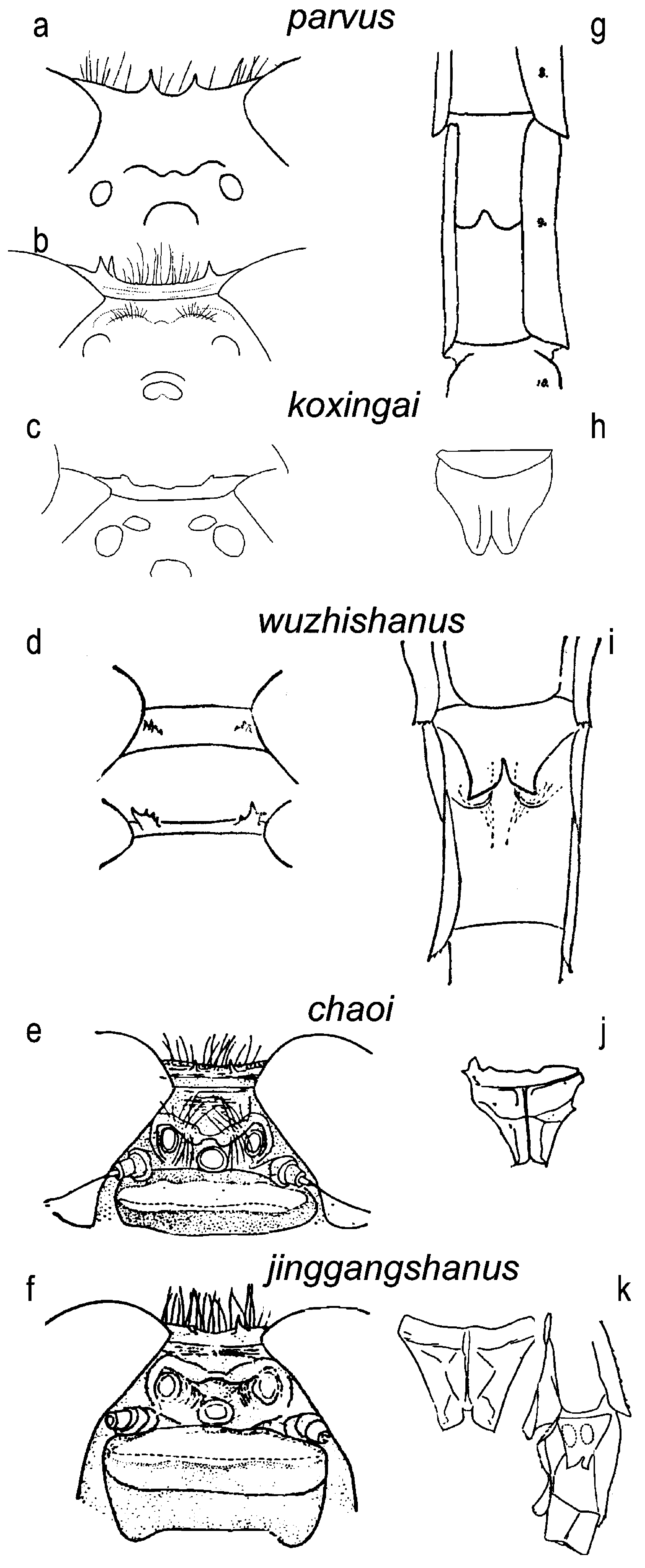
Differential diagnosis. The unique cerci, set closely but curving outside each other in dorsal view and furnished apically with a strong upward-directed tooth, two small black terminal tubercles and two small ventral teeth, provide the best feature differentiating males of the new species from other species of Euthygomphus (hitherto considered in either Anisogomphus or Merogomphus ). Among those species, E. schorri shows more similarity to E. koxingai which has a similar profile outline of the distal penis segment (V4) ( Fig. 1 View FIGURE 1 l-m), the dorsal stripes of synthorax isolated from the collar stripes, and the cerci similarly arched in lateral view ( Fig. 1 View FIGURE 1 f), but they are straight in dorsal view, have pointed tips directed behind and are furnished only with a tight series of fine, indistinct black ventroapical knobs ( Fig 1 View FIGURE 1 e–f).
The penis distal segment (V4) has a complicated shape ( Fig. 11 View FIGURE 11 j), which, however, could be derived from a much simpler shape of E. yunnanensis by strong expansion of the proximal and especially ventrodistal margins and elevation of the lateral margins to make a central groove, while the dorsal keels and dorsodistal margin are similar in both species.
The long, closely set occipital spines are a unique feature of the females of the new species. Their position is more or less as in the syntype of E. parvu s, where they are, however, very short ( Fig. 2 View FIGURE 2 a), while they are set more broadly (ca halfway between the centre and ends of the occiput) in E. chaoi ( Fig. 2 View FIGURE 2 e) and E. jinggangshanus ( Fig. 2 View FIGURE 2 f) and much more broadly (at the eyes) in E. koxingai ( Fig. 2 View FIGURE 2 c), E. wuzhishanus ( Fig. 2 View FIGURE 2 d), and the Malay specimen of E. parvus in Lieftinck (1964) ( Fig. 2 View FIGURE 2 b).
The shape of the female vulvar scale is also characteristic for the species, with its lobes touching each other for quite a distance and pointed apically, with straight outer and rounded inner margins. E. wuzhishanus ( Fig. 2 View FIGURE 2 i) and E. jinggangshanus ( Fig. 2 View FIGURE 2 k), known only by females, have a vulvar scale with a similar shape of its lobes, but they are apically pointed and not spined, and the space between them is broader in E. wuzhishanus ( Fig. 2 View FIGURE 2 i) and much broader in E. jinggangshanus ( Fig. 2 View FIGURE 2 k).
At the brook below Buu Sraa Watervall, the new species was found side by side with a superficially similar E. yunnanensis but differs from it, besides the structure of the penis, cerci and vulvar scale, by the dorsal thoracic stripes separated from the collar stripes, a black (vs with a yellow spot) occiput in males, presence of antehumeral stripe in females, and a slightly larger size.
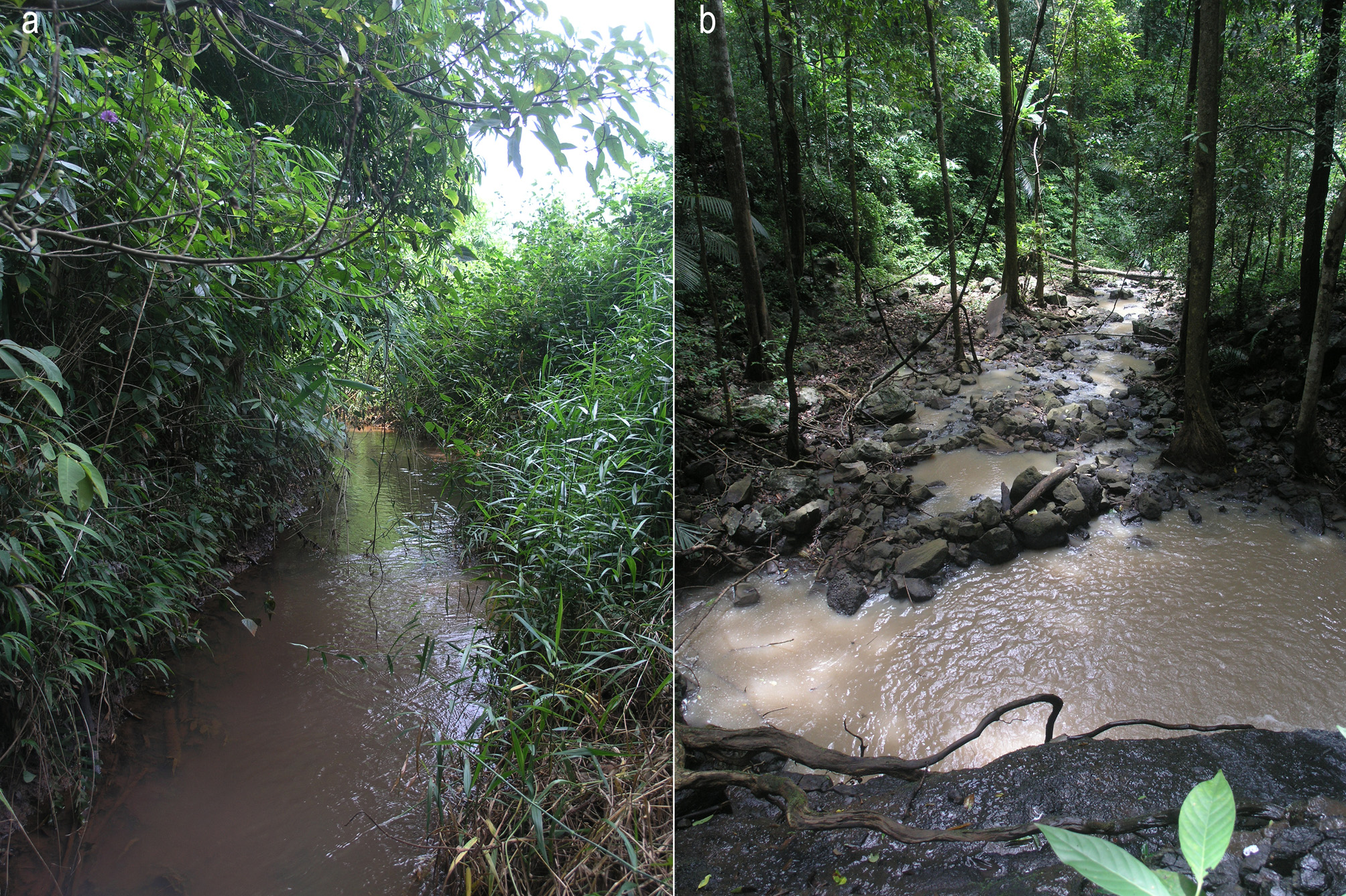
Habitat. The type locality, conventionally nicknamed ‘Culminicola Rivulet’, is a rivulet 2–3 m wide flowing in a gentle valley at about 700 m a.s.l. on the eastern part of the Central Plateau of the Annamense Mts. ( Fig. 12 View FIGURE 12 a) The plateau is clad with moist savannah but the rivulet is partly hidden in a forest strip occupying its valley. A male and female of A. schorri were collected when perched on tall riparian herbage/shrubbery at a forest-shaded slow reach with a gravel/silt bottom. The Odonata assemblage found at this rivulet was rich and included also Vest al is gracilis, Euphaea masoni inouei, Heliocypha biforata , H. perforata limbata, Agriocnemis femina, Mortonagrion aborense, Pseudagrion pruinosum, P. rubriceps, Copera marginipes, Prodasineura autumnalis , P. doisuthepensis, Pseudocopera ciliata, Burmagomphus asahinai, Brachydiplax farinosa, Neurothemis fluctuans, N. fulvia, Onychothemis culminicola, Orthetrum chrysis, O. luzonicum, Rhyothemis plutonia, R. triangularis, Tetrathemis platyptera, Tholymis tillarga, Trithemis aurora , T. festiva , and Zygonyx iris malayana (totally 27 species).
The second locality, nicknamed ‘Loringae brook’, is the right tributary of the main river just upstream the wellknown and impressive Buu Sraa Waterfall. It descends to the deep valley shaded by evergreen forest, although the valley itself is mostly surrounded by plantations. This brook has its own quite high, shady waterfall. The female of E. schorri was collected just below this waterfall ( Fig. 12 View FIGURE 12 b), while a male was photographed on 13 vi 2014 ( Fig. 8 View FIGURE 8. A ) above it. They landed mostly on fronds of gingers and small understorey palms. It was the same brook where E. yunnanensis were collected (above the waterfall). Besides, this brook is the type locality of Asiagomphus reinhardti Kosterin & Yokoi, 2016 ( Kosterin & Yokoi 2016) . The Odonata assemblage was found to be even richer than at the type locality and included also V. gracilis, E. masoni inouei, Aristocypha fulgipennis, H. biforata , H. perforata limbata, Philoganga loringae, Rhinagrion hainanense, Pseudagrion pruinosum, Coeliccia poungyi, C. marginipes , C. vittata , P. autumnalis , P. doisuthepensis, Protosticta grandis, Protosticta sp. cf. caroli, Gynacantha subinterrupta, Tetracanthagyna waterhousei, Gomphidictinus perakensis , Lamelligomphus castor , Leptogomphus baolocensis , Macrogomphus kerri, Idionyx thailandica, Macromidia cf. genialis, Macromia septima, Brachydiplax farinosa, Cratilla lineata calverti, Lathrecista asiatica, Neurothemis fulvia , N. intermedia atalanta, Orthetrum chrysis, O. glaucum , O. luzonicum , O. triangulare , T. platyptera , T. aurora , and Z. iris malayana (totally 38 species).
Distribution. The species is so far known from two close localities in Mondulkiri Province in eastern Cambodia. It is also expected from the same Central Plateau of the Annamense Mts. in southern Vietnam, as the border is just 17 km east of one of the known localities


| RMNH |
National Museum of Natural History, Naturalis |
No known copyright restrictions apply. See Agosti, D., Egloff, W., 2009. Taxonomic information exchange and copyright: the Plazi approach. BMC Research Notes 2009, 2:53 for further explanation.
|
Kingdom |
|
|
Phylum |
|
|
Class |
|
|
Order |
|
|
Family |
|
|
Genus |