
Dushia Corrêa, 1963
|
publication ID |
https://doi.org/ 10.11646/zootaxa.4691.4.2 |
|
publication LSID |
lsid:zoobank.org:pub:875FB4E7-148A-4AC2-904C-174416B11256 |
|
persistent identifier |
https://treatment.plazi.org/id/03B387A4-FFF3-CC26-FF73-C6BEFC3FFEC2 |
|
treatment provided by |
Plazi |
|
scientific name |
Dushia Corrêa, 1963 |
| status |
|
Genus Dushia Corrêa, 1963
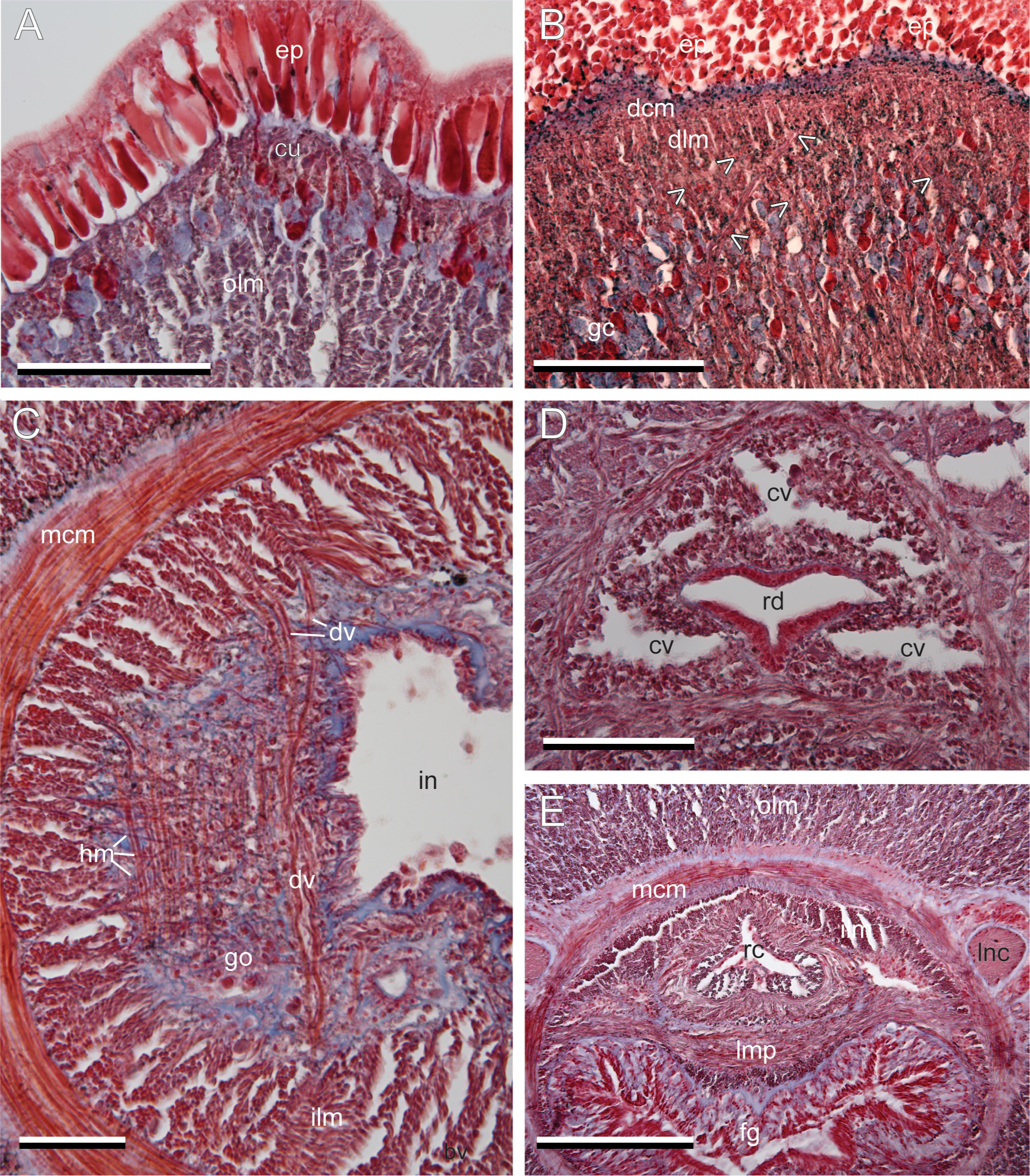
Diagnosis. Heteronemerteans with uniformly colored body (typically black, but also brown and orange) and white cephalic tip on both dorsal and ventral surfaces; with horizontal, lateral cephalic furrows; with caudal cirrus; dermal circular muscles external to dermal longitudinal muscles, the two separated by some diagonal fibers present as a lattice; connective tissue between cutis and outer longitudinal musculature (olm) at a minimum; dorsoventral muscles in body-wall inner longitudinal muscle layer in intestinal region, but not in outer longitudinal muscle layer; proboscis with typical heterotype musculature ( Chernyshev 2015) with two unequal muscle crosses; two lateral and one dorsal precerebral blood lacunae; neurocord cells present in dorsal portion of dorsal cerebral ganglia; no ocelli.
Etymology. Dushia appears to be a feminine latinized form derived from “ Dushi Korsu, ” which in Papiamentu, a local language in Curaçao, means “sweet Curaçao ” (Cynthia Santos, pers. comm. to MLS).
Type species. Herein fixed under Article 70.3.2 of the International Code of Zoological Nomenclature ( International Commission on Zoological Nomenclature 1999) as Dushia wijnhoffae Schwartz & Norenburg sp. nov., which was misidentified as Meckelia atra Girard, 1851 in the original designation by Corrêa (1963: 43).
Composition. The genus currently includes Dushia wijnhoffae Schwartz & Norenburg sp. nov. and Dushia nigra ( Stimpson, 1855) species complex comb. nov. The latter contains the following four nominal species: Meckelia nigra Stimpson, 1855 , Meckelia rubella Stimpson, 1855 , Micrura formosana Yamaoka, 1939 , and Micrura japonica Iwata, 1952 . Circumscription and name allocation within this species complex would require genetic analyses of specimens from wide geographic area in the Southwest Pacific, including the type localities of these four nominal species (see below).
Remarks. The genera Cerebratulus and Micrura lack definitive diagnoses, which makes it problematic to accurately place heteronemertean taxa into either of these nominal genera. When Corrêa (1963) established Dushia , she distinguished it from Cerebratulus on the basis of two features; Dushia lacked a caudal cirrus and does not have sharp lateral margins as in many Cerebratulus . Our observation on additional specimens from the Caribbean strongly suggests that Corrêa’s (1963) material was not intact, having lost the tail together with the caudal cirrus. While the presence or absence of a caudal cirrus often appears to be moderately clade-specific, current evidence suggests that it has either been gained or lost multiple times, and is not informative for generic placement as traditionally used ( Schwartz & Norenburg 2001; Schwartz 2009; Puerta et al. 2010).
Putative and nominal Cerebratulus spp. with sharp lateral margins were examined by Schwartz (2009). These had dorsoventral musculature in the outer longitudinal muscle layer (olm), i.e., external to the lateral nerve cords, or additional circular muscles below the cutis and even into the olm ( Schwartz 2009). Neither of these features is found in Dushia .
Dushia also can be separated from Micrura , although there has not been a comprehensive redescription of Micrura fasciolata Ehrenberg, 1828 , the type for the genus. Serial transverse sections of M. fasciolata collected in Sweden reveal a proboscis with “two muscle layers” in a traditional sense ( Schwartz & Norenburg 2001; Schwartz 2009), which corresponds to the modified heterotype without external muscle crosses and outer longitudinal musculature (consisting of four muscle layers, Chernyshev 2015), whereas the Dushia spp. examined here have a “threelayered” (in a traditional sense) proboscis with two muscle crosses, which actually consists of five layers ( Chernyshev 2015). The number of proboscis muscle layers and their positional relationship also appear to be homoplastic among Heteronemertea but they can be phylogenetically informative for less inclusive clades ( Schwartz 2009).
The presence or absence of neurocord cells has been used to distinguish Cerebratulus and Micrura from other genera. Neurocord cells and other neural cell types were first described in detail by Bürger (1895, 1897 –1907) and further refined by Thompson (1908); their definitions are used for identification here. At minimum, most heteronemertean species possess Bürger type I and II neural cells, whereas the presence of Bürger type III and neurocord cells is considered unique to certain taxa including Cerebratulus , Parvicirrus , and Polybranchiorhynchus ( Schwartz 2009). Neurocord cells in heteronemerteans are identified by their large size, over 20 µm, a pyriform shape with a substantial spherical to sub-spherical nucleus situated proximally in the cell ( Thompson 1908). Thompson (1908) observed that i) the cytoplasm tends to be clear when stained with hematoxylin and eosin; ii) they are found in the medial portions of the dorsal ganglia, and the dorsomedial portions of the ventral ganglia in one to seven pairs; and iii) occasionally, neurocord cells may also be found in proximal portions of the lateral nerve cords as they leave the ventral ganglia.
Large cells found in the dorsal ganglia of the Dushia spp. ( Figs 3B View FIGURE 3 , 6C View FIGURE 6 ) fit Thompson’s (1908) description of neurocord cells, except that the cytoplasm tends to be stained pink in this material. These cells, however, do not fit the definition of Bürger type III cells, which are smaller and can be seen adjacent to the neurocord cells. Many of the neurocord cells appear as though they suffer from shrinkage, perhaps accounting for their darker staining with hematoxylin. Caution should be taken when interpreting neural cell types because they are subject to fixation artifact and inherently stain poorly with general staining protocols. Neurocord cells also appear to be a homoplastic feature among heteronemerteans ( Schwartz 2009), but also among other nemertean groups; e.g., monostiliferans ( Maslakova & Norenburg 2008) and some reptantic polystiliferans (Kirsteur 1973, and references therein) also have neurocord-like cells.
Longitudinal and oblique sections of the cutis revealed between the dermal circular muscles and the dermal longitudinal muscles a layer of diagonal muscle fibers organized in a cross-hatch or lattice pattern ( Figs 2B View FIGURE 2 , 4B View FIGURE 4 ). It is possible that this layer has been overlooked in other heteronemerteans because internal examination usually only involves observations from transverse sections. Using confocal laser scanning microscopy, Chernyshev (2010) recorded such dermal diagonal muscle fibers in Cerebratulus marginatus auct. non Renier, 1804, Kulikovia alborostrata (Takakura, 1898) , K. torquata ( Coe, 1901) , Micrura kulikovae Chernyshev, 1992 , Micrura sp., Nipponomicrura uchidai (Yamaoka, 1940) , and Valenciniidae gen. et sp. indet.


No known copyright restrictions apply. See Agosti, D., Egloff, W., 2009. Taxonomic information exchange and copyright: the Plazi approach. BMC Research Notes 2009, 2:53 for further explanation.