
Sphingius hainan, Zhang, Feng, Fu, Jian-Ying & Zhu, Ming-Sheng, 2009
|
publication ID |
https://doi.org/ 10.5281/zenodo.191649 |
|
DOI |
https://doi.org/10.5281/zenodo.6219746 |
|
persistent identifier |
https://treatment.plazi.org/id/03B3E139-EF32-8C03-FF5F-FF53FB29A5C3 |
|
treatment provided by |
Plazi |
|
scientific name |
Sphingius hainan |
| status |
sp. nov. |
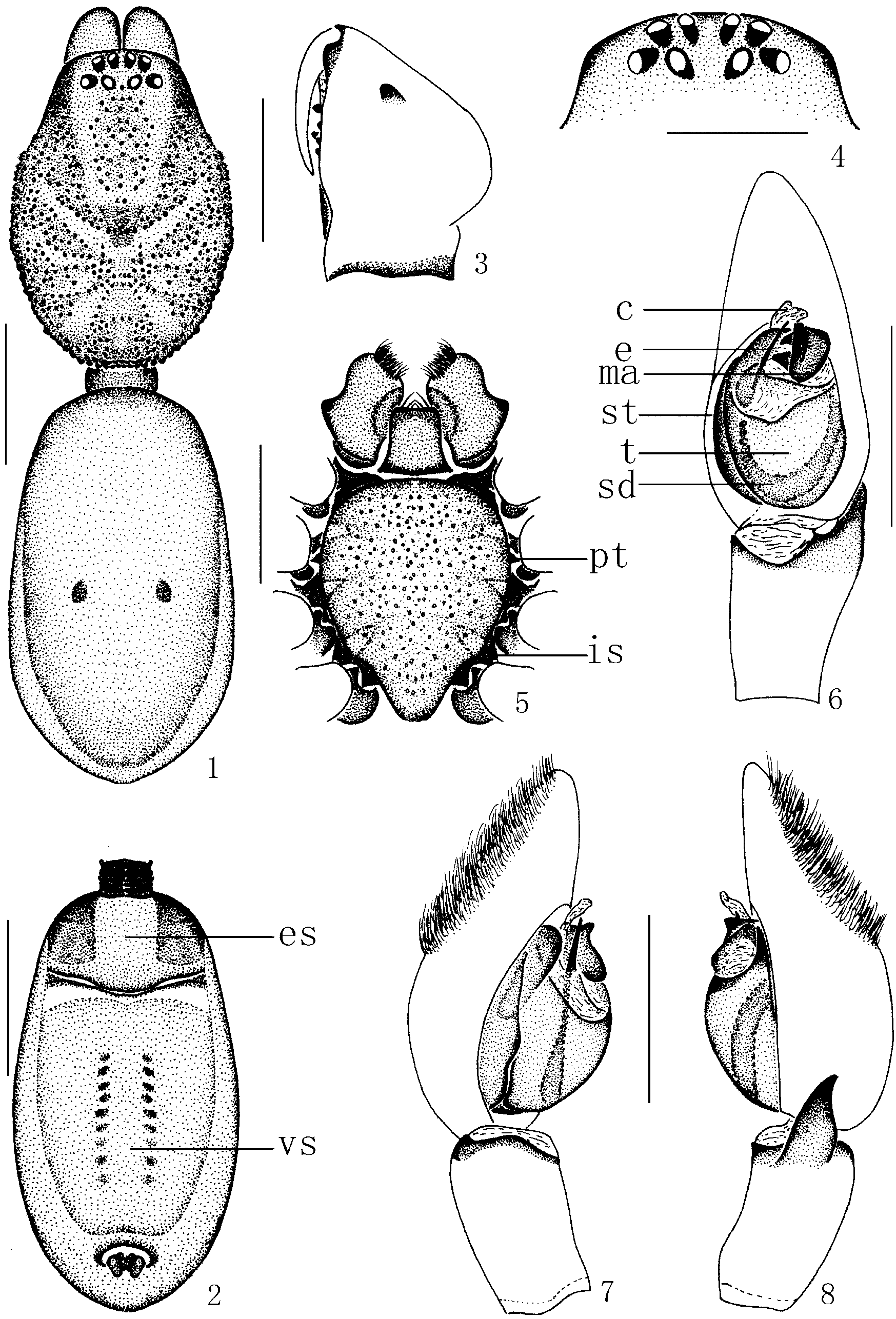
Sphingius hainan View in CoL sp. nov.
( Figs. 1–8 View FIGURES 1 – 8 )
Type material. Holotype male, CHINA: Hainan Province, Changjiang County, Mt. Bawangling [N 19.3°, E 109.1°], November 5, 2008, M. S. Zhu leg. ( MHBU) GoogleMaps .
Diagnosis. The new species is very similar to Sphingius vivax (Thorell, 1897) (cf. Deeleman-Reinhold, 2001, 494, figs. 840–842) in carapace (both with many small granulations) and palpal organs, but can be easily distinguished from the latter by having a broader base of tibial apophysis; a distal anterior tubercle on the chelicerae; a membranous conductor arising apical, while S. vivax lacks conductor.
Etymology. The species name is a noun in apposition, derived from the type locality.
Description. Male (holotype). Total length 5.62: carapace 2.47 long, 2.02 wide; abdomen 3.15 long, 1.62 wide. Carapace ovoid in dorsal view, deep reddish brown, with numerous small granulations, lateral and posterior margins with angular granulations. Eyes in two transverse rows; AER slightly recurved, PER straight or slightly recurved in dorsal view and longer than AER ( Fig. 4 View FIGURES 1 – 8 ). Eye diameters: AME 0.07, ALE 0.08, PME 0.08, PLE 0.07. Eye interdistances: AME–AME 0.10, AME–ALE 0.09, PME–PME 0.10, PME– PLE 0.11; MOA 0.19 long, front width 0.16, back width 0.21. Chelicerae with three promarginal and two retromarginal teeth, anterior side with a tubercle ( Fig. 3 View FIGURES 1 – 8 ). Endites brown, longer than wide, constricted at middle on lateral margin, anterior edge with clear serrula and scopula ( Fig. 5 View FIGURES 1 – 8 ). Labium slightly rectangular, anterior margin with a slight concavity centrally ( Fig. 5 View FIGURES 1 – 8 ). Sternum light brown, shield-shaped, covered with sparse granulations, posterior margin slightly extending between coxae IV, lateral margin with precoxal triangles and intercoxal sclerites. Space above the coxae and below the carapace with longitudinal, sclerotized pleural bars. Legs brown, anterior tibiae and metatarsi spineless, tarsi I–III almost as long as metatarsi. Leg spination: femora I-II with one small dorsal spines, tibia III v2-2 -2, p0-1-1, r0-0-1; metatarsus III v2 -0-0; tibia IV v1-1 -1, p0-0-1, r0-0-1, metatarsus IV p0-1-0, v2-1 -0, r0-1-0. Leg formula: 4123 ( Table 1).
Femur Patella Tibia Metatarsus Tarsus Total I 1.85 0.63 1.35 1.38 1.35 6.56 II 1.58 0.81 1.26 0.90 0.85 5.40 III 1.48 0.54 0.95 1.04 0.99 5.00 IV 1.90 0.72 1.62 1.94 1.30 7.48 Abdomen ( Fig. 1 View FIGURES 1 – 8 ) ovoid, dark brown, light brown centrally; dorsal scutum covering nearly all, and dorsum with one pair of muscular impression on middle part. Venter of abdomen yellow brown ( Fig. 2 View FIGURES 1 – 8 ), epigastric scutum tripartite (to some degree, at least in the color) divided into a central plate and two lateral plates, postgenital scutum relatively small, about two thirds of abdomen length, venter with two rows of longitudinal lines of spots.
Male palp ( Figs. 6–8 View FIGURES 1 – 8 ) with short and broad retrolateral tibial apophysis, beak-shaped at distal end. Genital bulb longitudinally pear-shaped in ventral view ( Fig. 6 View FIGURES 1 – 8 ), tegulum rounded at base; sperm duct a distinct, long loop encircling the tegulum, often originating from upper part of tegulum; subtegulum small, darker than tegulum, visible prolaterally ( Figs. 6–7 View FIGURES 1 – 8 ); embolus filiform, thin and short, emerging from tegulum prolaterally, and reaching beyond tip of tegulum ( Fig. 6 View FIGURES 1 – 8 ); conductor membranous, apical, behind the embolus; median apophysis slighty blocky, on distal-retrolateral sector of tegulum.
Female. Unknown.
Distribution. Presently known only from the type locality, Mt. Bawangling, China.
No known copyright restrictions apply. See Agosti, D., Egloff, W., 2009. Taxonomic information exchange and copyright: the Plazi approach. BMC Research Notes 2009, 2:53 for further explanation.
|
Kingdom |
|
|
Phylum |
|
|
Class |
|
|
Order |
|
|
Family |
|
|
Genus |