
Echinoderes samwisei, Grzelak & Sørensen, 2022
|
publication ID |
https://doi.org/ 10.5852/ejt.2022.844.1949 |
|
publication LSID |
lsid:zoobank.org:pub:193EDD91-B24D-455C-B8AA-8133586A00A1 |
|
DOI |
https://doi.org/10.5281/zenodo.7225531 |
|
persistent identifier |
https://treatment.plazi.org/id/9D00BABD-1183-40FA-BF7D-0F54E304DCD7 |
|
taxon LSID |
lsid:zoobank.org:act:9D00BABD-1183-40FA-BF7D-0F54E304DCD7 |
|
treatment provided by |
Felipe |
|
scientific name |
Echinoderes samwisei |
| status |
sp. nov. |
Echinoderes samwisei View in CoL sp. nov.
urn:lsid:zoobank.org:act:9D00BABD-1183-40FA-BF7D-0F54E304DCD7
Figs 20–22 View Fig View Fig View Fig ; Tables 14–15
Diagnosis
Echinoderes with spines in middorsal position on segments 4, 6 and 8, and spines in lateroventral positions on segments 6 to 9. Tubes present in lateroventral positions on segment 5, lateral accessory positions on segment 8, and laterodorsal positions on segment 10. A protuberance-like structure emerges between segments 10 and 11 in middorsal position.
Etymology
The species name refers to Samwise Gamgee, one of the main characters in the novel “ The Fellowship of the Ring ”, the first volume of J.R.R. Tolkien’s “ The Lord of the Rings ”. Samwise was a hobbit from the Shire, Frodo Baggins’ best friend and one of the most loyal members of the Fellowship of the Ring.
Material examined
Holotype
NEW ZEALAND • ♂; Hikurangi Slope, stn TAN1004/38; 41.5937° S, 175.8532° E; 1121 m b.s.l.; Apr. 2010; NIWA TAN1004 Voyage; soft sediment; NIWA-159418 . Mounted for LM in Fluoromount G on HS slide.
GoogleMapsParatypes GoogleMaps
NEW ZEALAND • 1 ♀, 3 ♂♂; Hikurangi Slope, stn TAN1004/4; 41.6837° S, 175.6642° E; 1046 m b.s.l.; Apr. 2010; NIWA TAN1004 Voyage; soft sediment; 1 ♀ NHMD-917299 , 1 ♂ NHMD-917300 , GoogleMaps 2 ♂♂ NIWA-159419 to 159420. GoogleMaps 1 ♀ and 2 ♂♂ mounted for LM in Fluoromount G on glass slides, 1 ♂ mounted as holotype GoogleMaps • 1 ♂; Pahaua Canyon , stn TAN1004/12; 41.5508° S, 175.7250° E; 1350 m b.s.l.; Apr. 2010; NIWA TAN1004 Voyage; soft sediment; NHMD-917301 . Mounted for LM in Fluoromount G on glass slide GoogleMaps • 1 ♂; same collection data as for holotype; NIWA-159421. Mounted as holotype GoogleMaps • 1 ♀; Seamount 310 , stn TAN1004/72; 41.3657° S, 176.1958° E; 985 m b.s.l.; Apr. 2010; NIWA TAN1004 Voyage; soft sediment; NHMD-917302 . Mounted for LM in Fluoromount G on glass slide GoogleMaps .
Additional material
NEW ZEALAND • 1 ♀; same collection data as for holotype; personal reference collection of MVS. Mounted for SEM GoogleMaps • 1 ♀; Hikurangi Slope, stn TAN1004/44; 41.5258° S, 175.8003° E; 728 m b.s.l.; Apr. 2010; NIWA TAN1004 Voyage; soft sediment; personal reference collection of MVS. Mounted for SEM GoogleMaps • 1 ♀; Honeycomb Canyon, stn TAN1004/62; 41.4760° S, 175.9477° E; 1171 m b.s.l.; Apr. 2010; NIWA TAN1004 Voyage; soft sediment; personal reference collection of MVS. Mounted for SEM GoogleMaps • 1 ♂; Seamount 310, stn TAN1004/72; 41.3657° S, 176.1958° E; 985 m b.s.l.; Apr. 2010; NIWA TAN1004 Voyage; soft sediment; personal reference collection of MVS. Mounted for SEM GoogleMaps .
Description
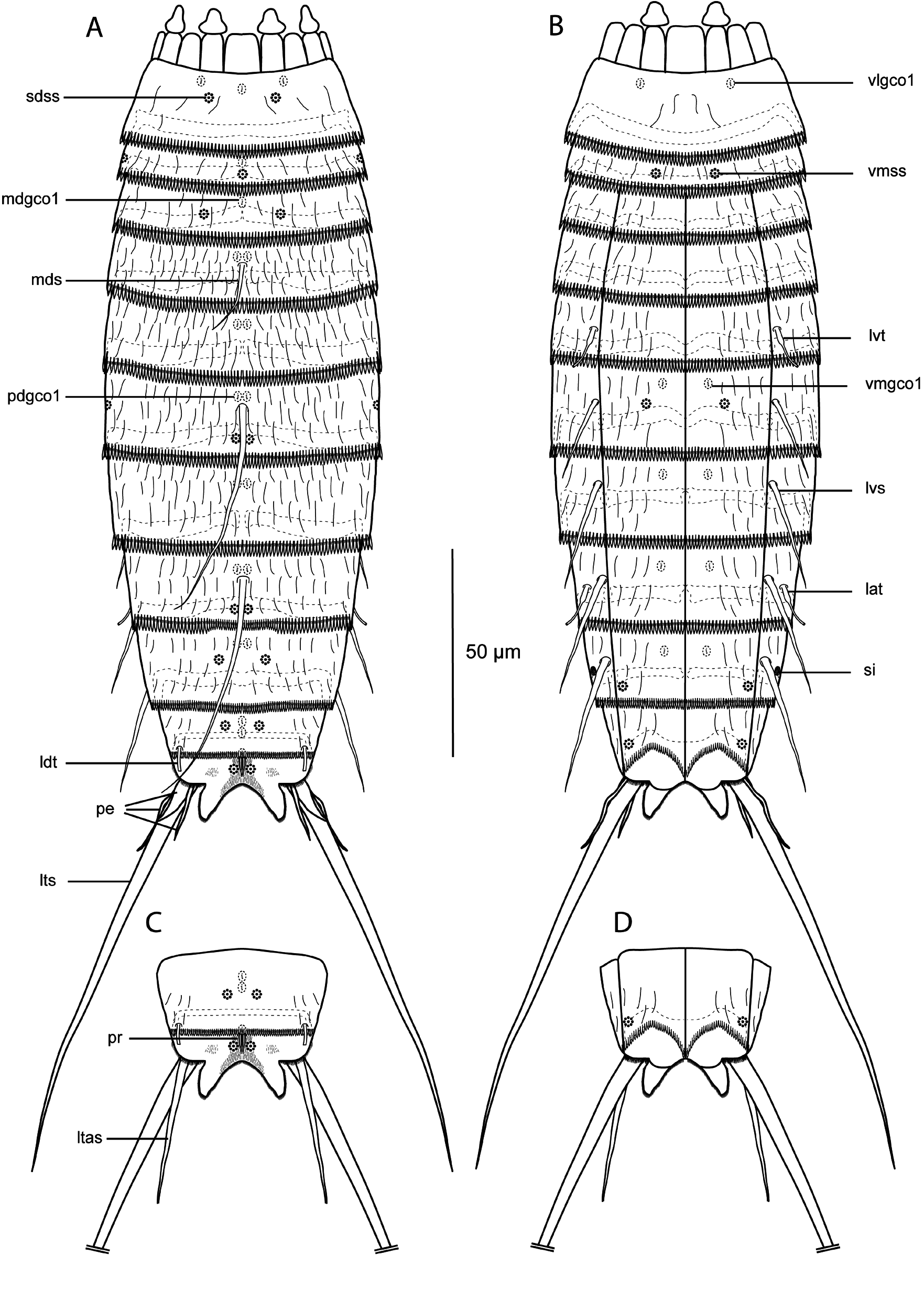
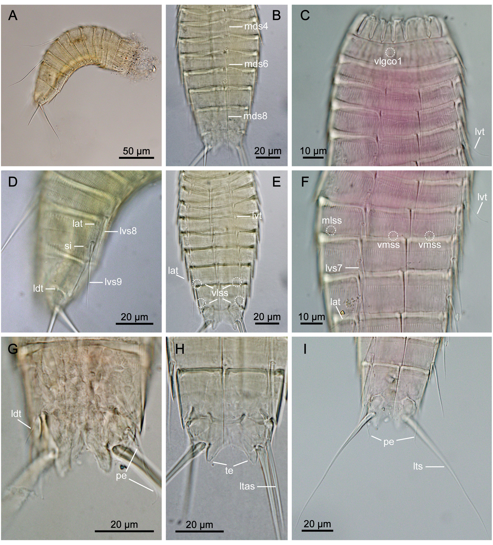
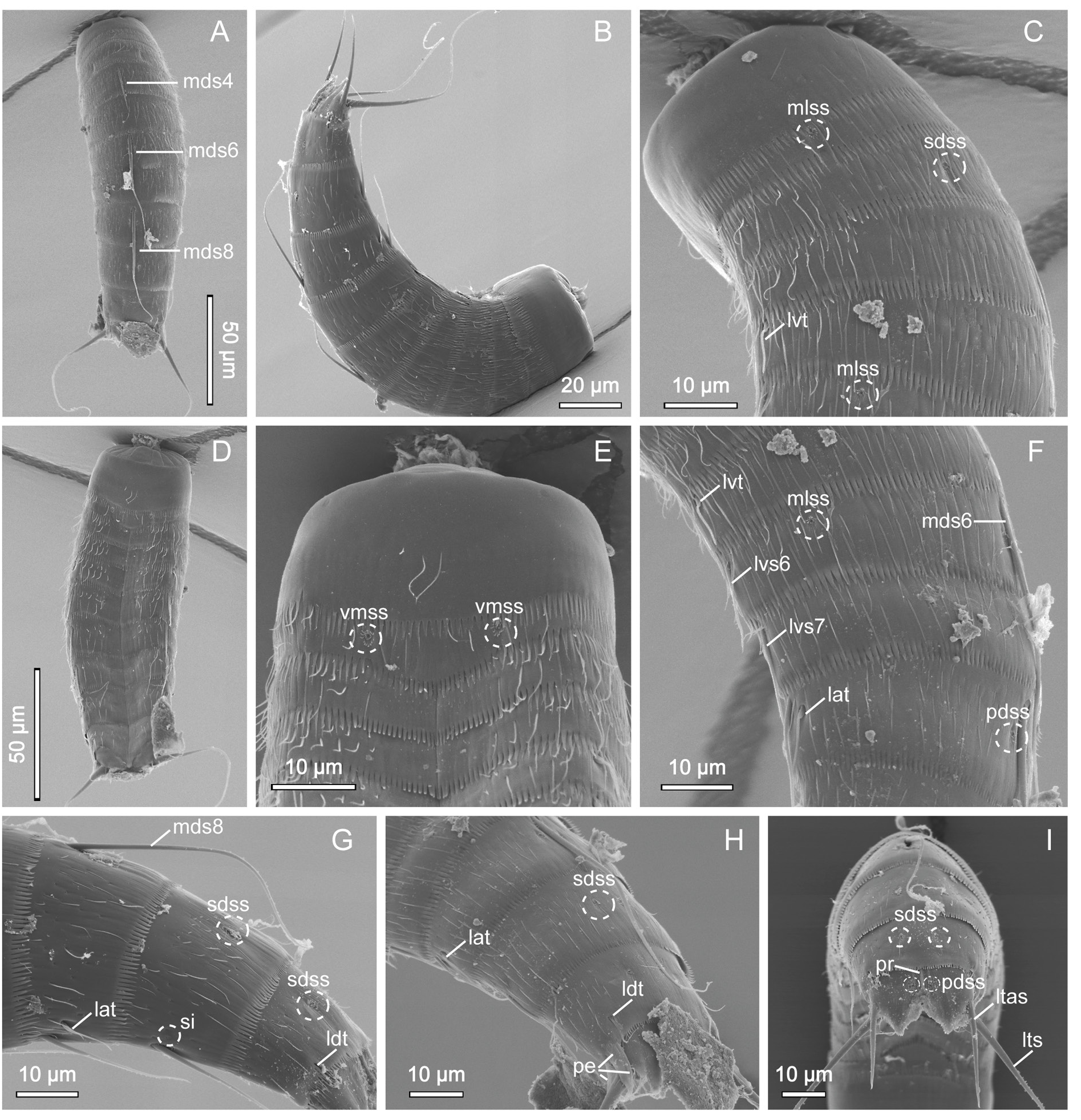
GENERAL. Adults with head, neck and eleven trunk segments ( Figs 20–22 View Fig View Fig View Fig ). Overview of measurements and dimensions in Table 14. Distribution of cuticular structures, i.e., sensory spots, glandular cell outlets, spines and tubes, summarized in Table 15. No details regarding scalid arrangement and morphology could be provided, because introverts of all specimens mounted for SEM fully or partially retracted.
NECK. With 16 placids. Midventral placid broadest, 10 µm in width and 13 µm in length, whereas all others narrower, measuring 5 µm in width at bases and 12 µm in length, similar in size ( Figs 21C View Fig , 22D View Fig ). Trichoscalid plates well developed.
SEGMENT 1. Consists of complete cuticular ring. Sensory spots located on anterior half of segment, in subdorsal positions.Glandular cell outlet type 1 present in middorsal, subdorsal and ventrolateral positions ( Figs 20A–B View Fig , 21C View Fig ). Segment almost completely hairless ( Fig. 22B–E View Fig ). Posterior segment margin almost straight, forming pectinate fringe. Fringe with well-developed, relatively long tips, homogenous along segment margin ( Fig. 22C, E View Fig ).
SEGMENT 2. Consists of complete cuticular ring, without tubes. Sensory spots present in middorsal, midlateral and ventromedial positions ( Figs 20A–B View Fig , 22B, E View Fig ). Glandular cell outlets type 1 present in middorsal position ( Figs 20A–B View Fig , 21B View Fig ). Other structures not observed. Pachycyclus of anterior segment margin of regular thickness, without interruption. Secondary pectinate fringe present near anterior segment margin of this and following segments, but covered by preceding segment. Fairly long, single cuticular hairs sparsely scattered around segment. Posterior segment margin almost straight; pectinate fringe tips as on preceding segment.
SEGMENT 3. Present segment, and eight remaining ones, consist of one tergal and two sternal plates ( Figs 20A–B View Fig , 21C, E–F View Fig , 22D View Fig ). Pachycyclus of anterior segment margin of regular thickness, interrupted at tergosternal and midsternal junctions and middorsally, on this and following seven segments. Segment with middorsal glandular cell outlet type 1 and subdorsal sensory spots only; no other traits observed. Cuticular hairs more densely distributed across tergal plate than on preceding segment, except for narrow hairless line in laterodorsal area; sternal plates with very few cuticular hairs; on this and following segments paraventral areas completely devoid of hairs. Posterior segment margin straight, terminating in pectinate fringe with fringe tips as on preceding segments.
SEGMENT 4. With spine in middorsal position; spine relatively short (23 µm), only slightly exceeding posterior segment margin ( Figs 20A View Fig , 22A View Fig ). Pair of glandular cell outlets type 1 present in paradorsal positions. No other traits observed. Segment otherwise as segment 3.
SEGMENT 5. With tubes in lateroventral positions ( Figs 20B View Fig , 21C, E–F View Fig , 22C, F View Fig ). Glandular cell outlets type 1 present in paradorsal positions only. No sensory spots or other structures present. Pectinate fringe of posterior segment margin and cuticular hairs as on preceding segment.
SEGMENT 6. With spines in middorsal and lateroventral positions ( Figs 20A–B View Fig , 21A View Fig , 22B View Fig ). Sensory spots present in paradorsal, midlateral and ventromedial positions ( Figs 20A–B View Fig , 21F View Fig , 22C, F View Fig ). Glandular cell outlets type 1 present in paradorsal and ventromedial positions ( Figs 20A–B View Fig , 21F View Fig ). Tips of pectinate fringe of posterior segment margin as on preceding segments. Segment otherwise as segment 5.
SEGMENT 7. With spines in lateroventral positions, and glandular cell outlets type 1 in paradorsal and ventromedial positions ( Figs 20A–B View Fig , 21F View Fig ). Sensory spots not observed. Cuticular hairs covering as on preceding segment.
SEGMENT 8. With spines in middorsal and lateroventral positions, and tubes in lateral accessory positions ( Figs 20A–B View Fig , 21D–F View Fig , 22F–H View Fig ). Middorsal spine long, reaching posterior part of segment 10 ( Figs 21A– B View Fig , 22A View Fig ). Sensory spots present in paradorsal positions only. Glandular cell outlets type 1 in paradorsal and ventromedial positions. Pectinate fringe of posterior segment margin as on preceding segment, except slightly shorter and narrower fringe tips along paradorsal and subdorsal areas of segment margin.
SEGMENT 9. With spines in lateroventral positions ( Figs 20B View Fig , 21D View Fig ). Sensory spots located in subdorsal and ventrolateral positions; subdorsal pair situated close to paradorsal area ( Figs 20A–B View Fig , 21E View Fig , 22G–H View Fig ). Glandular cell outlets type 1 present in paradorsal and ventromedial positions. Small, rounded sieve plates located in lateral accessory positions. Cuticular hair covering and pectinate fringe as on preceding segment.
SEGMENT 10. With laterodorsal tubes located near posterior segment margin; tubes well developed in both sexes, but slightly longer in males ( Figs 21G View Fig , 22G–H View Fig ). Sensory spots present in subdorsal and ventrolateral positions ( Fig. 22I View Fig ). Glandular cell outlets type 1 present as two middorsal ones. Cuticular hairs scarcer than on preceding segment. Central part of tergal plate devoid of hairs; short cuticular hairs lightly scattered on lateral halves only. Posterior segment margin of tergal plate straight, with much shorter fringe and narrower tips than those on preceding segment; margins of sternal plates extend midventrally, reaching posterior margin of terminal segment ( Fig. 22D View Fig ).
SEGMENT 11. With pair of lateral terminal spines ( Fig. 20C View Fig ). Females with relatively strong, stout lateral terminal accessory spines ( Figs 21H View Fig , 22I View Fig ). Males with three pairs of penile spines; dorsal ones of medium length, ventral ones long and relatively thin, whereas median ones markedly thicker, conical and stout ( Figs 20A–B View Fig , 21G, I View Fig 22H View Fig ). Sensory spots present in paradorsal positions ( Figs 20A, C View Fig , 22I View Fig ). Glandular cell outlet type 1 present in middorsal position. Middorsal protuberance-like structure extends from intersegmentary joint ( Figs 20A, C View Fig , 22I View Fig ). Segment devoid of cuticular hairs, but with dense covering of relatively long hair-like extensions in paradorsal area, as well as small patches of shorter ones in subdorsal area. Short fringes covering margins of tergal and sternal plates. Tergal extensions short and triangular ( Figs 21H View Fig , 22I View Fig ). Sternal extensions do not extend beyond tergal extensions.
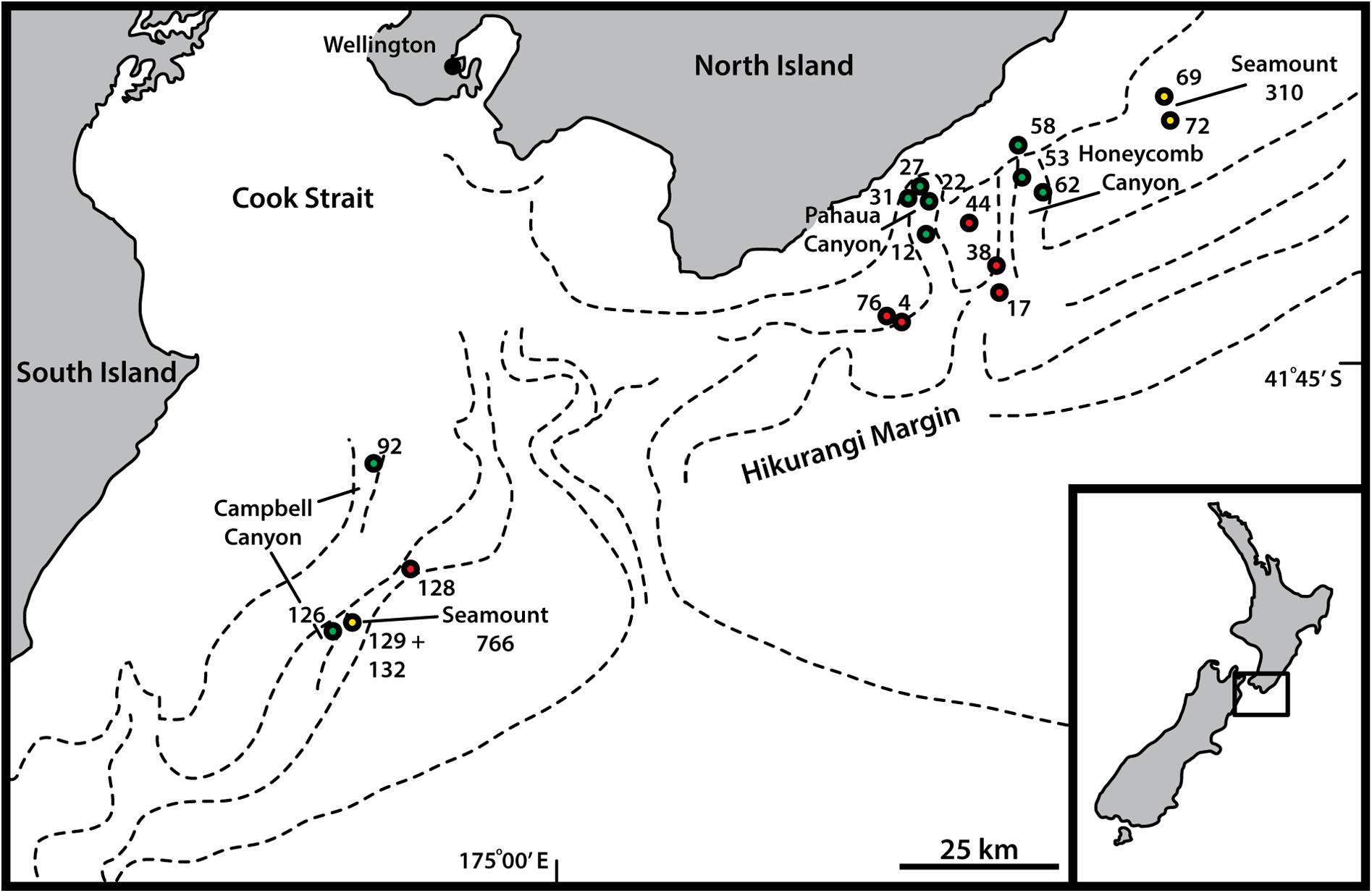
Distribution
Hikurangi Margin, from slope, through canyon, and seamount habitats, 728–1350 m b.s.l. See Fig. 1 View Fig for a geographic overview of stations and Table 1 View Table 1 for station and specimen information.
Taxonomic remarks on Echinoderes samwisei sp. nov.
The spine pattern with middorsal spines on segments 4, 6 and 8 and lateroventral spines on segments 6 to 9 is very common within Echinoderes , and is shared with 33 species (Yamasaki et al. 2020). Nevertheless, among these only E. anniae Sørensen et al., 2018 , E. meteorensis Yamasaki et al., 2018 , and an undescribed species, Echinoderes sp. 3 , from Senghor Seamount reported by Yamasaki et al. (2019) resemble E. samwisei sp. nov. by lacking tubes on segment 2 ( Sørensen et al. 2018; Yamasaki et al. 2018 c, 2019). Thus, none of these species can be confused with E. samwisei . Echinoderes anniae and E. meteorensis differ from E. samwisei in having several glandular cell outlets type 2 – structures not observed in E. samwisei . Moreover, both species are characterized by very long lateral terminal spines and a lack of tubes on segments 5 and 8. Echinoderes samwisei differs from Echinoderes sp. 3 in Yamasaki et al. (2019) as well, because the latter lacks tubes on segment 8 and possesses characteristic long and spinose tergal extensions, whereas the new species has lateral accessory tubes on segment 8 and relatively short tergal extensions.
Moreover, E. samwisei sp. nov. is generally characterized by a low number of cuticular structures. The new species not only lacks glandular cell outlets type 2, but also its number of sensory spots and glandular cell outlets type 1 is relatively low, as compared with other species. It is indeed possible to miss certain glandular cell outlets type 1, in particular in species with an extraordinary thin cuticle, but the availability of several specimens for SEM examination makes it less likely that any sensory spots have been overlooked. The available SEM specimens were generally in a good condition and relatively clean, which allowed for a thorough examination using SEM. In spite of this, we observed only 13 pairs of sensory spots on the trunk.
Therefore, the combination of spine and tube patterns, together with the lack of tubes or glandular cell outlets type 2 on segment 2 and the relatively ‘simple’ overall appearance of the trunk, makes E. samwisei sp. nov. unique among its congeners.
No known copyright restrictions apply. See Agosti, D., Egloff, W., 2009. Taxonomic information exchange and copyright: the Plazi approach. BMC Research Notes 2009, 2:53 for further explanation.
|
Kingdom |
|
|
Phylum |
|
|
Class |
|
|
Order |
|
|
Family |
|
|
Genus |