
Conorete undetermined
|
publication ID |
https://doi.org/ 10.1111/zoj.12138 |
|
persistent identifier |
https://treatment.plazi.org/id/03B48793-FFB2-F86A-CF51-FC023DC52D29 |
|
treatment provided by |
Carolina |
|
scientific name |
Conorete undetermined |
| status |
SP. |
CONORETE POURTALESI SP. NOV. ( FIGS 2 View Figure 2 , 3 View Figure 3 ; TABLE 1)
Type material: Holotype: USNM 1231335 View Materials , MOV ‘ Johnson Sea Link II ’, dive 3817, 07 Aug. 2010, Alligator bioherm 1635, Pourtales Terrace , Florida, 24°42.2806′ N, 80°30.6811′ W, 198 m, 70% ethanol. GoogleMaps
Comparative material: Conorete mucronatum ( Wilson, 1904, originally Eurete erectum mucronatum ), co-types, USNM 008488 View Materials , USFS Albatross , stn 3358, 24 Feb. 1891, Gulf of Panama, 6°30′ N, 81°44′ W, 1015 m, four specimens, ethanol GoogleMaps .
Diagnosis: Conorete with pinular rays of dermal hexactins styloid in form, rarely bushy; all atrialia as macrospined pentactins. Microscleres mainly oxyhexasters with discohexasters and onychohexasters as minor components.
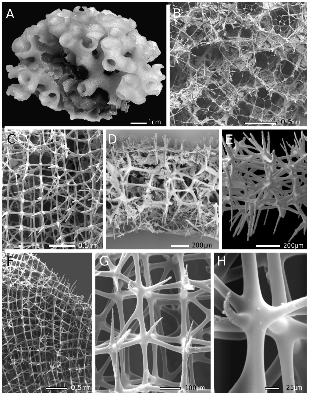
Description: General body form of the single known specimen is clathrate–globular, an ovoid mass of branching and anastomosing tubes with overall dimensions 9.8 × 7.0 × 6.4 cm ( Fig. 2A View Figure 2 ). Length of internodal components of tubes approximates tube diameter in the compact form. External tube diameter is 7.4–9.5– 14.3 ± 1.4 mm (n = 24), internal diameter is 4.5–6.6– 9.4 ± 1.3 mm (n = 14), and wall thickness is 1.2–1.5 mm. Tube surfaces appear fairly smooth to the naked eye, but under a dissection microscope it bears shallow pits and poorly defined ridges ( Fig. 2B View Figure 2 ). Removal of tissues shows that these shallow pits are not canal apertures but merely irregularities in the surface dictyonalia ( Fig. 2C, F View Figure 2 ); skeletal channels are not present. Spiculation of outer and inner surfaces of the tubes differ dramatically: outer surfaces are ornament- ed with projecting distal rays of hexactine dermalia, uncinate tips, and scopules, whereas the smooth inner surfaces are defined by tangential rays of pentactine atrialia ( Fig. 2D View Figure 2 ). Colour, when preserved, is light brown. Symbionts consist of a dark-brown branching system of tentaculate hydrozoans with polyps opening on both inner and outer tube surfaces. The known distribution of the species is a single depth, 198 m, at a single location off southern Florida on the Pourtales Plateau ( Fig. 1 View Figure 1 ).
The skeletal framework (for data see Table 1) is a typical euretoid dictyonal type, of between three or four meshes in thickness ( Fig. 2D–E View Figure 2 ). Longitudinal strands are present in the primary layer; they curve gently at low angles to both dermal and atrial surfaces. Irregularly arranged dictyonalia form thin patchy cortices on older areas of both surfaces. Beams are mainly smooth, with a few low spines occurring irregularly on beams or nodes ( Fig. 2G–H View Figure 2 ); nodes are not swollen. Dictyonal meshes are primarily rectangular, often square. Spurs of both surfaces are long, mainly straight, rough, and sharp-pointed ( Fig. 2E, G View Figure 2 ). No small oxyhexactins are appended to the framework.
Megascleres are surficial hexactins and pentactins, tylo- and subtyloscopules, and uncinates. Dermalia are mainly finely rough hexactins ( Fig. 3A View Figure 3 ), with distal ray more or less differentiated by larger spines. These distal rays are occasionally bushy and pinular, in the sense of differing in form from other rays, but not in the sense of being similar to a small fir tree. Other rays are tapered and end in abruptly sharp tips. The mean dimensions of rays are: distal, 67 × 6.8 μm; tangential, 130 × 6.6 μm; and proximal, 162 × 6.7 μm. A few dermalia (<1%) are pentactins. Atrialia are all pentactins, sparsely ornamented with macrospines and with the missing distal ray represented only by a nub ( Fig. 3B View Figure 3 ). Atrialia tangential rays, 140 × 8.7 μm; atrialia proximal rays, 196 × 9.0 μm. Scopules are mostly geniculate tyloscopules, with between two and five distally spined tines ending in large caps ( Fig. 3C View Figure 3 1 View Figure 1 ; 60%), common non-geniculate tyloscopules, with between three and five rough tines ending in smaller caps ( Fig. 3C3 View Figure 3 ; 34%), and uncommon subtyloscopules ( Fig. 3C View Figure 3 2 View Figure 2 ; 6 View Figure 6 %), with three or four thin smooth tines bearing small
Table 1. List of recognizable and unrecognizable
Hexactinellida species names reported from the area encompassing the West Indies , Caribbean Sea, and Gulf of Mexico, with accepted junior synonyms excluded
AMPHIDISCOSIDA RECOGNIZABLE, VALID View in CoL : 5 Hyalonema kenti ( Schmidt, 1880: 65, as Asconema View in CoL ) Hyalonema schmidti Schulze, 1899: 9 View in CoL Hyalonema toxeres Thomson, 1873: 248 View in CoL Pheronema annae Leidy, 1868: 10 View in CoL Poliopogon amadou Thomson, 1873: 29 View in CoL
LYSSACINOSIDA RECOGNIZABLE, VALID View in CoL : 11 Asconema foliata View in CoL (Fristedt, 1887: 413 as Hyalonema View in CoL ) Calycosoma validum Schulze, 1899: 27 View in CoL Euplectella jovis Schmidt, 1880: 60 View in CoL Euplectella suberea Thomson, 1876: 93 View in CoL Hertwigia falcifera Schmidt, 1880: 62 View in CoL Heterotella pomponae Reiswig, 2000: 573 View in CoL Lophocalyx oregoni Menshenina et al., 2007: 455 View in CoL Regadrella phoenix Schmidt, 1880: 61 View in CoL Rhabdopectella tintinnus Schmidt, 1880: 62 View in CoL Sympagella nux Schmidt, 1870: 15 View in CoL Vazella pourtalesi ( Schmidt, 1870: 14 as Holtenia )
HEXACTINOSIDA RECOGNIZABLE, VALID : 9 Aphrocallistes beatrix Gray, 1857: 115 Claviscopulia facunda ( Schmidt, 1870: 16 as Farrea ) Cyrtaulon sigsbeei ( Schmidt, 1880: 58 as Volvulina ) Dactylocalyx pumiceus Stutchbury, 1841: 87 View in CoL Dactylocalyx subglobosus Gray, 1867: 506 View in CoL Farrea occa Bowerbank, 1862: 1118 Iphiteon panicea Bowerbank, 1869: 76 View in CoL Lefroyella decora Thomson, 1877: 403 Myliusia callocyathus Gray, 1859: 439 View in CoL
AULOCALYCOIDA RECOGNIZABLE, VALID : 2 Cyathella lutea Schmidt, 1880: 46 Rhabdodictyum delicatum Schmidt, 1880: 46
LYCHNISCOSIDA RECOGNIZABLE, VALID View in CoL : 3 Lychnocystis superstes View in CoL ( Schmidt, 1880: 51 as Cystispongia ) Neoaulocystis grayi View in CoL (Bowerbank, 1869: 335 as Myliusia View in CoL ) Scleroplegma lanterna View in CoL ( Schmidt, 1880: 50 as Auloplegma )
UNRECOGNIZABLE BUT VALID NAMES: 20 Dactylocalyx crispus Schmidt, 1870: 19 Dactylocalyx potatorum Schmidt, 1880: 53 View in CoL Deanea virgultosa Bowerbank, 1875: 275 View in CoL Diaretula cornu Schmidt, 1879: 45 View in CoL Diaretula muretta Schmidt, 1880: 46 View in CoL Farrea aculeata Bowerbank, 1875: 561 Farrea fistulata Bowerbank, 1875: 276 Farrea gassioti Bowerbank, 1875: 272 Farrea inermis Bowerbank, 1876: 536 Farrea infundibuliformis Carter, 1873: 448 Farrea laevis Bowerbank, 1875: 278 Farrea parasitica Bowerbank, 1875: 279 Farrea perarmata Bowerbank, 1876: 538 Farrea pocillum Bowerbank, 1875: 273 Farrea robusta Bowerbank, 1875: 562 Farrea spinifera Bowerbank, 1875: 558 Myliusia conica View in CoL ( Schmidt, 1880: 57 as Scleroplegma View in CoL ) Myliusia seriatum ( Schmidt, 1880: 57 as Scleroplegma View in CoL ) Rhabdostauridium retortula Schmidt, 1880: 59 Scleroplegma herculeum Schmidt, 1880: 57
smooth terminal swellings. Scopules with a total length
of 304 μm are very abundant on the dermal surface but are absent from the atrial surface; a few subtyloscopules occur in subatrial position between atrialia, but do not extend to the surface membrane. Uncinates ( Fig 3D View Figure 3 ) are relatively small for Euretidae , with a mean length of 763 μm and a mean width of 4.7 μm, but their barbs and brackets are well developed; the anterior end is rather bushy.
Microscleres are all spherical, composed mainly of oxyhexasters (71%), accompanied by common discohexasters (26%), and rare onychohexasters (4%). Oxyhexasters with a mean diameter of 73.5 μm are robust, with between two and four sharply pointed straight terminals borne on each primary ray; all surfaces are covered with fine recurved spines ( Fig. 2E View Figure 2 ). Discohexasters are similar to oxyhexasters, but are smaller and with marginally spined discs at terminal ray ends ( Fig. 3F View Figure 3 ). Onychohexasters have between two and five small claws emanating perpendicularly from the distal ends of terminal rays. Individual microscleres with mixed oxy- and onycho-tips are common; terminal rays from a single primary ray often differ in form ( Fig. 3G View Figure 3 ). Disco- and onychohexasters are 55.0 μm in mean diameter.
Etymology: The species name ‘ pourtalesi ’ commemorates Louis François de Pourtalès (1824–1880), a student and close associate of Alexander Agassiz who togeth- er carried out the early explorations of the West Indian marine fauna. Pourtalès’ historical importance is reflected in the name of the collection location: Pourtales Terrace.
Remarks: The new specimen with hexactine dermalia and macrospined atrialia clearly belongs to the genus Conorete as diagnosed in Systema Porifera ( Reiswig & Wheeler, 2002). The genus presently contains three recognized species: the type species, Conorete erectum ( Schulze, 1899) , containing four subspecies, all from the tropical eastern Pacific; Conorete mucronatum ( Wilson, 1904) , from the same region; and Conorete gordoni Reiswig & Kelly, 2011 , from the Kermadec Ridge north of New Zealand. All specimens of the first two species have columnar form, basically elongate tubular stems with lateral tubular branches in a spiral pattern. The body is usually simple, flute-like, but occasionally undergoes secondary branching without anastomoses. This contrasts with the globular form of the new species with extensive anastomoses of tubes. The body form of the third species, C. gordoni , remains unknown, but the small elongate tubule fragment with lateral oscula is consistent with the columnar form of the first two species, and is inconsistent with the tightly branching globular form of the new species. In
spiculation all three present species have bushy pinules
of surface hexactins, whereas the new species differs in having mainly sparsely spined styloid pinules. The new species shares oxyhexasters as the major microsclere with C. mucronatum and C. gordoni , but differs from C. mucronatum in having some discohexasters ( C. mucronatum has none); it differs from C. gordoni in having tyloscopules as the main scopule form ( C. gordoni has mainly subtyloscopules of quite different shape). These differences justify the recognition of the new specimen as a new species, here designated C. pourtalesi sp. nov. This addition to the genus Conorete is the first member known from the Atlantic basin; it expands the distribution of the genus Conorete , which has previously been restricted to the eastern and western coasts of the equatorial and the southern Pacific Ocean.
Framework type, as farreoid or euretoid, has been an element of considerable interest in this genus since the description of its first species, Conorete (then Eurete ) erectum by Schulze (1899). He noted that the terminal tube ends had a single-layered frame, like the condition in Farrea , and unlike the general threedimensional non-layered framework of Eurete . Most later workers did not comment on the framework of Conorete specimens, but Wilson (1904) noted that occasional areas of one-layered frame occurred in C. erectum tubuliferum , and Reiswig & Kelly (2011) reported areas of onelayered farreoid framework in C. gordoni . In our review of C. mucronatum co-type fragments we did not have access to terminal tube ends, but the framework is definitely two-layered, with the second layer being a mirror image of the primary atrial layer, and completely in register with it. This is consistent with Reid’s (1964) definition of an expanded farreoid framework and inconsistent with his definition of a non-layered euretoid framework; however, our detailed review of the framework of the new species shows it to be three-dimensional and non-layered, with longitudinal strands curving to both dermal and atrial surfaces, completely consistent with Reid’s description of a euretoid framework. Unfortunately, we do not have undamaged terminal tube ends of this specimen to assess whether those areas are one-layered or three-dimensional. More detailed surveys of primary framework details in various members of Euretidae and Farreidae are needed to develop a better understanding of the range of framework form in the genera of these families, and how they and individual species might be related by shared patterns.
SUBFAMILY CHONELASMATINAE SCHRAMMEN, 1912 GENUS VERRUCOCOELOIDEA REID, 1969
Restricted synonymy: Verrucocoeloidea Reid, 1969: 485 ; Finks et al. 2011: 73.
Diagnosis: Cylindrical to funnel-shaped euretids with short lateral tubes in quincuncial, linear–longitudinal, or irregular arrangement; each lateral tube sometimes with an axillary oscule on the upper side, in addition to the terminal oscule; lateral tubes confluent with an internal system of plexiform (and spiral?) longitudinal tubes or irregular subdivisions of the atrial cavity formed by ingrowths of the atrial wall that may be only partially skeletalized; a cortex with ostia may be developed on the dermal side. Loose spicules include pentactins, hexactins, tyloscopules, strongyloscopules, uncinates, oxyhexasters, and discohexasters. (From Finks et al., 2011, emended.)
Type species: Verrucocoeloidea burtoni Reid, 1969 .
Remarks: The original diagnosis of Reid (1969) was emended by Finks et al. (2011) to include a fossil species with main body form as a cylindrical tube. Finks et al. divided the genus into two subgenera, Verrucocoeloidea Reid, 1969 and Euretella Finks et al., 2011 , to accommodate the recent type species, V. burtoni , with funnelform body in the first, and their new cylindrical Eocene species, Verrucocoeloidea corallina , in the second. Here we do not deal with the subgenera diagnoses formed by Finks et al. (2011), as we are not involved with fossil material.


No known copyright restrictions apply. See Agosti, D., Egloff, W., 2009. Taxonomic information exchange and copyright: the Plazi approach. BMC Research Notes 2009, 2:53 for further explanation.
|
Kingdom |
|
|
Phylum |
|
|
Class |
|
|
Order |
|
|
Family |
|
|
Genus |
Conorete undetermined
| Reiswig, Henry M. & Dohrmann, Martin 2014 |
LYSSACINOSIDA
| Schulze FE 1899: 27 |
| Schmidt O 1880: 60 |
| Schmidt O 1880: 62 |
| Schmidt O 1880: 61 |
| Schmidt O 1880: 62 |
| Schmidt O 1870: 15 |
| Schmidt O 1870: 14 |
AMPHIDISCOSIDA
| Schulze FE 1899: 9 |
| Schmidt O 1880: 65 |
AULOCALYCOIDA
| Schmidt O 1880: 46 |
| Schmidt O 1880: 46 |
LYCHNISCOSIDA
| Schmidt O 1880: 51 |
| Schmidt O 1880: 50 |
HEXACTINOSIDA
| Schmidt O 1880: 58 |
| Schmidt O 1870: 16 |
| Gray JE 1867: 506 |
| Stutchbury S 1841: 87 |