
Hippasa madraspatana Gravely, 1924
|
publication ID |
https://doi.org/10.11646/zootaxa.5230.2.1 |
|
publication LSID |
lsid:zoobank.org:pub:D4803049-9F65-4885-943E-0B0A3A084677 |
|
DOI |
https://doi.org/10.5281/zenodo.7554987 |
|
persistent identifier |
https://treatment.plazi.org/id/03B487A7-F450-CE1B-5DDB-FE5EB862FC1D |
|
treatment provided by |
Plazi (2023-01-20 07:43:41, last updated 2024-11-26 08:51:57) |
|
scientific name |
Hippasa madraspatana Gravely, 1924 |
| status |
|
Hippasa madraspatana Gravely, 1924 View in CoL View at ENA
Figs 1C–D, H View FIGURE 1 , 23–26 View FIGURE 23 View FIGURE 24 View FIGURE 25 View FIGURE 26 , 40 View FIGURE 40
Hippasa madraspatana Gravely, 1924: 595 View in CoL , fig. 1J (♂ ♀). Tikader & Malhotra 1980: 289, figs 93–96 (♂ ♀). Sen et al. 2015: 46, plate XIV, figs 177–182 (♂). Dhali et al. 2017: 68, plate XXIII, figs 306–311 (♂). Caleb 2020: 15725, figs 11H–L, 26J (♀).
Type material. Syntypes ♂ and ♀ from INDIA: Tamil Nadu: Chennai (=Madras) (13°10'N, 79°56'E; 19 m alt.), 6 September 1921, F.H. Gravely leg., repository NZC-ZSI, Kolkata (5224/H2), examined GoogleMaps .
Topotype material examined. INDIA: Tamil Nadu: Chennai: Tambaram: Madras Christian College campus (12°55'16.05''N, 80°07'19.42''E; 41 m alt.), 10 December 2018, M.S. Pradeep & A. V. Mathew leg. GoogleMaps , from web on ground, by hand: 1 ♂, 1 subadult ♂, 3 ♀♀ (1 with egg sac), 1 subadult ♀ ( ADSH5950281 View Materials ) .
Other material examined. INDIA: Tamil Nadu: Salem: Yercaud (11°46'N, 78°12'E; 1420 m alt.), 28 May 2019, M.S. Pradeep & A. V. Mathew leg. GoogleMaps , from web on roadside mud embankment, by hand: 2 ♀♀ ( ADSH5950282 View Materials ) .
Diagnosis. Males of H. madraspatana are closely related to the males of H. agelenoides as both share large, anterior and triangular mesal arms of tegular apophysis and ventrally visible, triangular tegular process, but can be distinguished from the latter by less developed lamellate process of tegulum (vs. well-developed in H. agelenoides ), anterior arm of tegular apophysis long, with slightly curved distal part (vs. comparatively short, with abruptly curved distal part in H. agelenoides ), conductor broad (vs. comparatively narrow in H. agelenoides ) and long, embolus with ventral membranous sheath and sharp distal curvature (vs. embolus short, with slight curvature and lacking membranous sheath in H. agelenoides ) (compare Figs 25B, D View FIGURE 25 , 26A, C View FIGURE 26 with 7B, D, 8A, C). Females of H. madraspatana are similar to the females of H. deserticola as both share short spermathecal stalks and oval spermathecae, but can be distinguished from the latter by median plate without posterior scape (vs. present in H. deserticola ) and vulva with accessory glands (vs. apparently absent in H. deserticola ) (compare Figs 25F–G View FIGURE 25 , 26D–E View FIGURE 26 with Marusik & Nadolny 2021: fig. 3A–B, D, F–G).
Supplementary description. Male in ethanol (ADSH5950281; Fig. 24A–F View FIGURE 24 ). Carapace, clypeus, chelicerae brownish; endites, labium, sternum, leg and pedipalp segments pale brownish; carapace with broad white lateral bands ( Fig. 24A View FIGURE 24 ); eye region brownish with black shades; leg and pedipalp segments with black annulations and patches, particularly on femora, patellae and tibiae; dorsum of opisthosoma medially with a broad pale brown patch bordered by narrow white longitudinal bands that are discontinuous posteriorly; sides of opisthosoma, spinnerets blackish; venter of opisthosoma greyish black. Carapace medially clothed with fine black appressed setae. Thoracic fovea reddish, long (0.46), straight, longitudinal ( Fig. 24A View FIGURE 24 ). Chelicerae dorsally clothed with moderately long setae; inner and outer surfaces provided with stridulatory files; promargin provided with a series of long setae with bend tips, pro- and retromargins with three teeth ( Fig. 24E View FIGURE 24 ). Sternum provided with thick covering of black setae, without a median longitudinal band ( Fig. 24C View FIGURE 24 ). Opisthosoma elongate-ovoid, hirsute ( Fig. 24A View FIGURE 24 ). Spinnerets hirsute ( Fig. 24D View FIGURE 24 ). Legs long, slender, hirsute, spinose; metatarsi lack scopulae; tarsi with reduced scopulae ( Fig. 24F View FIGURE 24 ). Body length 6.42. Carapace 3.41 long, 2.62 wide. Opisthosoma 3.01 long, 1.96 wide. Eye diameters and interdistances: ALE 0.10, AME 0.11, PLE 0.21, PME 0.23; AME–ALE 0.05, AME–AME 0.11, AME–PME 0.15, PLE–PLE 0.63, PME–PLE 0.24, PME–PME 0.15. Clypeus height at AMEs 0.16, at ALEs 0.13. Length of chelicerae 1.35. Measurements of pedipalp and legs: pedipalp 3.76 [1.25, 0.56, 0.76, 1.19], I 7.54 [2.00, 0.94, 1.51, 1.88, 1.21], II 7.88 [2.12, 0.95, 1.52, 2.12, 1.17], III 8.20 [2.26, 0.94, 1.60, 2.23, 1.17], IV 11.22 [2.71, 0.98, 2.27, 3.66, 1.60]. Leg formula: 4321. Spination of pedipalp: femur pld 1 do 3 rld 1, patella pld 1 do 2, tibia pld 1 do 1 plv 1, tarsus/cymbium pld 1 plv 2; legs: femur I pld 2 do 3 rld 3, II pld 4 do 3 rld 3, III pld 3 do 3 rld 3, IV pld 3 do 3 rld 1; patellae I–IV pld 1 do 2 rld 1; tibia I pl 1 plv 3 do 1 rl 2 rld 1 rlv 3, II pl 1 pld 1 plv 1 do 1 rl 1 rld 1 rlv 3, III pl 1 pld 1 plv 3 do 1 rld 3 rlv 1, IV plv 3 do 1 rl 1 rld 2 rlv 2; metatarsus I pld 2 plv 3 rld 3 rlv 3 vt 1, II pld 3 plv 3 rld 3 rlv 3 vt 1, III pld 3 plv 3 rld 3 rlv 3 vt 1, IV pld 3 plv 3 rld 3 rlv 4 vt 1; tarsi I–IV spineless. Pedipalp ( Figs 25A–D View FIGURE 25 , 26A–C View FIGURE 26 ): segments hirsute; cymbium proximally wide, gradually narrowing towards apex, without apical claw-like macrosetae, distoventrally provided with long hairs with bend tips ( Fig. 25A, C View FIGURE 25 ). Tegulum large, occupying more than half of the ventral side of bulb ( Figs 25A–B View FIGURE 25 , 26A–B; T View FIGURE 26 ); tegular groove less evident ventrally, with large lamellate process of tegulum partly visible in ventral view with a narrow tegular stalk ( Fig. 26A–B View FIGURE 26 ; LT, TG, TS). Subtegulum small, subglobular, posteroprolaterally located ( Figs 25A–B View FIGURE 25 , 26A View FIGURE 26 ; ST). Palea small, roughly rectangular, less sclerotised ( Figs 25D View FIGURE 25 , 26A, C View FIGURE 26 ; Pa). Synembolus short, narrow, less curved, arising on ventroprolateral margin of palea, with smoothly rounded tip ( Figs 25D View FIGURE 25 , 26C View FIGURE 26 ; Se). Tegular process broad, triangular ( Figs 25B View FIGURE 25 , 26A View FIGURE 26 ; RTP). Tegular apophysis with long, anterior arm having slight retrolateral curvature and angular tip, with short retrolaterally directed mesal arm having broad base and angular tip ( Figs 25B–C View FIGURE 25 , 26A–B View FIGURE 26 ; TA, AT, MT). Conductor large, hyaline, lying behind embolus, with a retrolateral bent ( Figs 25B–C View FIGURE 25 , 26A–B; C View FIGURE 26 ). Embolus thin, moderately long, with broad embolic base, ventrally provided with membranous sheath-like extension, with C-shaped distal curvature, with rounded tip ( Figs 25D View FIGURE 25 , 26C; E View FIGURE 26 , EB, MS).
Female in ethanol (ADSH5950281; Fig. 24G–L View FIGURE 24 ). Like the male, except by the following: carapace, eye region, clypeus, endites, labium, sternum, leg and palp segments pale brownish; carapace and eye region medially clothed with black and white appressed setae; dorsum of opisthosoma medially with a broad creamy-white patch that bears an anterior brown marking; venter creamy-white with black shades. Thoracic fovea slightly long (0.48) ( Fig. 24G View FIGURE 24 ). Body length 6.79. Carapace 3.16 long, 2.44 wide. Opisthosoma 3.63 long, 2.41 wide. Eye diameters and interdistances: ALE 0.10, AME 0.11, PLE 0.16, PME 0.18; AME–ALE 0.07, AME–AME 0.12, AME–PME 0.17, PLE–PLE 0.58, PME–PLE 0.21, PME–PME 0.24. Clypeus height at AMEs 0.16, at ALEs 0.15. Length of chelicerae 1.26. Measurements of palp and legs: palp 3.40 [1.10, 0.54, 0.68, 1.08], I 7.48 [2.05, 1.01, 1.47, 1.70, 1.25], II 7.54 [2.16, 1.06, 1.47, 1.70, 1.15], III 8.12 [2.16, 1.04, 1.54, 2.11, 1.27], IV 11.24 [2.85, 1.13, 2.24, 3.42, 1.60]. Spination of palp: femur pld 1 do 4, tibia pl 1 plv 1 rld 1, tarsus pl 1 pld 1 plv 1 rl 1 rlv 1; legs: femur I pld 2 do 3 rld 2, II pld 3 do 3 rld 3, III pld 2 do 3 rld 3; tibia I pl 1 pld 1 do 1 rl 1 rld 1 rlv 3, II pl 1 pld 1 plv 1 do 2 rl 1 rld 1 rlv 3, III pl 1 pld 1 plv 3 do 2 rl 1 rld 1 rlv 1, IV pl 1 pld 1 plv 3 do 2 rl 1 rld 1 rlv 2; metatarsus I pld 2 plv 3 rl 1 rld 1 rlv 3 vt 1, II pld 3 plv 3 rld 2 rlv 3 vt 1. Genitalia ( Figs 25E–G View FIGURE 25 , 26D–E View FIGURE 26 ): epigyne clothed in bushy setae ( Fig. 25E View FIGURE 25 ), with broadly triangular median plate having irregular margin and short conical lateral plates ( Figs 25F View FIGURE 25 , 26D View FIGURE 26 ; MEP, LEP). Spermathecal stalks short, wavy ( Figs 25G View FIGURE 25 , 26E View FIGURE 26 ; SS). Accessory glands nearly oval, with short stalk arising from the base of spermathecal stalks ( Fig. 26E View FIGURE 26 ; AG). Spermathecae oval ( Figs 25G View FIGURE 25 , 26E; S View FIGURE 26 ). Fertilization ducts anteriorly directed, diverging ( Fig. 26E View FIGURE 26 ; FD).
Variation. Female (n=3): body length 6.52–6.79.
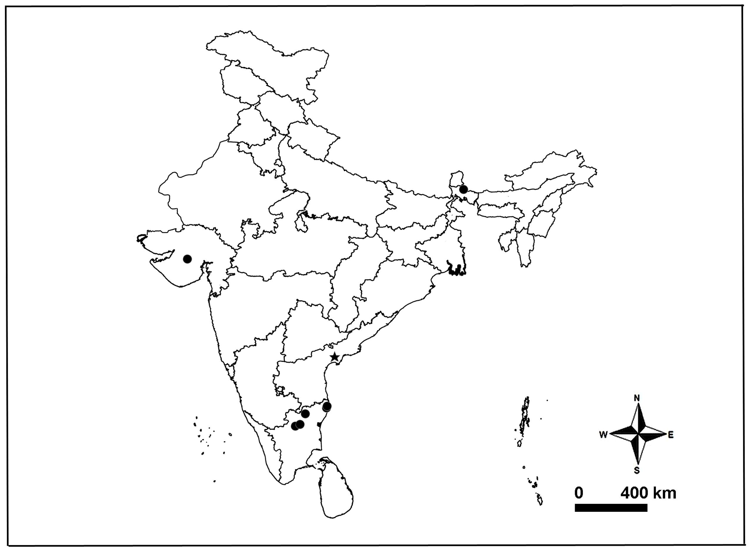
Distribution. India: Gujarat, Tamil Nadu, West Bengal ( Gravely 1924; Tikader & Malhotra 1980; Sen et al. 2015; present data) ( Fig. 40 View FIGURE 40 ).
Remarks. The NZC-ZSI collection has one glass bottle for H. madraspatana , labeled as ‘Types’ (5224/H2), containing pieces of prosoma, opisthosoma, chelicerae, left pedipalp and a few leg segments, all are in bad condition ( Fig. 23A–E View FIGURE 23 ).
Caleb, J. T. D. (2020) Spider (Arachnida: Araneae) fauna of the scrub jungle in the Madras Christian College campus, Chennai, India. Journal of Threatened Taxa, 12, 15711 - 15766. https: // doi. org / 10.11609 / jott. 5758.12.7.15711 - 15766
Dhali, D. C., Saha, S. & Raychaudhuri, D. (2017) Litter and ground dwelling spiders (Araneae: Arachnida) of reserve forests of Dooars, West Bengal. World Scientific News, 63, 1 - 242.
Gravely, F. H. (1924) Some Indian spiders of the family Lycosidae. Records of the Indian Museum, Calcutta, 26, 587 - 613. https: // doi. org / 10.26515 / rzsi / v 26 / i 6 / 1924 / 162654
Marusik, Y. M. & Nadolny, A. A. (2021) Redescription of Hippasa deserticola, the northernmost species of Hippasa (Aranei: Lycosidae), with taxonomic notes on other species of the genus. Zoosystematica Rossica, 30, 222 - 235. https: // doi. org / 10.31610 / zsr / 2021.30.2.222
Sen, S., Dhali, D. C., Saha, S. & Raychaudhuri, D. (2015) Spiders (Araneae: Arachnida) of Reserve Forests of Dooars: Gorumara National Park, Chapramari Wildlife Sanctuary and Mahananda Wildlife Sanctuary. World Scientific News, 20, 1 - 339.
Tikader, B. K. & Malhotra, M. S. (1980) Lycosidae (Wolf-spiders). Fauna India, Araneae, 1, 248 - 447.
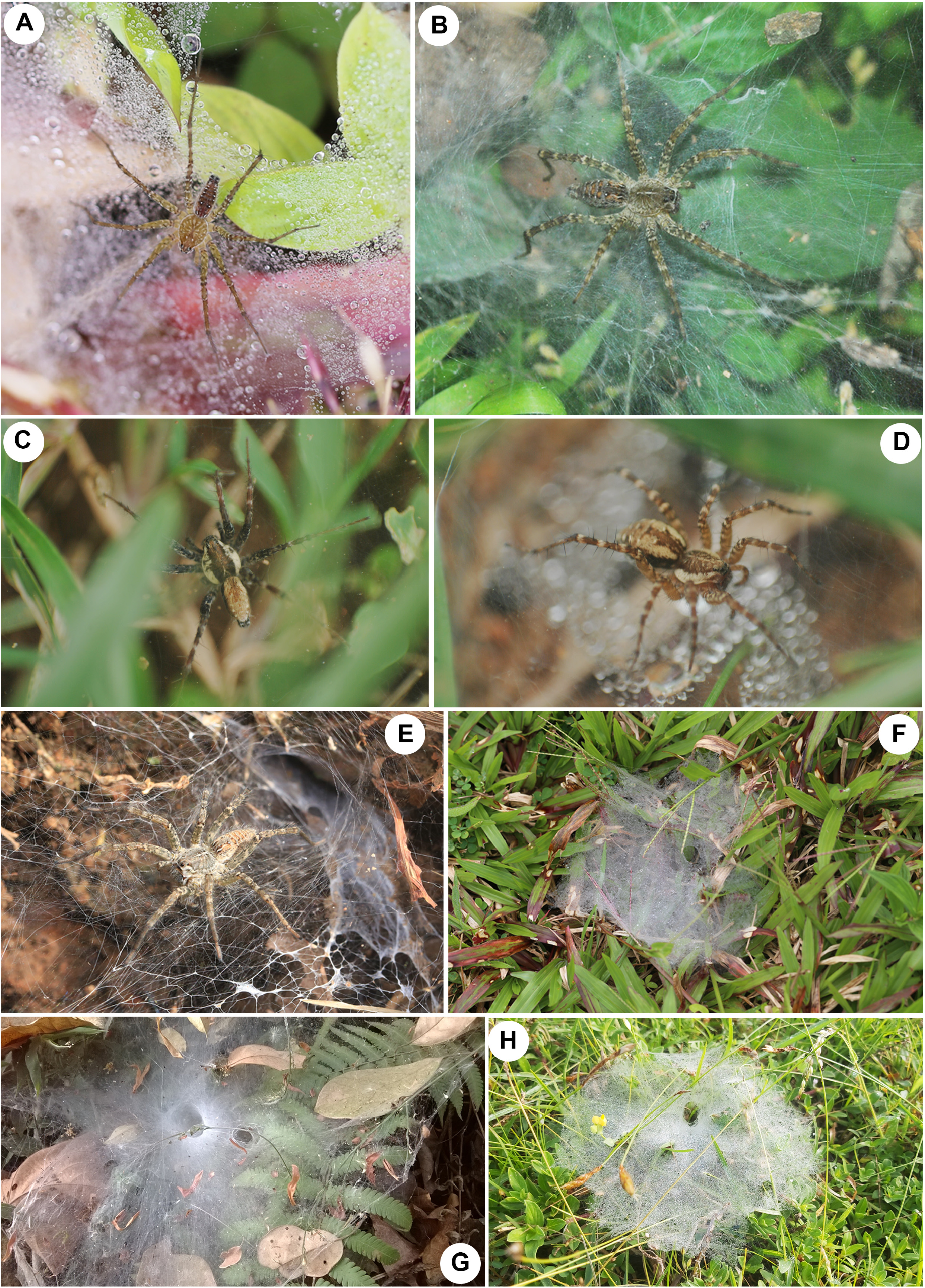
FIGURE 1. Field photographs of Hippasa spp. and their webs. A–B, F Hippasa agelenoides (Simon, 1884): A male; B female; F web. C–D, H Hippasa madraspatana Gravely, 1924: C male; D female; H web. E Hippasa pantherina Pocock, 1899, female. G web of Hippasa lycosina Pocock, 1900. Figures not to scale. Photo credits: A, F–G Pradeep M. Sankaran, B–D, H John T. D. Caleb, E, Jimmy Paul.
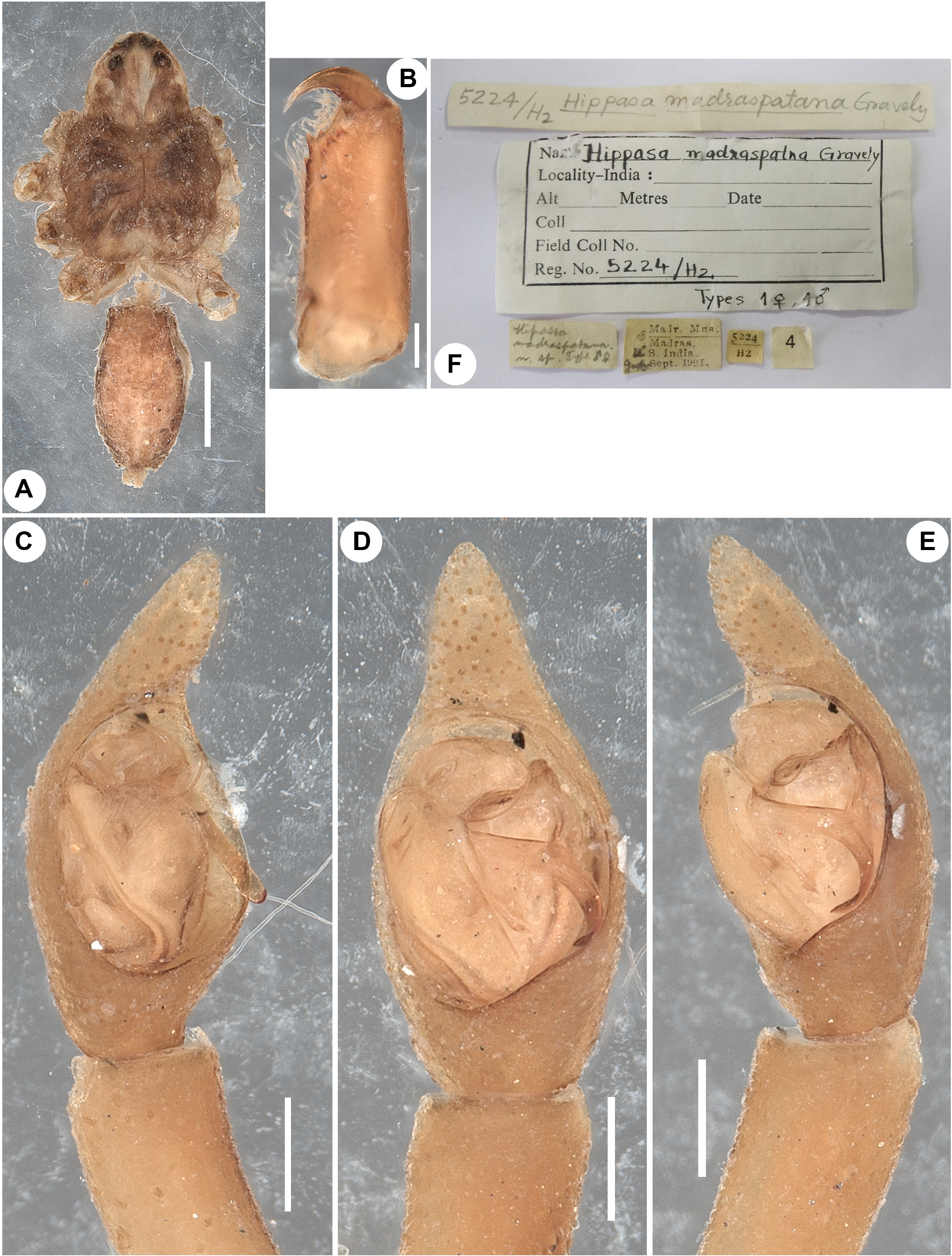
FIGURE 23. Hippasa madraspatana Gravely, 1924, syntype male (NZC-ZSI 5224/H2).A habitus, dorsal view. B left chelicera, retrolateral view. C–E left pedipalp: C prolateral view; D ventral view; E retrolateral view. F labels from the syntype tube. Scale bars: A, 1 mm; B–E, 0.2 mm.
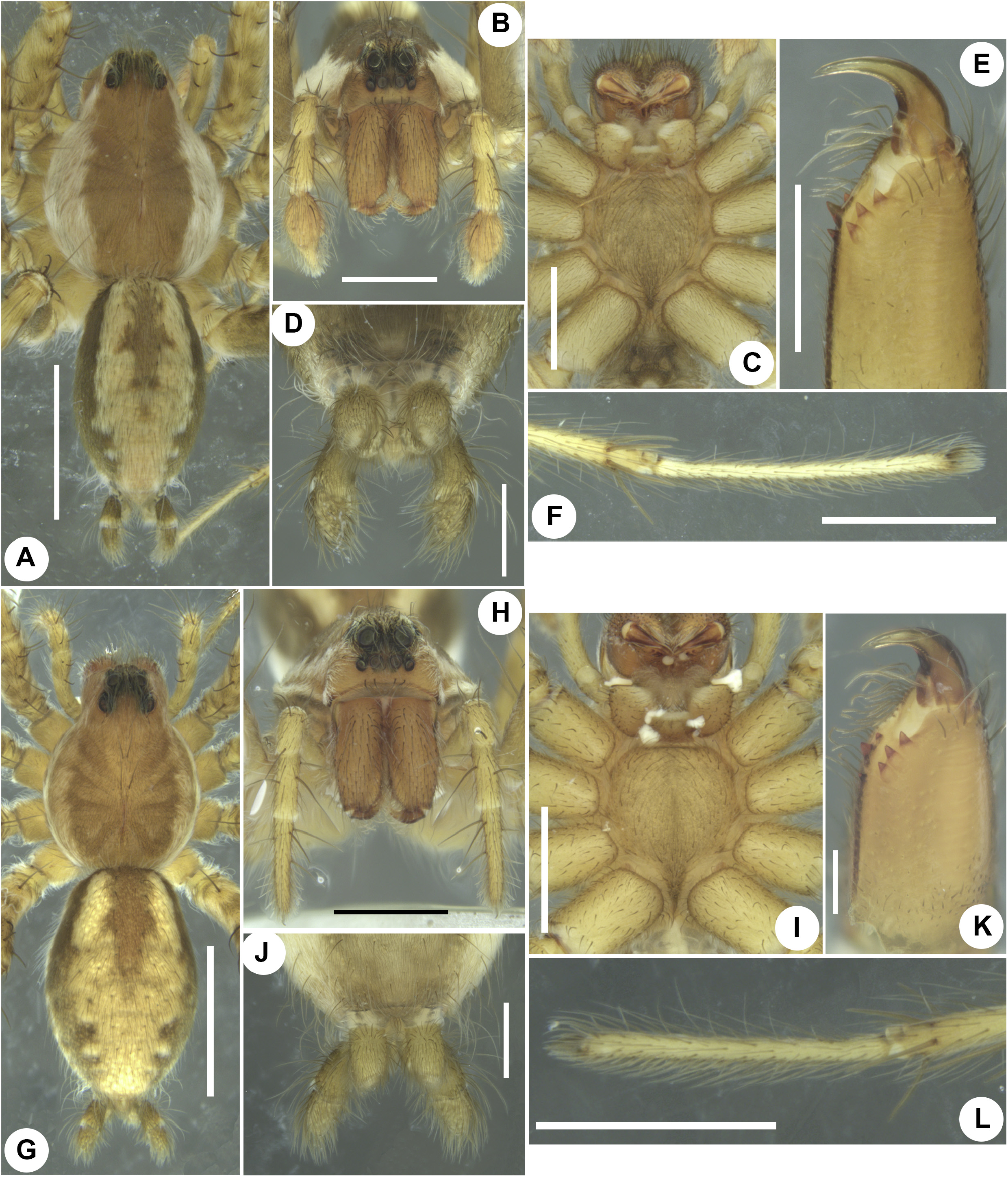
FIGURE 24. Hippasa madraspatana Gravely, 1924, male (A–F) and female (G–L) (ADSH5950281). A, G habitus, dorsal view. B, H same, frontal view. C, I prosoma showing endites, labium, sternum, ventral view. D, J spinnerets, ventral view. E, K left chelicera, retrolateral view. F, L left tarsus IV showing reduced scopula, lateral view. Scale bars: A, G, 2 mm; B–C, F, H–I, 1 mm; D–E, J, L, 0.5 mm; K, 0.2 mm.
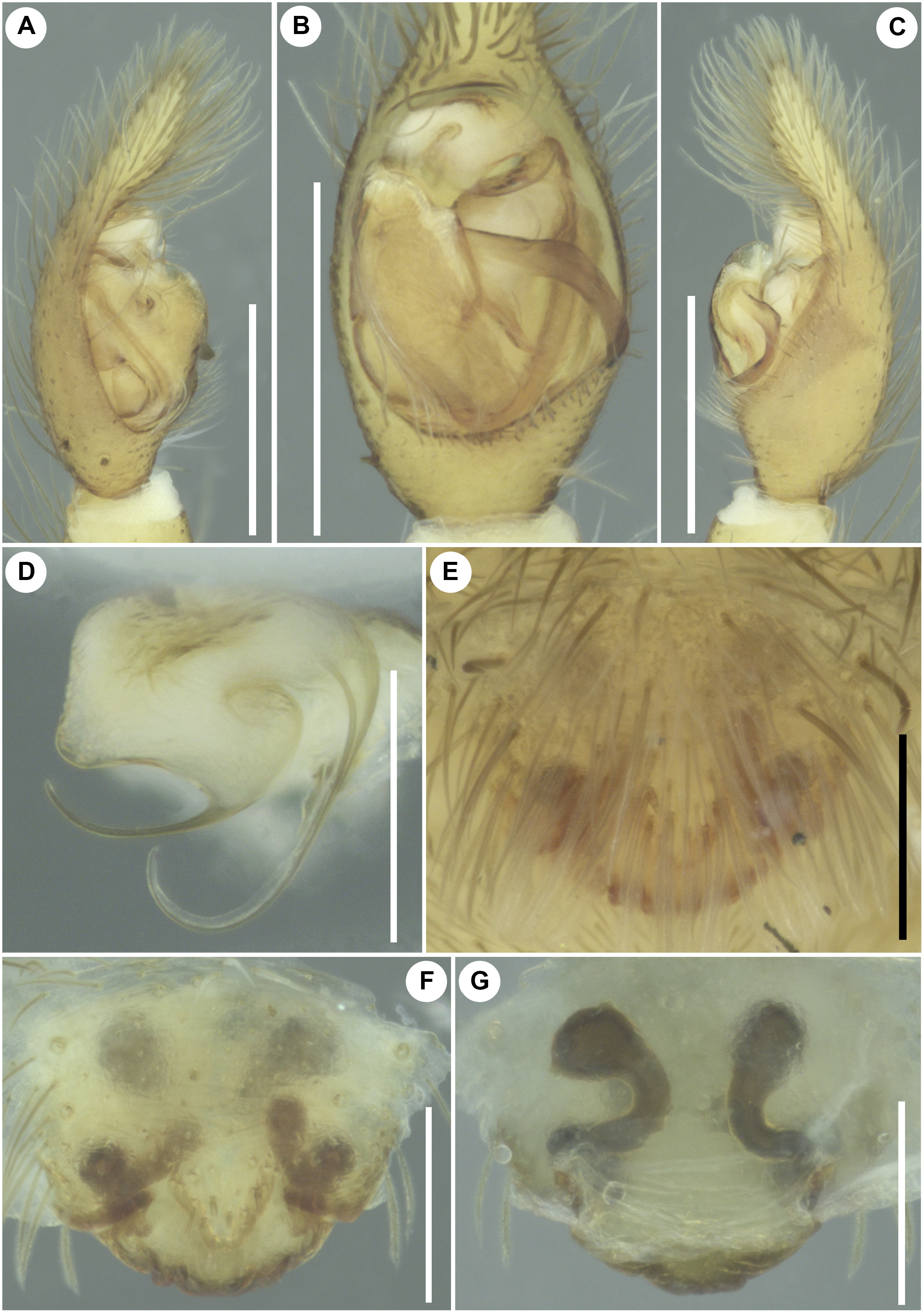
FIGURE 25. Hippasa madraspatana Gravely, 1924, male and female genitalia (ADSH5950281). A–C male left pedipalp, D male right pedipalp: A prolateral view; B ventral view; C retrolateral view; D embolic division, ventral view. E–G female genitalia: E intact epigyne, ventral view; F same, after clearing in KOH and removing bushy setae, ventral view; G vulva, dorsal view. Scale bars: A–C, 0.5 mm; D–G, 0.2 mm.
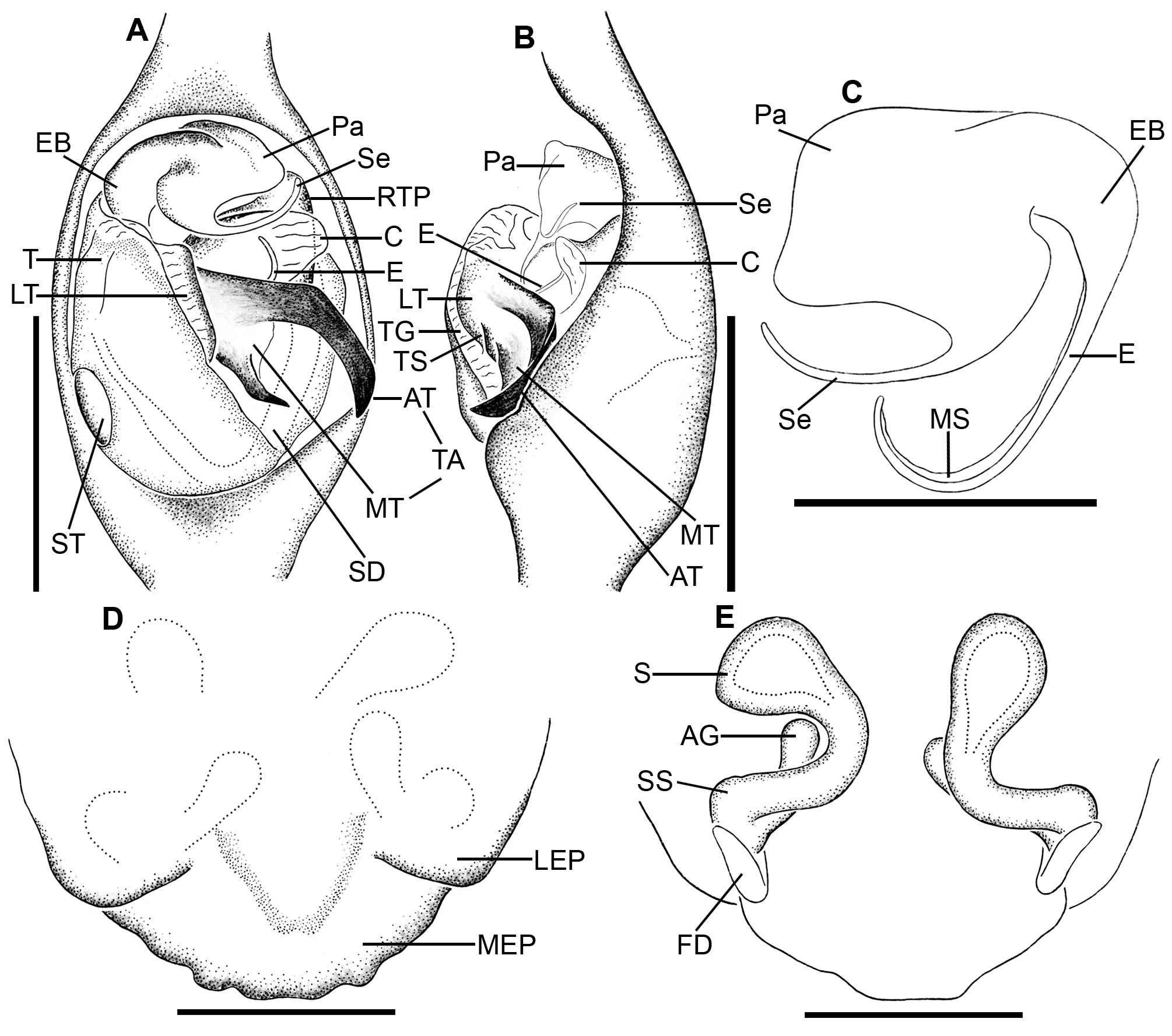
FIGURE 26. Hippasa madraspatana Gravely, 1924, male and female genitalia (ADSH5950281). A–B male left pedipalp, C male right pedipalp: A ventral view; B retrolateral view; C embolic division, ventral view. D–E female genitalia: D epigyne, ventral view; E vulva, dorsal view. Abbreviations: AG accessory gland; AT anterior arm of tegular apophysis; C conductor; E embolus; EB embolic base; FD fertilization duct; LEP lateral epigynal plate; LT lamellate process of tegulum; MEP median epigynal plate; MS membranous sheath; MT mesal arm of tegular apophysis; Pa palea; RTP retrolateral process of tegulum; S spermatheca; Se synembolus; SD sperm duct; SS spermathecal stalk; ST subtegulum; T tegulum; TA tegular apophysis; TG tegular groove; TS tegular stalk. Scale bars: A–B, 0.5 mm; C–E, 0.2 mm.
| V |
Royal British Columbia Museum - Herbarium |
No known copyright restrictions apply. See Agosti, D., Egloff, W., 2009. Taxonomic information exchange and copyright: the Plazi approach. BMC Research Notes 2009, 2:53 for further explanation.
|
Kingdom |
|
|
Phylum |
|
|
Class |
|
|
Order |
|
|
Family |
|
|
Genus |
Hippasa madraspatana Gravely, 1924
| SANKARAN, PRADEEP M. & CALEB, JOHN T. D. 2023 |
Hippasa madraspatana
| Caleb, J. T. D. 2020: 15725 |
| Dhali, D. C. & Saha, S. & Raychaudhuri, D. 2017: 68 |
| Sen, S. & Dhali, D. C. & Saha, S. & Raychaudhuri, D. 2015: 46 |
| Tikader, B. K. & Malhotra, M. S. 1980: 289 |
| Gravely, F. H. 1924: 595 |