
Hippasa lycosina Pocock, 1900
|
publication ID |
https://doi.org/10.11646/zootaxa.5230.2.1 |
|
publication LSID |
lsid:zoobank.org:pub:D4803049-9F65-4885-943E-0B0A3A084677 |
|
DOI |
https://doi.org/10.5281/zenodo.7554981 |
|
persistent identifier |
https://treatment.plazi.org/id/03B487A7-F454-CE24-5DDB-FF5ABC07FE95 |
|
treatment provided by |
Plazi (2023-01-20 07:43:41, last updated 2024-11-26 08:51:57) |
|
scientific name |
Hippasa lycosina Pocock, 1900 |
| status |
|
Hippasa lycosina Pocock, 1900 View in CoL
Figs 1G View FIGURE 1 , 19–22 View FIGURE 19 View FIGURE 20 View FIGURE 21 View FIGURE 22 , 39 View FIGURE 39
Hippasa lycosina Pocock, 1900: 250 View in CoL (♀). Gravely 1924: 593, fig. 1B (♀)
Hippasa mahabaleshwarensis Tikader & Malhotra, 1980: 285 View in CoL , figs 85–89 (♂ ♀), synonymised by Song (1987) (for complete list of references, see World Spider Catalog 2022).
Type material. H. lycosina . Syntypes 2 ♀♀ from INDIA: Maharashtra: Nashik (=Nasik) (20°04'N, 73°36'E; 625 m alt.), date unknown, Millet leg., repository NHM (1899), not examined (illustrations of this species given in Tikader & Malhotra (1980: figs 91–92), who studied the types are diagnostic and were used for comparative purpose). H. mahabaleshwarensis GoogleMaps . Holotype ♀ from INDIA: Maharashtra: Satara: Mahabaleshwar (17°56'N, 73°31'E; 151 m alt.); 26 March 1976, B.K. Tikader leg., repository NZC-ZSI (4651/18), examined GoogleMaps . Paratypes 3 ♀♀ and allotypes 2 ♂♂, with the same data as holotype, examined GoogleMaps .
Other material examined. INDIA: Karnataka: Chikmagalur: Mullayanagiri Peak (13°23'N, 75°43'E; 1894 m alt.), 18 February 2014, M.S. Pradeep leg., from web on ground, by hand: 1 ♂, 1 ♀ ( ADSH595026 View Materials ) GoogleMaps .
Diagnosis. Males of H. lycosina are most similar to the males of H. albopunctata Thorell, 1899 as both share a short anterior arm of tegular apophysis and ventrally visible subtegulum, but can be separated from the latter by anterior arm of tegular apophysis with prolaterally oriented tip (vs. posteriorly in H. albopunctata ), and broad mesal arm of tegular apophysis with rounded tip (vs. narrow, thorn-like in H. albopunctata , compare Figs 19F–G View FIGURE 19 , 21A–B View FIGURE 21 with Alderweireldt & Jocqué 2005: fig. 14). Females are similar to the females of H. himalayensis as both share a large epigynal atrium, but can be separated from the latter by a widely triangular epigynal atrium (vs. narrowly triangular in H. himalayensis ), and spherical spermathecae (vs. peanut-shaped in H. himalayensis , compare Figs 20F–G View FIGURE 20 , 21D–E View FIGURE 21 with Fig. 14C–D View FIGURE 14 ).
Supplementary description. Male in ethanol (ADSH595026; Fig. 19A–D View FIGURE 19 ). Carapace pale yellow coloured, with a median straight white stripe extending from PMEs up to rear end of fovea, with paired lateral longitudinal white bands extending along the entire length of carapace, medially clothed with fine black appressed setae, with a few scattered erect spine-like setae restricted to cephalic part. Eye region, clypeus, sternum pale yellow coloured; chelicerae, endites, labium, leg and pedipalp segments, and spinnerets pale brownish; dorsum of opisthosoma black, sides and venter creamy-white; leg and pedipalp segments with black annulations and patches. Thoracic fovea reddish, long (1.38), straight, longitudinal ( Fig. 19A View FIGURE 19 ). Thoracic part laterally black. Chelicerae dorsally clothed with moderately long setae; inner and outer surfaces provided with stridulatory files; promargin provided with a series of long setae with bend tips, pro- and retromargins with three teeth ( Fig. 19C View FIGURE 19 ). Sternum provided with thick covering of black setae, with a broad median longitudinal black band ( Fig. 19B View FIGURE 19 ). Opisthosoma elongate-ovoid, hirsute ( Fig. 19A View FIGURE 19 ); cardiac area marked with a reddish brown patch bordered by narrow longitudinal creamy-white bands; dorsum anteriorly provided with two pairs of white lateral stripes, with a few scattered white spots and patches, medioposteriorly with a few transverse roughly W-shaped bands; sides with thin black streaks; venter medially with paired, longitudinal broad bands of chalk white spots. Spinnerets hirsute ( Fig. 19D View FIGURE 19 ). Legs long, slender, hirsute, spinose; metatarsi I–II with distal and all tarsi with complete scopulae, all well-developed. Body length 16.02. Carapace 7.93 long, 5.75 wide. Opisthosoma 8.09 long, 4.62 wide. Eye diameters and interdistances: ALE 0.32, AME 0.34, PLE 0.44, PME 0.50; AME–ALE 0.09, AME–AME 0.16, AME–PME 0.21, PLE–PLE 1.35, PME–PLE 0.51, PME–PME 0.42. Clypeus height at AMEs 0.34, at ALEs 0.34. Length of chelicerae 3.92. Measurements of pedipalp and legs: pedipalp 11.65 [4.13, 1.77, 2.73, 3.02], I 29.76 [7.36, 3.40, 6.70, 8.06, 4.24], II 28.40 [7.41, 3.12, 6.24, 7.82, 3.81], III 26.58 [7.19, 2.90, 5.56, 7.50, 3.43], IV 36.20 [8.83, 3.22, 7.90, 11.78, 4.47]. Leg formula: 4123. Spination of pedipalp: femur pld 1 do 4, patella spineless, tibia pl 1 pld 1 do 2, tarsus/cymbium spineless; legs: femur I pld 2 do 3 rld 3, II–III pld 3 do 3 rld 3, IV pld 3 do 3 rld 1; patellae I–IV pld 1 do 2 rld 1; tibia I pl 1 pld 1 plv 3 rl 1 rld 1 rlv 3, II pl 1 pld 1 plv 2 rl 2 rlv 3, III pl 1 pld 1 plv 3 do 2 rl 1 rld 1 rlv 2, IV pl 1 pld 1 plv 3 do 2 rl 1 rld 1 rlv 3; metatarsus I pld 2 plv 3 rld 2 rlv 3 vt 1, II pld 3 plv 3 rld 2 rlv 3 vt 1, III pld 3 plv 3 rld 3 rlv 3 vt 1, IV pld 3 plv 3 rld 3 rlv 4 vt 1; tarsi I–IV spineless. Pedipalp ( Figs 19E–H View FIGURE 19 , 23A–C View FIGURE 23 ): segments hirsute; cymbium proximally wide, gradually narrowing towards apex, without apical claw-like macrosetae, distoventrally provided with long hairs with bend tips ( Fig. 19G View FIGURE 19 ). Tegulum large, occupying more than half of the ventral side of bulb ( Figs 19E–G View FIGURE 19 , 21A–B; T View FIGURE 21 ). Subtegulum small, subglobular, posteroprolaterally located ( Figs 19E–F View FIGURE 19 , 21A View FIGURE 21 ; ST). Palea small, roughly rectangular, less sclerotised ( Fig. 19F, H View FIGURE 19 , 21A, C View FIGURE 21 ; Pa). Synembolus short, narrow, C-shaped, arising on ventroprolateral margin of palea, with smoothly rounded tip ( Figs 19H View FIGURE 19 , 21C View FIGURE 21 ; Se). Tegular process short, irregular, visible only in retrolateral view ( Figs 19G View FIGURE 19 , 21B View FIGURE 21 ; RTP). Tegular apophysis with short, flat, wide anterior arm having prolaterally directed rounded tip and short retrolaterally directed mesal arm ( Figs 19F–G View FIGURE 19 , 21A–B View FIGURE 21 ; TA, AT, MT). Conductor large, hyaline, lying behind embolus, masking tegular process, with a retrolateral bent ( Figs 19F–G View FIGURE 19 , 21A–B; C View FIGURE 21 ). Embolus thin, masked entirely by distal part of tegulum, moderately long, with U-shaped curved tip, with broad embolic base ( Figs 19H View FIGURE 19 , 21C; E View FIGURE 21 , EB).
Female in ethanol (ADSH595026; Fig. 20A–D View FIGURE 20 ). Like the male, except by the following: carapace, clypeus, sternum, leg and palp segments, spinnerets pale brownish; chelicerae, endites, labium brownish; venter of opisthosoma lacks bands of chalk white spots. Thoracic fovea slightly short (1.25) ( Fig. 20A View FIGURE 20 ). Body length 17.99. Carapace 8.17 long, 6.04 wide. Opisthosoma 9.82 long, 5.58 wide. Eye diameters and interdistances: ALE 0.26, AME 0.28, PLE 0.42, PME 0.45; AME–ALE 0.21, AME–AME 0.26, AME–PME 0.32, PLE–PLE 1.50, PME–PLE 0.55, PME–PME 0.41. Clypeus height at AMEs 0.42, at ALEs 0.34. Length of chelicerae 3.72. Measurements of palp and legs: palp 10.31 [3.55, 1.73, 2.29, 2.74], I 24.22 [6.95, 3.14, 5.40, 5.67, 3.06], II 23.79 [6.99, 3.04, 5.05, 5.48, 3.23], III 22.94 [6.72, 2.89, 4.80, 5.76, 2.77], IV 32.66 [8.73, 3.20, 7.13, 9.71, 3.89]. Spination of palp: femur pld 1 do 3 rld 1, patella pld 1 do 2, tibia pl 1 pld 1 rld 1, tarsus pl 1 pld 2 rl 1 rlv 1; legs: femur II pld 3 do 2 rld 3, IV pld 2 do 3 rld 1; tibia I pl 1 pld 1 plv 3 rl 2 rlv 3, II pl 1 pld 1 plv 2 rl 1 rld 1 rlv 3, III pl 1 pld 1 plv 3 do 2 rl 1 rld 1 rlv 1, IV pl 1 pld 1 plv 3 do 2 rl 1 rld 2 rlv 1. Genitalia ( Figs 20E–G View FIGURE 20 , 21D–F View FIGURE 21 ): epigyne clothed in bushy setae ( Fig. 20E View FIGURE 20 ), with broadly triangular median and narrow lateral plates ( Figs 20F View FIGURE 20 , 21D View FIGURE 21 ; MEP, LEP); median plate with large atrium leading to triangular hood internally ( Figs 20F–G View FIGURE 20 , 21D–E; H View FIGURE 21 ). Spermathecal stalks with irregular wide proximal and narrow tubular distal parts ( Figs 20G View FIGURE 20 , 21E–F View FIGURE 21 ; SS). Accessory glands globular, without stalk arising distolateral to spermathecal stalks ( Fig. 21F View FIGURE 21 ; AG). Spermathecae globular ( Figs 20G View FIGURE 20 , 21E; S View FIGURE 21 ). Fertilization ducts anteriorly directed, diverging ( Figs 20G View FIGURE 20 , 21E View FIGURE 21 ; FD).
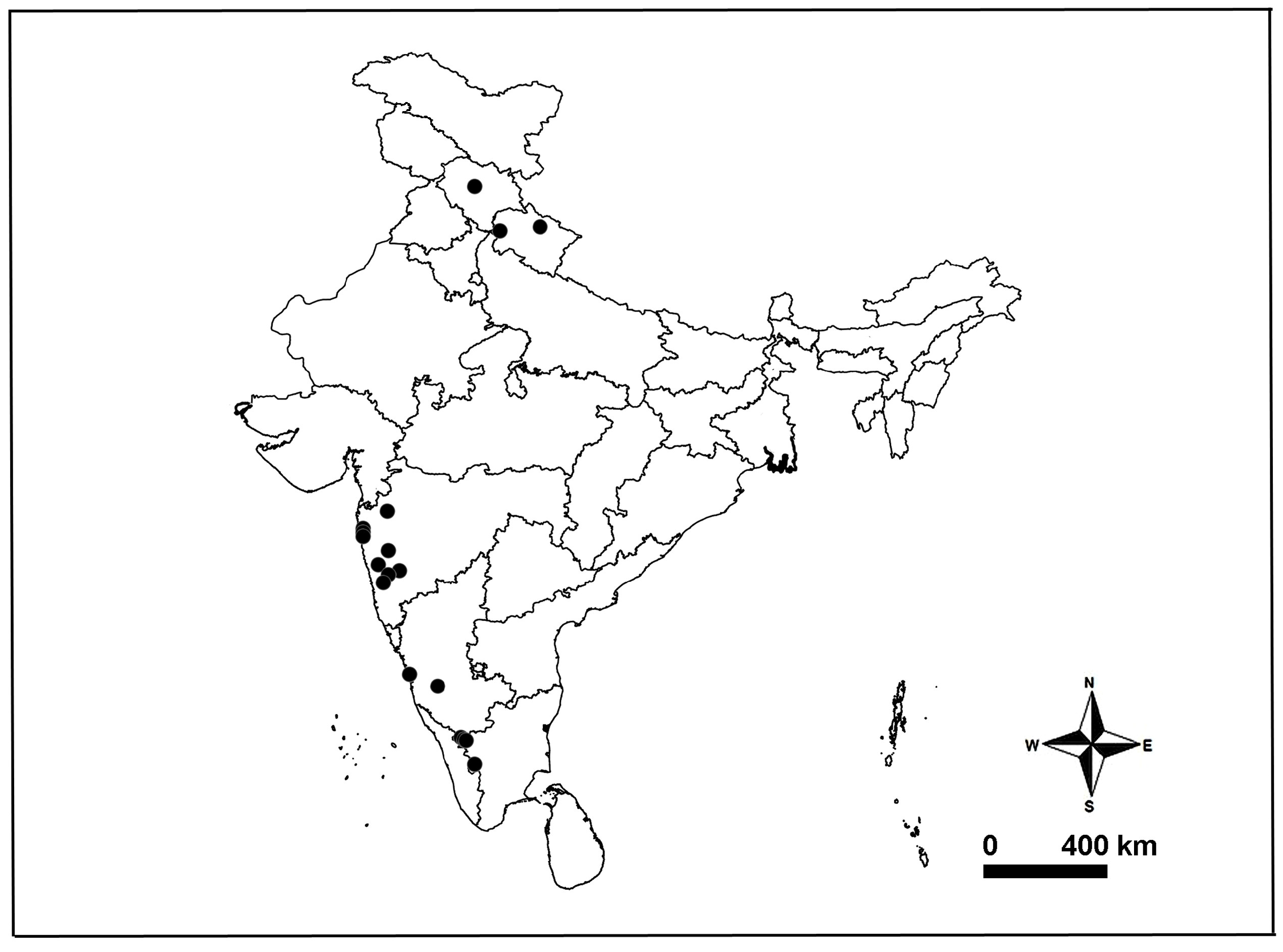
Distribution. China, Laos and India: Himachal Pradesh, Karnataka, Maharashtra, Tamil Nadu, Uttarakhand ( Pocock 1900; Gravely 1924; Tikader & Malhotra 1980; Yin & Wang 1980; Hu 1984; Chen & Gao 1990; Jäger & Praxaysombath 2011; Wang et al. 2015; Ahmed et al. 2015; Marusik & Nadolny 2021; present data) ( Fig. 39 View FIGURE 39 ).
Remarks. We examined the types of Hippasa mahabaleshwarensis Tikader & Malhotra, 1980 and confirmed its synonymy with H. lycosina as proposed by Song (1987, compare Figs 19F–G View FIGURE 19 , 21A–B View FIGURE 21 with Fig. 22B–C View FIGURE 22 ).
Ahmed, J., Satam, Y., Khalap, R. & Mohan, K. (2015) First record of Portia albimana (Simon, 1900) from Maharashtra, Mumbai (Araneae: Salticidae: Spartaeinae). Peckhamia, 129.1, 1 - 6.
Alderweireldt, M. & Jocque, R. (2005) A taxonomic review of the Afrotropical representatives of the genus Hippasa (Araneae, Lycosidae). Journal of Afrotropical Zoology, 2, 45 - 68.
Chen, X. E. & Gao, J. C. (1990) The Sichuan farmland spiders in China. Sichuan Science and Technology Publishing House, Chengdu, 226 pp.
Gravely, F. H. (1924) Some Indian spiders of the family Lycosidae. Records of the Indian Museum, Calcutta, 26, 587 - 613. https: // doi. org / 10.26515 / rzsi / v 26 / i 6 / 1924 / 162654
Hu, J. L. (1984) The Chinese spiders collected from the fields and the forests. Tianjin Science and Technology Press, Tianjn, 482 pp.
Jager, P. & Praxaysombath, B. (2011) Spiders from Laos with forty-three new records and first results from the provinces Bolikhamsay and Champasak (Arachnida: Araneae). Acta Arachnologica, 60, 9 - 31. https: // doi. org / 10.2476 / asjaa. 60.9
Marusik, Y. M. & Nadolny, A. A. (2021) Redescription of Hippasa deserticola, the northernmost species of Hippasa (Aranei: Lycosidae), with taxonomic notes on other species of the genus. Zoosystematica Rossica, 30, 222 - 235. https: // doi. org / 10.31610 / zsr / 2021.30.2.222
Pocock, R. I. (1900) The fauna of British India, including Ceylon and Burma. Arachnida. Taylor and Francis, London, 279 pp. https: // doi. org / 10.5962 / bhl. title. 48423
Song, D. X. (1987) Spiders from agricultural regions of China (Arachnida: Araneae). Agriculture Publishing House, Beijing, 376 pp.
Thorell, T. (1899) Araneae Camerunenses (Africae occidentalis) quas anno 1891 collegerunt Cel. Dr Y. Sj ˆ stedt aliique. Bihang till Kongliga Svenska Vetenskaps-Akademiens Handlingar, 25, 1 - 105.
Tikader, B. K. & Malhotra, M. S. (1980) Lycosidae (Wolf-spiders). Fauna India, Araneae, 1, 248 - 447.
Wang, L. Y., Li, Z. X., Zhou, K. X. & Zhang, Z. S. (2015) Redescription of three Hippasa species from China (Araneae: Lycosidae), with a proposed species group-division and diagnosis. Zootaxa, 3974 (2), 231 - 244. https: // doi. org / 10.11646 / zootaxa. 3974.2.7
World Spider Catalog (2022) World Spider Catalog. Version 23.5. Natural History Museum Bern, Bern. Available from: http: // wsc. nmbe. ch (accessed 26 November 2022) https: // doi. org / 10.24436 / 2
Yin, C. M. & Wang, J. F. (1980) Descriptions of three new species of Hippasa (Araneae, Lycosidae) from China. Journal of Hunan Teachers College, Natural Science Edition, 1980, 55 - 60.
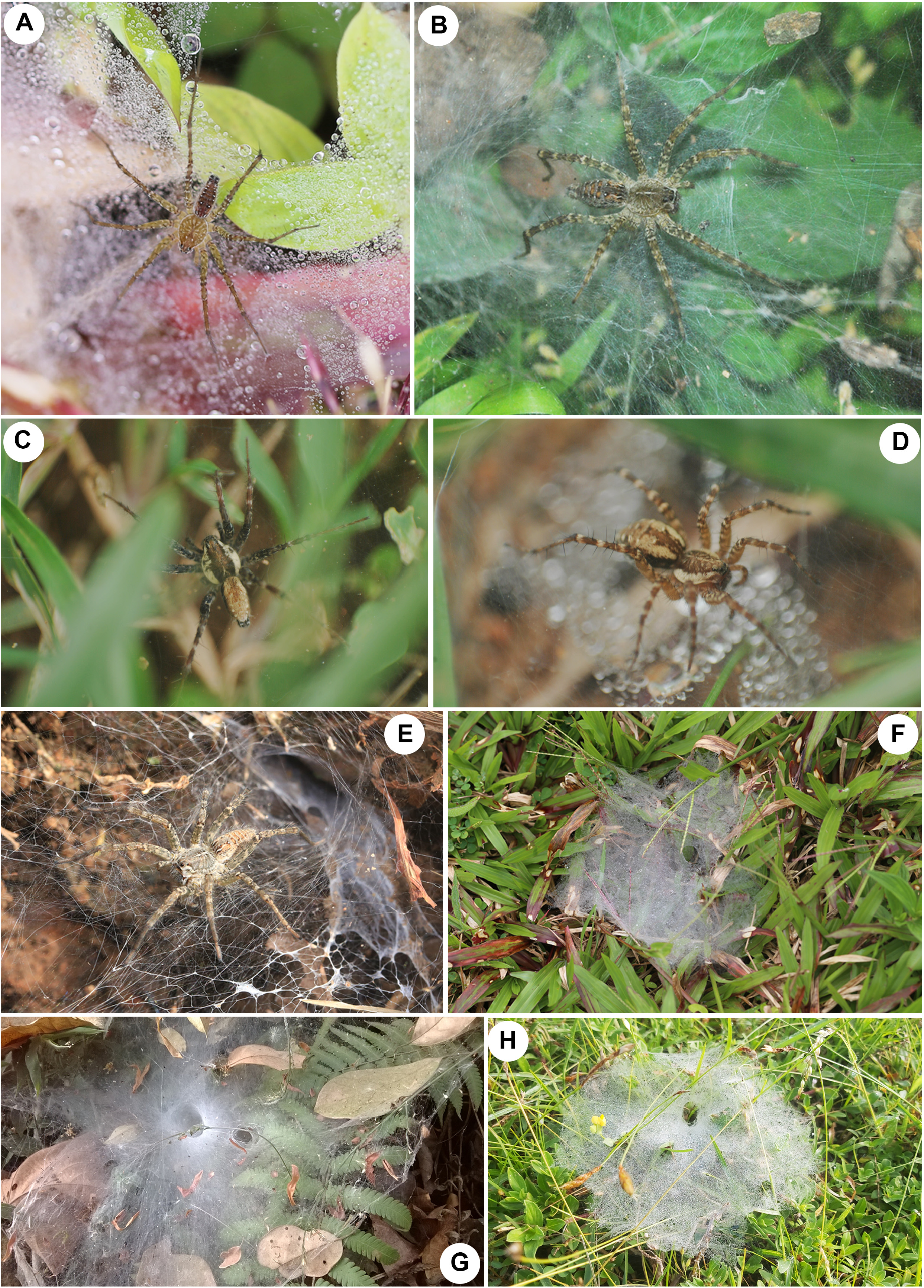
FIGURE 1. Field photographs of Hippasa spp. and their webs. A–B, F Hippasa agelenoides (Simon, 1884): A male; B female; F web. C–D, H Hippasa madraspatana Gravely, 1924: C male; D female; H web. E Hippasa pantherina Pocock, 1899, female. G web of Hippasa lycosina Pocock, 1900. Figures not to scale. Photo credits: A, F–G Pradeep M. Sankaran, B–D, H John T. D. Caleb, E, Jimmy Paul.
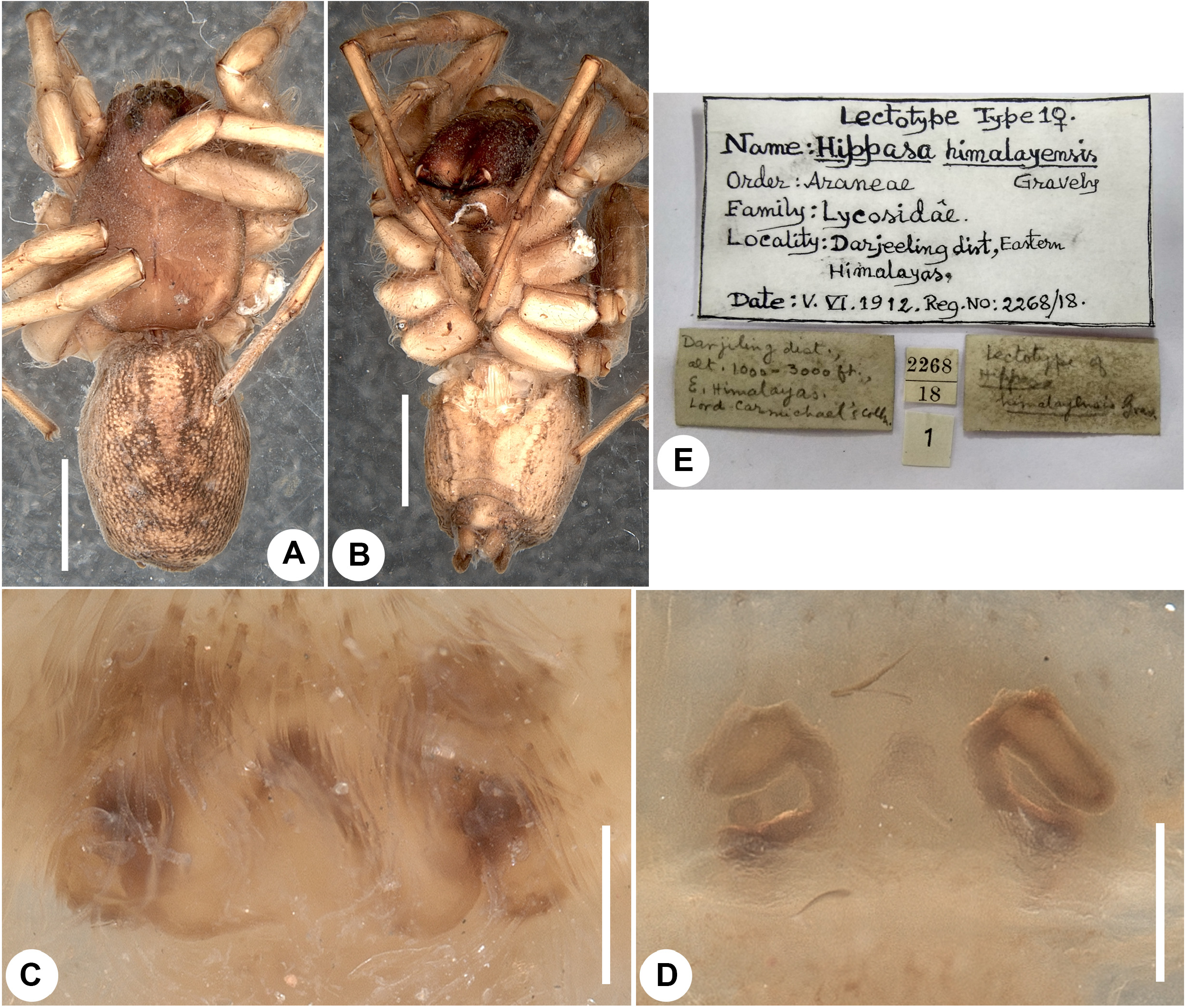
FIGURE 14. Hippasa himalayensis Gravely, 1924, lectotype female (NZC-ZSI 2268/18). A habitus, dorsal view. B same, ventral view. C epigyne, ventral view. D vulva, dorsal view. E labels from type bottle. Scale bars: A–B, 2 mm; C, 0.2 mm; D, 0.5 mm.
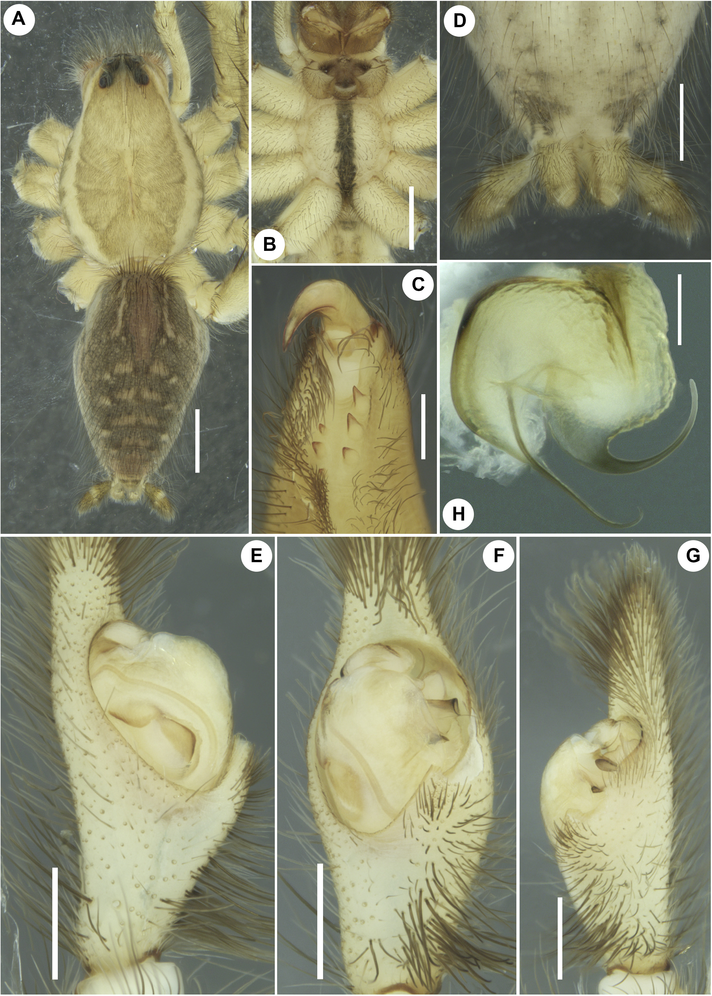
FIGURE 19. Hippasa lycosina Pocock, 1900, male (ADSH595026). A habitus, dorsal view. B prosoma showing sternum, ventral view. C left chelicera, ventral view. D spinnerets, ventral view. E–H left pedipalp: E prolateral; F ventral; G retrolateral; H embolic division, ventral view. Scale bars: A–B, 2 mm; C, E–G, 0.5 mm; D, 1 mm; H, 0.2 mm.
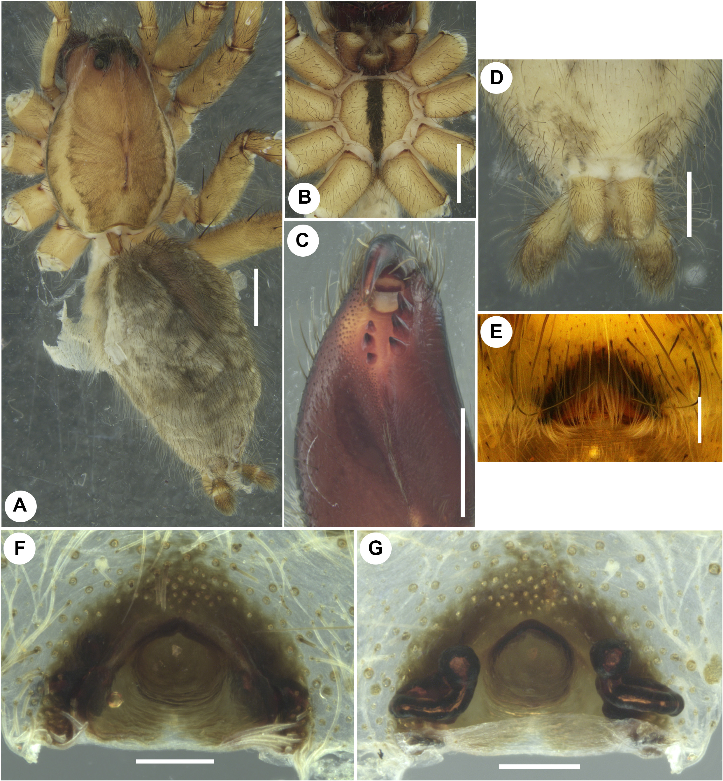
FIGURE 20. Hippasa lycosina Pocock, 1900, female (ADSH595026). A habitus, dorsoretrolateral view. B prosoma showing sternum, ventral view. C left chelicera, ventral view. D spinnerets, ventral view. E intact epigyne, ventral view; F same, after clearing in KOH and removing bushy setae, ventral view; G vulva, dorsal view. Scale bars: A–B, 2 mm; C–D, 1 mm; E–G, 0.2 mm.
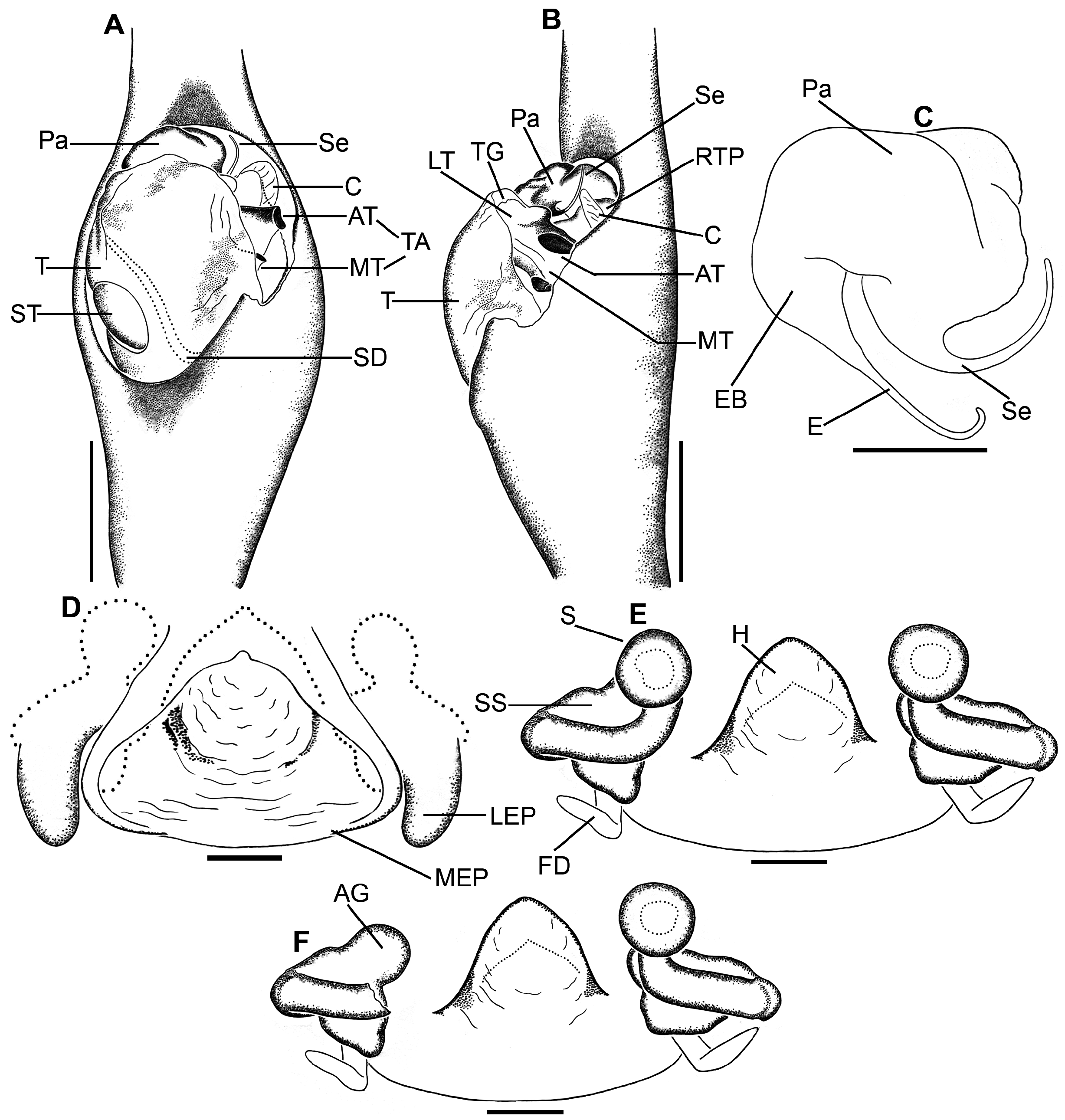
FIGURE 21. Hippasa lycosina Pocock, 1900, male and female genitalia (ADSH595026). A–C male left pedipalp: A ventral view; B retrolateral view; C embolic division, ventral view. D–E female genitalia: D epigyne, ventral view; E vulva, dorsal view; F same without left spermatheca showing accessory gland, dorsal view. Abbreviations: AG accessory gland; AT anterior arm of tegular apophysis; C conductor; E embolus; EB embolic base; FD fertilization duct; H hood; LEP lateral epigynal plate; MEP median epigynal plate; MT mesal arm of tegular apophysis; Pa palea; RTP retrolateral process of tegulum; S spermatheca; Se synembolus; SD sperm duct; SS spermathecal stalk; ST subtegulum; T tegulum; TA tegular apophysis. Scale bars: A–B, 0.5 mm; C–F, 0.2 mm.
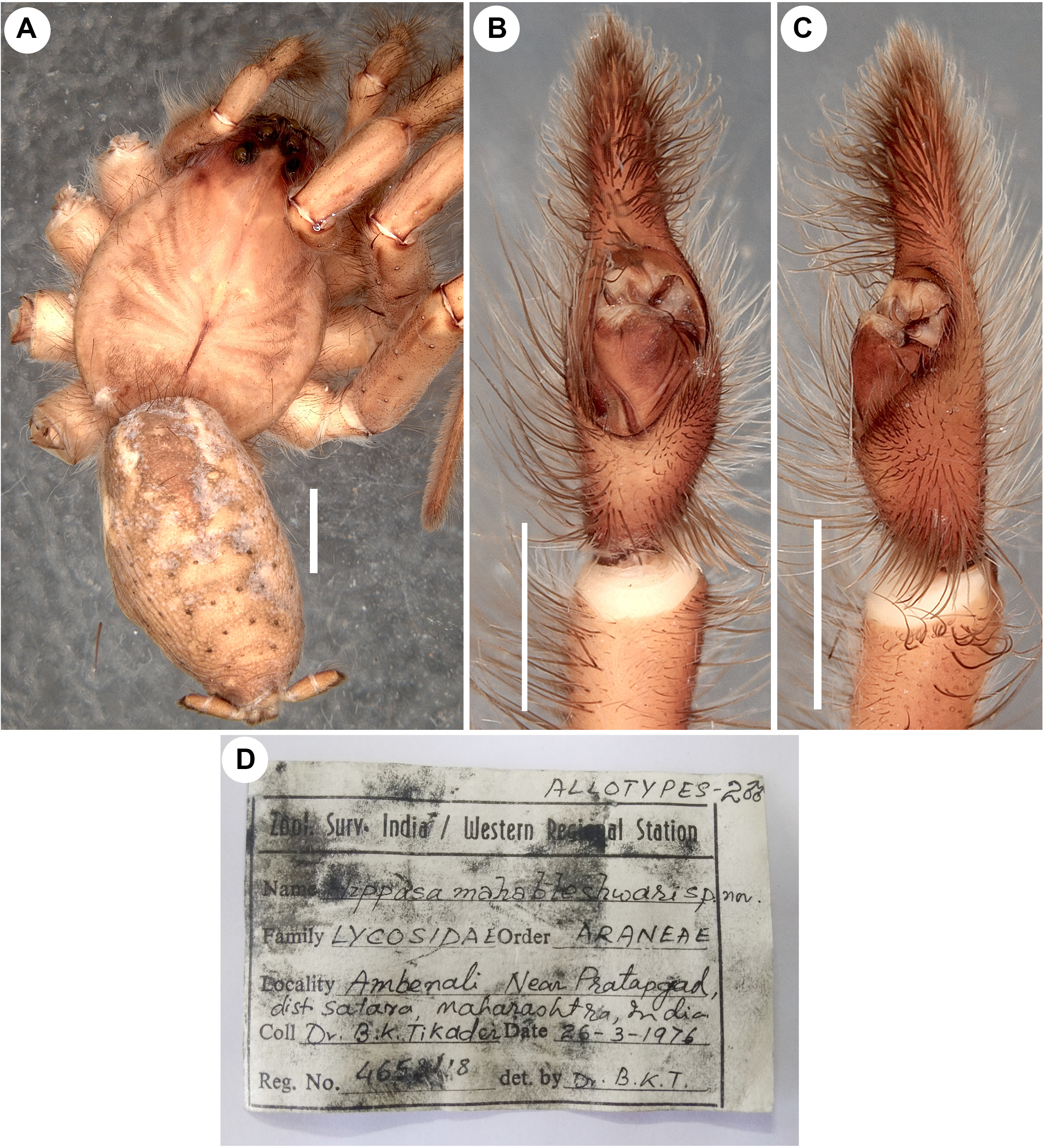
FIGURE 22. Hippasa lycosina Pocock, 1900, allotype male of Hippasa mahabaleshwarensis Tikader & Malhotra, 1980 (NZC- ZSI 4651/18). A habitus, dorsal view. B–C left pedipalp: B ventral view; C retrolateral view. D label from the allotype tube. Scale bars: A, 2 mm; B–C, 1 mm.
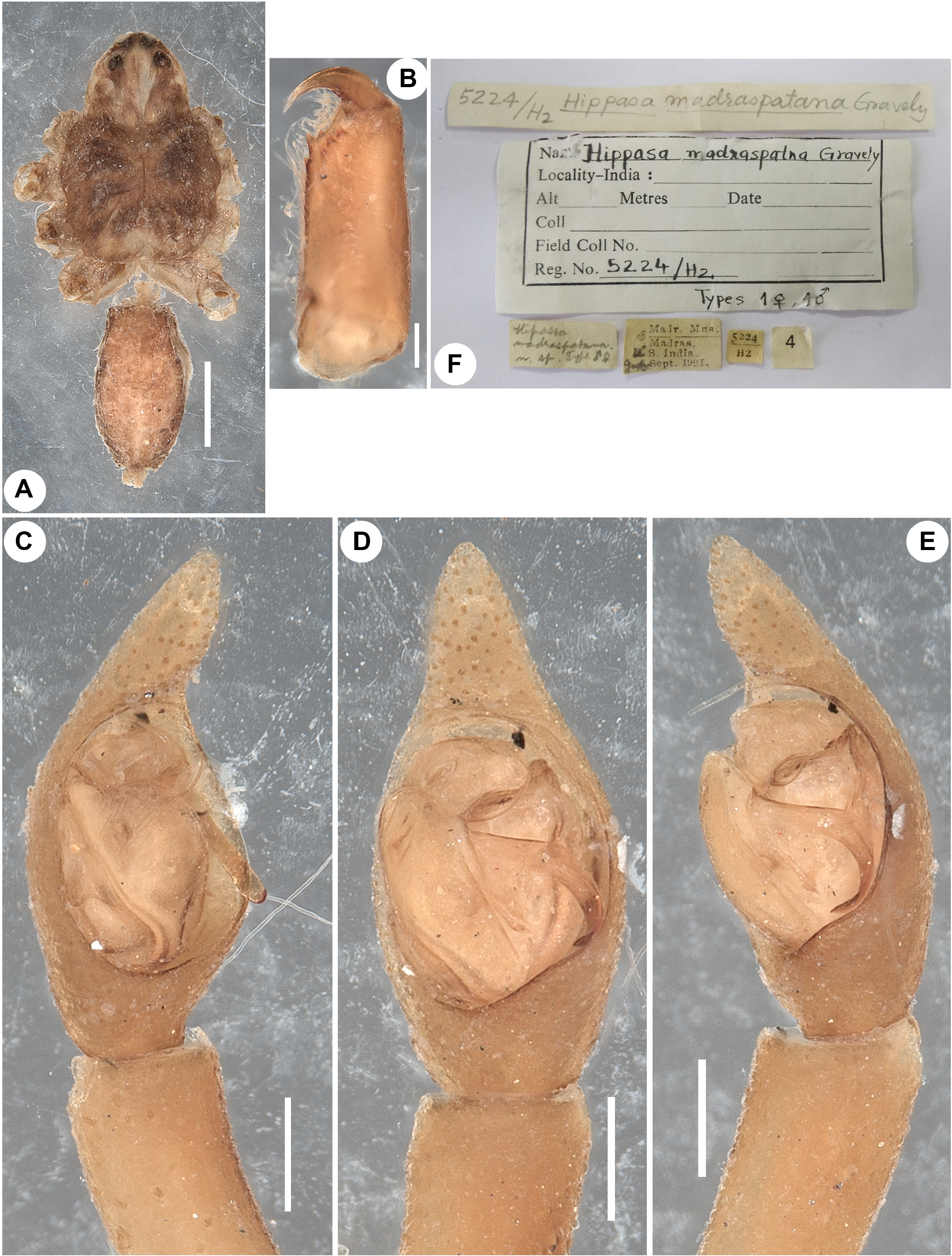
FIGURE 23. Hippasa madraspatana Gravely, 1924, syntype male (NZC-ZSI 5224/H2).A habitus, dorsal view. B left chelicera, retrolateral view. C–E left pedipalp: C prolateral view; D ventral view; E retrolateral view. F labels from the syntype tube. Scale bars: A, 1 mm; B–E, 0.2 mm.
No known copyright restrictions apply. See Agosti, D., Egloff, W., 2009. Taxonomic information exchange and copyright: the Plazi approach. BMC Research Notes 2009, 2:53 for further explanation.
|
Kingdom |
|
|
Phylum |
|
|
Class |
|
|
Order |
|
|
Family |
|
|
Genus |
Hippasa lycosina Pocock, 1900
| SANKARAN, PRADEEP M. & CALEB, JOHN T. D. 2023 |
Hippasa mahabaleshwarensis
| Tikader, B. K. & Malhotra, M. S. 1980: 285 |
Hippasa lycosina
| Gravely, F. H. 1924: 593 |
| Pocock, R. I. 1900: 250 |