
Hippasa loundesi Gravely, 1924
|
publication ID |
https://doi.org/ 10.11646/zootaxa.5230.2.1 |
|
publication LSID |
lsid:zoobank.org:pub:D4803049-9F65-4885-943E-0B0A3A084677 |
|
DOI |
https://doi.org/10.5281/zenodo.7554975 |
|
persistent identifier |
https://treatment.plazi.org/id/03B487A7-F458-CE2F-5DDB-FE39B8CCFD19 |
|
treatment provided by |
Plazi |
|
scientific name |
Hippasa loundesi Gravely, 1924 |
| status |
|
Hippasa loundesi Gravely, 1924 View in CoL
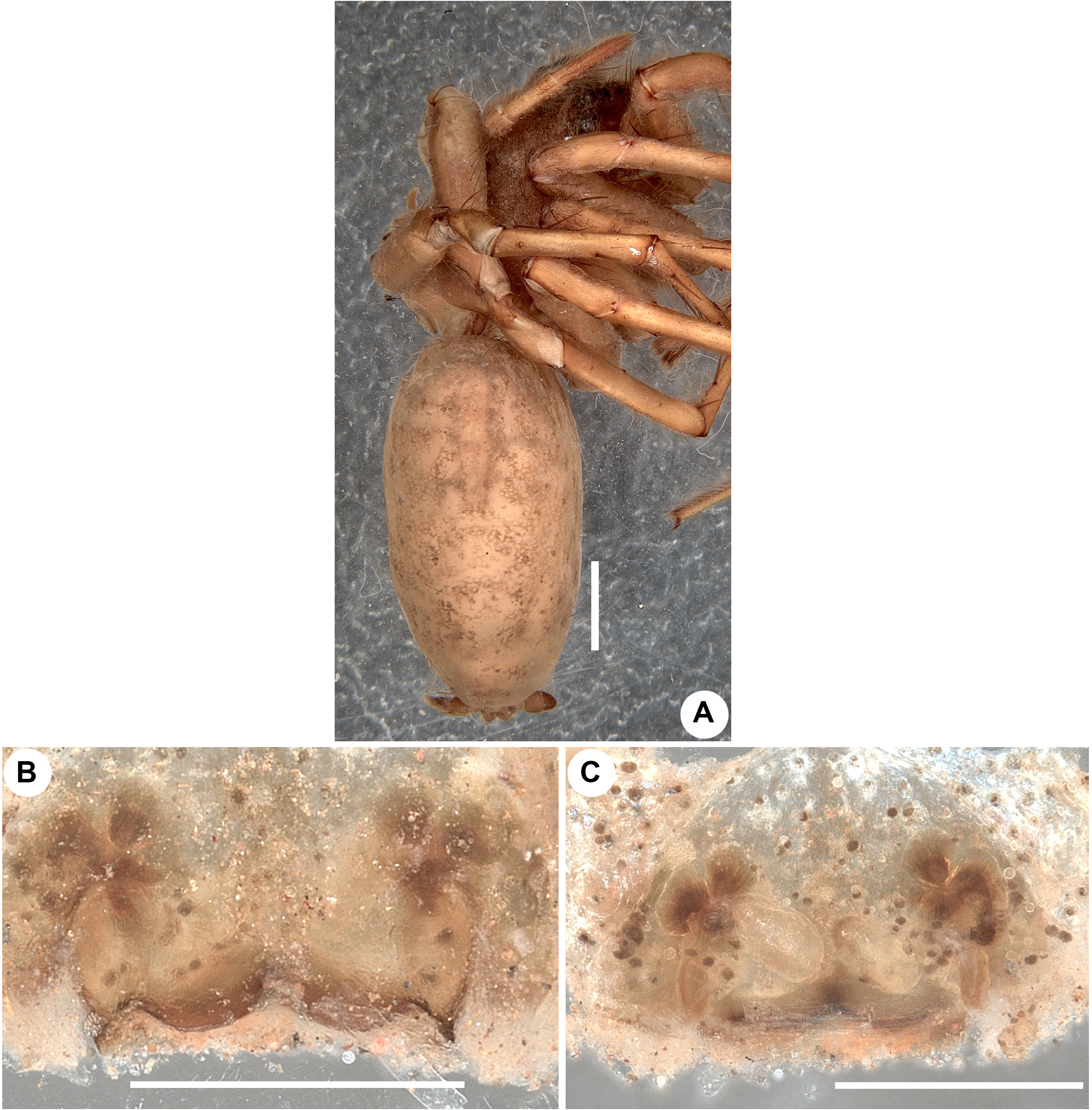
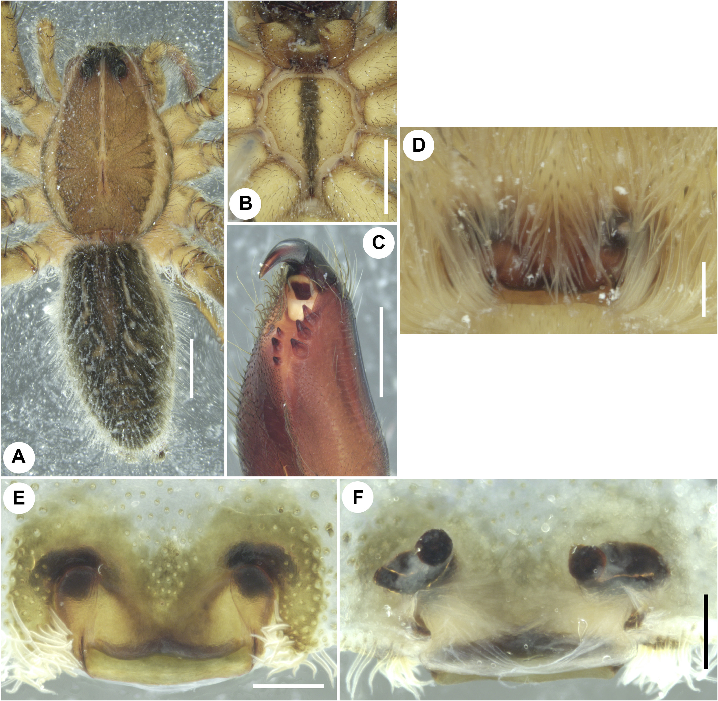
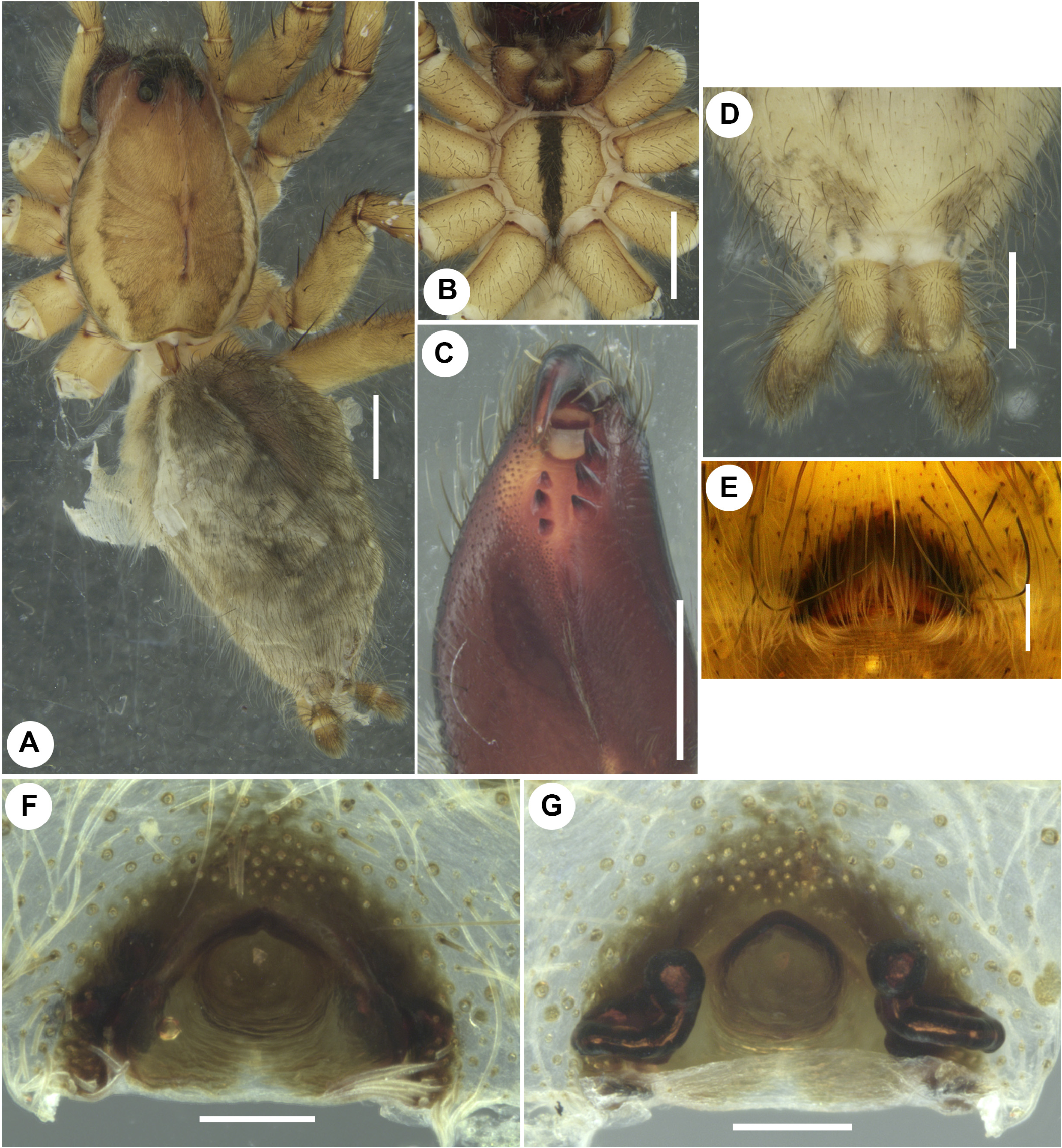
Figs 16–18 View FIGURE 16 View FIGURE 17 View FIGURE 18 , 38 View FIGURE 38
Hippasa loundesi Gravely, 1924: 594 View in CoL , fig. 1E (♀). Tikader & Malhotra 1980: 280, figs 77–79 (♀).
Type material. Holotype ♀ from INDIA: Tamil Nadu: Salem: Yercaud: Servarayan Hills (Shevaroy Hills) (11°46'N, 78°12'E; 1410 m alt.), date unknown, D. Loundes leg., repository NZC-ZSI (5225/H2), not found. GoogleMaps
Topotype material examined. INDIA: Tamil Nadu: Salem: Yercaud (11°46'N, 78°12'E; 1420 m alt.), 28 May 2019, M.S. Pradeep & A. V. Mathew leg. GoogleMaps , from web on roadside mud embankment, by hand: 2 ♀♀, 1 subadult ♀ ( ADSH5950251 View Materials ) .
Other material examined. INDIA: Tamil Nadu: Javadi Hills / Javadhu Hills / Jawadhi Hills / Jawadhu Hills : Nadur , 26-30 June 1929, H.S. Pruthi leg.: 1 ♀, 1 subadult ♀ ( NZC-ZSI 2229 /18) . Kerala: Pathanamthitta: Gavi (9°26'09.07''N, 77°09'56.78''E; 1192 m alt.), 21 December 2013, M.S. Pradeep leg., from web on grassland, by hand: 1 ♀ ( ADSH5950252 View Materials ) GoogleMaps .
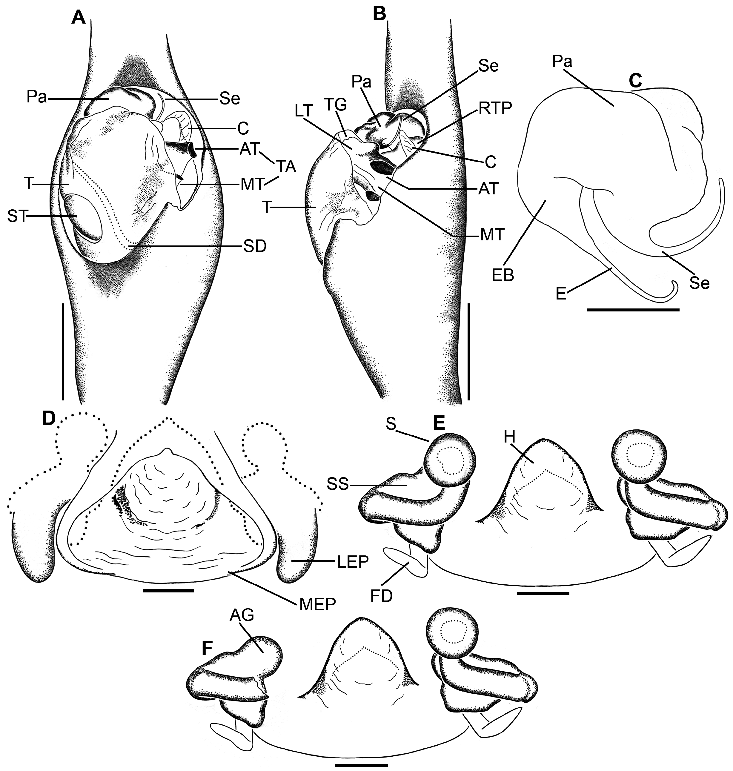
Diagnosis. Females of H. loundesi are similar to the females of H. lycosina as both share similar spermathecal stalks of vulva with wide proximal and tubular distal parts and large accessory glands, but can be separated from the latter by squarish median plate of epigyne (vs. triangular in H. lycosina ), median plate without atrium (vs. present in H. lycosina ) and median plate with lateral and posterior thickenings (vs. absent in H. lycosina , compare Figs 17E–F View FIGURE 17 , 18A–B View FIGURE 18 with Figs 20F–G View FIGURE 20 , 21D–F View FIGURE 21 ).
Supplementary description. Female in ethanol (ADSH5950251; Fig. 17A–C View FIGURE 17 ). Carapace medially pale brownish, with a median straight white stripe extending from PMEs to rear end of fovea, with paired lateral longitudinal white stripes extending along the entire length of carapace, medially clothed with fine, black, appressed setae, with a few scattered erect spine-like setae restricted to cephalic part. Eye region, clypeus, endites, labium, sternum pale brownish; chelicerae dark brownish; dorsum and sides of opisthosoma, and spinnerets blackish, venter of opisthosoma creamy-white; leg and palp segments pale brownish to brownish with black patches and annulations. Thoracic fovea reddish brown, long (0.99), straight, longitudinal ( Fig. 17A View FIGURE 17 ). Thoracic part laterally black. Cheliceral inner and outer surfaces with stridulatory files; promargin provided with a series of long setae with bend tips, both pro- and retromargins with three teeth ( Fig. 17C View FIGURE 17 ). Sternum with scattered, greyish black setae, with a broad, median longitudinal black band ( Fig. 17B View FIGURE 17 ). Opisthosoma elongate-ovoid, hirsute ( Fig. 17A View FIGURE 17 ); cardiac area marked with a brownish patch; dorsum anteriorly with a pair of white lateral stripes, with a few scattered white spots and patches, medioposteriorly with a few transverse, roughly W-shaped bands. Spinnerets hirsute. Legs long, slender, hirsute, spinose; all tarsi with complete and metatarsi I–II with distal scopulae, all well-developed. Palp segments with black patches and annulations; tarsus with single claw, distoventrally with bunch of long setae. Body length 17.33. Carapace 8.13 long, 5.73 wide. Opisthosoma 9.20 long, 4.88 wide. Eye diameters and interdistances: ALE 0.25, AME 0.29, PLE 0.51, PME 0.52; AME–ALE 0.23, AME–AME 0.22, AME–PME 0.42, PLE–PLE 1.24, PME–PLE 0.42, PME–PME 0.48. Clypeus height at AMEs 0.41, at ALEs 0.36. Length of chelicerae 3.38. Measurements of palp and legs: palp 9.95 [3.32, 1.64, 2.17, 2.82], I 19.93 [5.40, 2.57, 4.61, 4.89, 2.46], II 19.86 [5.40, 2.50, 4.28, 4.86, 2.82], III 19.39 [5.45, 2.24, 3.98, 5.22, 2.50], IV 27.34 [6.72, 2.53, 5.95, 8.59, 3.55]. Leg formula: 4123. Spination of palp: femur pld 1 do 4 rld 1, patella pld 1 do 2, tibia pl 2 rld 1, tarsus/cymbium pl 2 pld 1 rl 1 rld 1; legs: femur I pld 2 do 3 rld 3, II pld 3 do 3 rld 4, III pld 3 do 3 rld 3, IV pld 2 do 3 rld 1; patellae I–IV pld 1 do 2 rld 1; tibia I pl 1 pld 1 plv 3 rl 1 rlv 3, II pl 1 pld 1 plv 1 rl 2 rld 2 rlv 3, III pl 1 pld 1 plv 3 rl 1 rld 1 rlv 1, IV pl 1 pld 1 plv 3 do 1 rl 2 rld 1; metatarsus I pld 2 plv 3 rld 1 rlv 3 vt 1, II pld 2 plv 3 rl 1 rld 2 rlv 3, III–IV pld 3 plv 3 rld 3 rlv 3 vt 1; tarsi I–IV spineless. Genitalia ( Figs 17D–F View FIGURE 17 , 18A–B View FIGURE 18 ): epigyne clothed in bushy setae ( Fig. 17D View FIGURE 17 ), with squarish median and short lateral plates ( Figs 17E View FIGURE 17 , 18A View FIGURE 18 ; MEP, LEP); median plate with lateral and W-shaped posterior thickenings ( Figs 17E View FIGURE 17 , 18A View FIGURE 18 ). Spermathecal stalks with wide proximal and narrow tubular distal parts ( Figs 17F View FIGURE 17 , 18B View FIGURE 18 ). Accessory glands globular, without stalk, arising laterally on proximal part of spermathecal stalks ( Fig. 18B View FIGURE 18 ; AG). Spermathecae globular ( Figs 17F View FIGURE 17 , 18B; S View FIGURE 18 ). Fertilization ducts anteriorly directed, parallel ( Fig. 18B View FIGURE 18 ; FD).
Male. Unknown.
Variation. Female (n=3): body length 17.21–17.33.
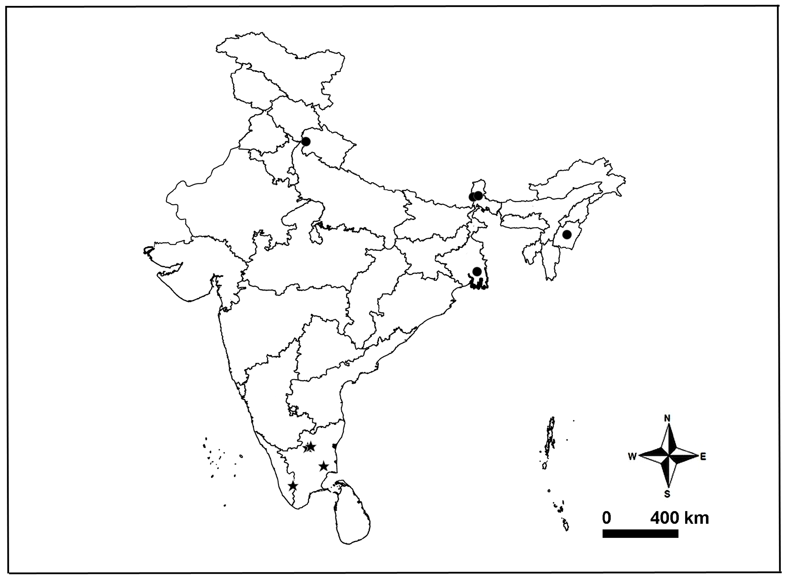
Distribution. India: Kerala (new record), Tamil Nadu ( Gravely 1924; Tikader & Malhotra 1980; present data) ( Fig. 38 View FIGURE 38 ).
Remarks. We were unable to trace the type of H. loundesi in the NZC-ZSI collection, which may either be lost or misplaced elsewhere in the collection. The NZC-ZSI collection currently has one glass bottle for this species, containing a female with intact genitalia and a subadult female specimens, both are in bad condition (2229/18). These specimens were collected by H. S. Pruthi and were determined as H. loundesi by T. B. Sinha, who studied the type of H. loundesi ( Sinha 1951) . Moreover, these specimens were collected from the Javadhu Hills in Tamil Nadu, which is ~150 Km away from the Servarayan Hills, the type-locality of H. loundesi ( Gravely 1924) . The NZC-ZSI collection has one more glass bottle labeled as ‘ Paralectotypes, H. loundesi ’. These specimens are misidentified and are of H. himalayensis .

| V |
Royal British Columbia Museum - Herbarium |
No known copyright restrictions apply. See Agosti, D., Egloff, W., 2009. Taxonomic information exchange and copyright: the Plazi approach. BMC Research Notes 2009, 2:53 for further explanation.