
Edessa (E.) urus Erichson, 1848
|
publication ID |
https://doi.org/ 10.11646/zootaxa.5240.1.1 |
|
publication LSID |
lsid:zoobank.org:pub:2FE467C1-EAC2-4E90-B673-CCE2CCA93C1D |
|
DOI |
https://doi.org/10.5281/zenodo.7871433 |
|
persistent identifier |
https://treatment.plazi.org/id/03B487C0-FFF8-252A-FF47-C785F88BF90B |
|
treatment provided by |
Plazi |
|
scientific name |
Edessa (E.) urus Erichson, 1848 |
| status |
|
Edessa (E.) urus Erichson, 1848
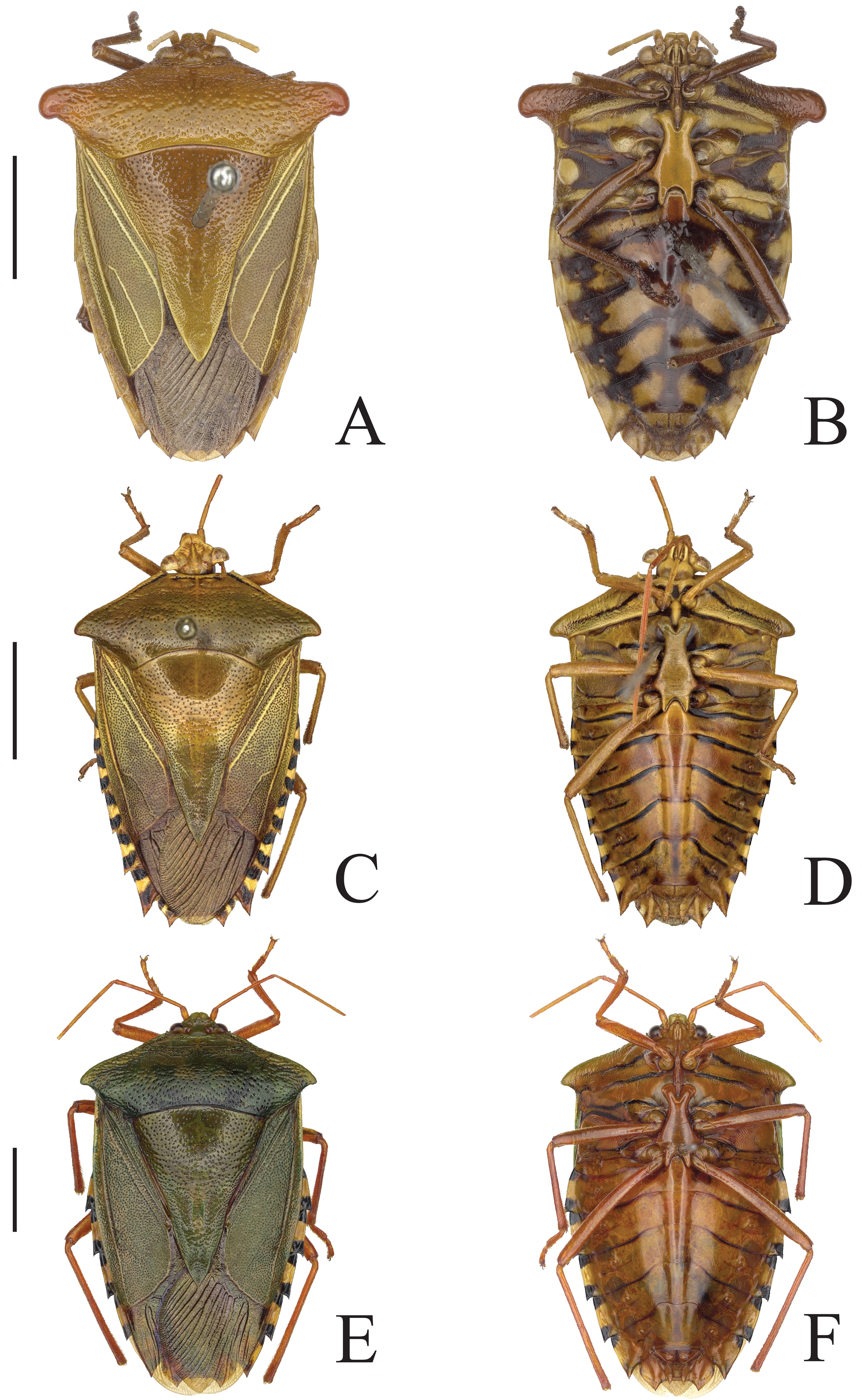
( Figs. 23 View FIGURE 23 , 29 E,F View FIGURE 29 , 32 A View FIGURE 32 )
Stoll Pun. f. 209.
Edessa urus Erichson, 1848: 610 ; Doesburg, 1991: 313; Silva et al., 2018: 425 View Cited Treatment .
Edessa dentata Dallas, 1851: 328 ; Stål, 1868: 36; Lethierry & Severin, 1893: 194; Kirkaldy, 1909: 165; Doesburg, 1991: 313. syn. nov.
Edessa excellens Walker, 1868: 446 ; Lethierry & Severin, 1893: 190; Kirkaldy, 1909: 157 syn. nov.
Edessa urus . Lectotype male. N. Stoll fig. 209, Pará, Sieber/ E. urus Hoffgg. [von Hoffmannsegg] (Stoll. fig. 209 Urus) ( MNKB). Designated by Doesburg (1991:313).
Edessa urus . Paralectotypes female. Same data, except von Hoffmannsegg’s label ( MNKB) .
Edessa dentata . Holotype male. Pará /50-2 ( BMNH). Examined.
Edessa excellens . Lectotype female. Villa Nova / 53 37 [Amazonas] ( BMNH). Examined.
Material examined. BRAZIL, Amazonas: 1♁, Res. Adolpho Ducke, 1-V-2006, A. L. Nunes ( UFPA); 1♀, Res. Adolpho Ducke, 8–10-VI-1989, M. S. Hoogmoed leg. ( RMNH); 1♀, Reserva Ducke, 4-06-76, Eduardo col. ( INPA); 1♁, Manacapuru, Manaus, III-1928, S. M. Klages ( Edessa lavata Breddin, 1903 , Comp. W. type Fernandes JAM 1999; Edessa dentata, Dallas 1851 , Comp. W. type Fernandes JAM 1999; Edessa urus Erichson 1848 , Comp. W. type Fernandes JAM 1999) ( CMNH); 1♀, Manaus, T. Federal-Rondônia, 6-IX-1966, Eduardo ( INPA); 1♁, Ceplac, Manaus, 28-VII-1977, I. S. Gorayeb col. ( MPEG); 1♁, E. Lo. Moult, Maneoro, Rio Madeira, Z. Amerika ( RMNH); Pará: 1♀, Altamira, Castelo dos Sonhos, área 28, 13-XI-2005, A. A. Pinheiro col. ( UFPA); Rondônia: 1♀, Porto Velho, 5-X-1978, J. Becker leg. ( Edessa urus Erichson 1848 , Comp. W. type Fernandes JAM 1999; Edessa excellens Walker, 1868 , Comp. W. type Fernandes JAM 1999 ( MNRJ).
Measurements (n= 9). Total length: 20.4–23.6; head length: 1.8–2.3; head width: 3.6–3.7; pronotum length: 3.7–4.1; pronotum width: 12.5–14.1; scutellum length: 9.5–10.9; scutellum width: 7.0–8.0; abdominal width: 12.0– 13.5; length antennomers: I: 1.0–1.0; II: 2.0–2.5; III: 1.5–2.0; IV: 4.5–5.0; V: 5.0.
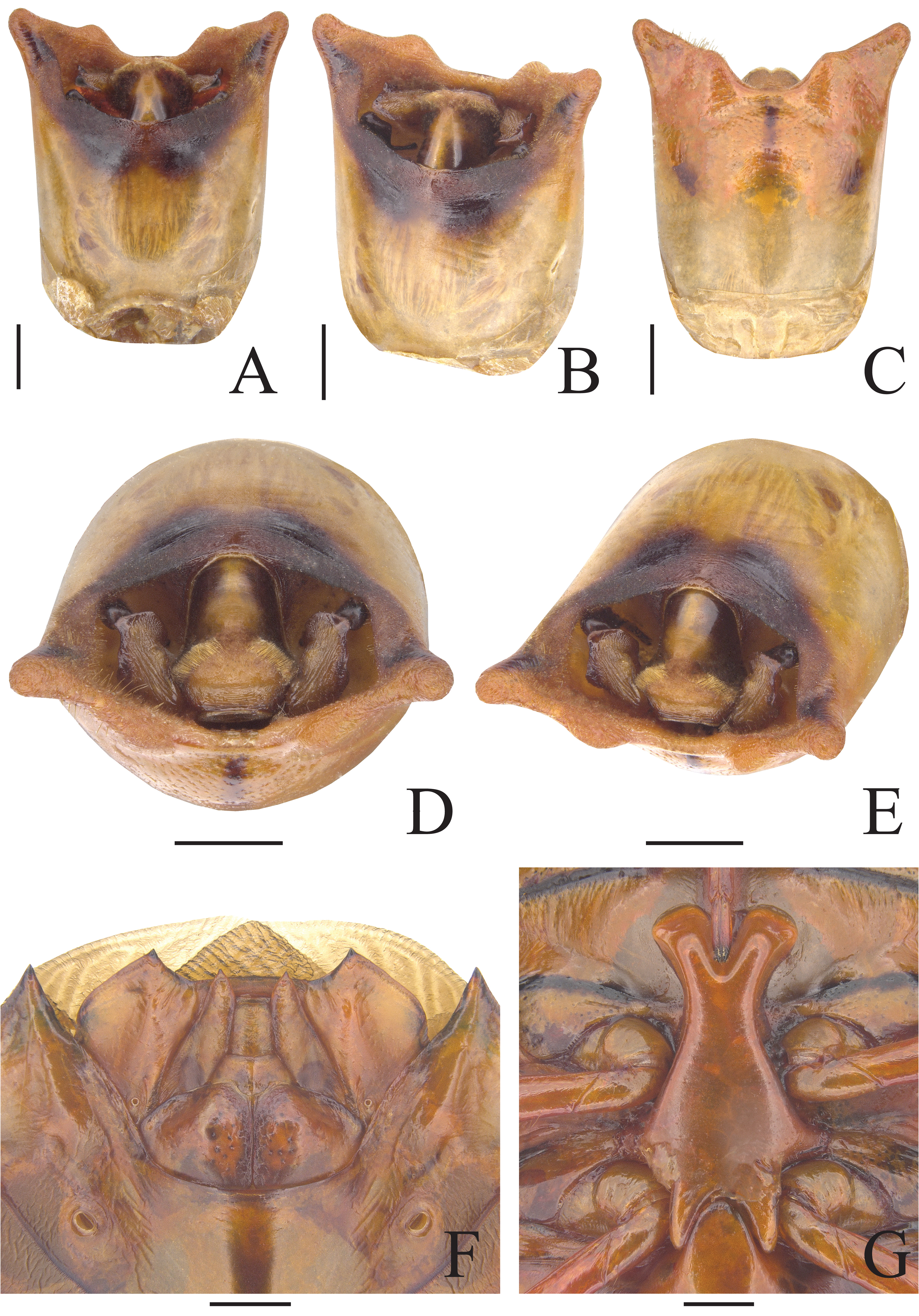
Diagnosis. Specimens large (20.4–23.6 mm). Dorsal surface green ( Fig. 29 E View FIGURE 29 ). Ventral surface dark yellow to brown with transversal black lines on thorax and abdomen ( Fig. 29 F View FIGURE 29 ). Antennae reddish brown ( Fig. 29 E View FIGURE 29 ). Pronotum with punctures black to brown ( Fig. 29 E View FIGURE 29 ); anterolateral margin and cicatrices with black punctures ( Fig. 29 E View FIGURE 29 ). Humeral angles short (2,0 times wider than long) ( Fig. 29 E View FIGURE 29 ), apices concolorous with surface ( Fig. 29 E View FIGURE 29 ). Scutellum with brown punctures ( Fig. 29 E View FIGURE 29 ); apex not reach end of corium ( Fig. 29 E View FIGURE 29 ). Corium with all veins concolorous with surface ( Fig. 29 E View FIGURE 29 ). Posterolateral angles of connexivum with apices black ( Fig. 29 E View FIGURE 29 ); connexival segments with concavities entirely covered by rectangular black spots and separated by a large yellow median spot ( Fig. 29 E View FIGURE 29 ); spots extending ventrally, triangular ( Fig. 29 F View FIGURE 29 ). Ventral surface. Thorax with black stripes ( Fig. 29 F View FIGURE 29 ); dark stripe of the propleuron almost reaching the dark spot of the humeral angle ( Fig. 29 F View FIGURE 29 ). Evaporatorium concolorous with thorax ( Fig. 29 F View FIGURE 29 ). Metasternal process ( Fig. 23 G View FIGURE 23 ) with anterior apex rounded and laterally little expanded, margin rounded; anterior face broadly excavated; anterior bifurcation receiving fourth rostral segment. Legs brown ( Fig. 29 F View FIGURE 29 ). Abdomen with spine of third segment acuminated ( Fig. 23 G View FIGURE 23 ). Intersegmental areas brown reaching ventral spots of connexivum ( Fig. 29 F View FIGURE 29 ). Pseudosutures concolorous with surface ( Fig. 29 F View FIGURE 29 ). Median longitudinal brown band restricted to last segment ( Fig. 29 F View FIGURE 29 ). Trichobotria one in line with spiracle and the other laterad. Posterolateral angles of segment VII on the same level the level of apices of laterotergites IX in females ( Fig. 23 F View FIGURE 23 ). Male genitalia, dorsal side of the pygophore with a suffused brown area occupying 1/3 of the surface ( Fig. 23 A,B,D,E View FIGURE 23 ). Posterolateral angle of the pygophore developed ( Fig. 23 A View FIGURE 23 ). Superior process of genital cup laminar, rectangular, thick; flattened, coarse and concave in posterior view; continuing ventrally in a crenulated high carina, ending in a small dentiform projection ( Fig. 23 B,E View FIGURE 23 ). Anterior half of proctiger brown ( Fig. 23 A,B,D,E View FIGURE 23 ). Ventral rim with long setae, but without lateral tufts ( Fig. 23 C View FIGURE 23 ). Female genitalia, valvifers VIII wrinkled ( Fig. 23 F View FIGURE 23 ). Laterotergites VIII with one dark spots on outer lateral margins ( Fig. 23 F View FIGURE 23 ).
Male genitalia ( Fig. 23 A–E View FIGURE 23 ): Parameres with brown margin, anterior lobe rounded; dorsal lobe rounded and subrectangular, little curved at the apex; posterior lobe rounded and subrectangular ( Fig. 23 B,D,E View FIGURE 23 ). Proctiger with subelliptical posterior face ( Fig. 23 D,E View FIGURE 23 ). Ventral rim with expansions little developed, rounded, concolorous with surface, the expansions not reaching beyond apices of posterolateral angles ( Fig. 23 C View FIGURE 23 ).
[...... Continues on page 60]
Female genitalia: Valvifers VIII with dark punctures; inner margins contiguous, with brown band and not divergent; distal margin forming distal U-shaped excavation, with brown band and arched. Laterotergites IX with apices acuminate passing the sclerite uniting laterotergites VIII ( Fig. 23 F View FIGURE 23 ).
Comments. The Edessa (E.) urus looks like E. (E.) alces , E. (E.) congrua , and E. (E.) sexdens . See comments of E. (E.) alces .
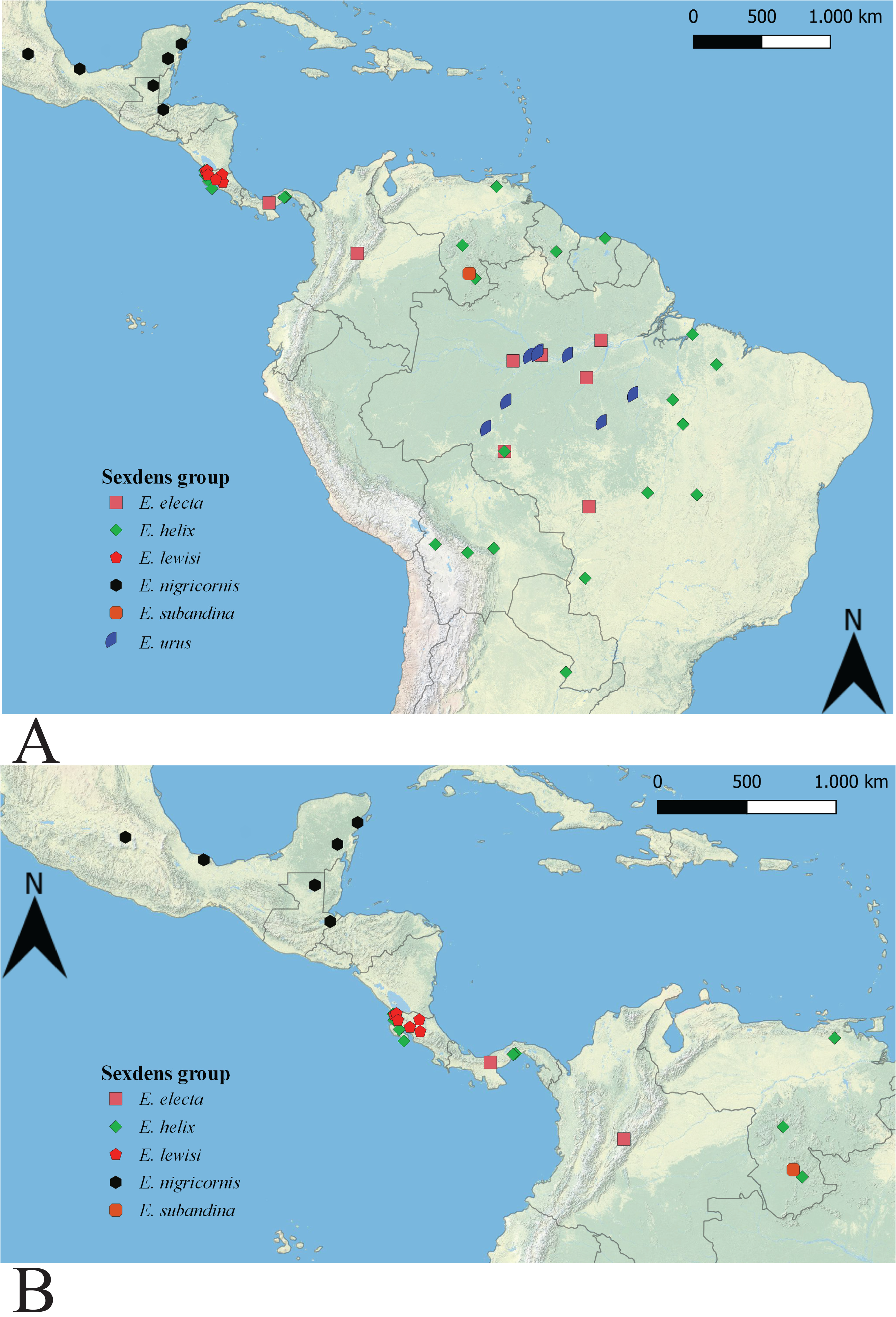
Distribution ( Fig. 32 A View FIGURE 32 ): BRAZIL: Pará, Amazonas, Rondônia.
No known copyright restrictions apply. See Agosti, D., Egloff, W., 2009. Taxonomic information exchange and copyright: the Plazi approach. BMC Research Notes 2009, 2:53 for further explanation.
|
Kingdom |
|
|
Phylum |
|
|
Class |
|
|
Order |
|
|
Family |
|
|
Genus |
Edessa (E.) urus Erichson, 1848
| Mendonça, Maria Thayane Da Silva, Silva, Valéria Juliete Da & Fernandes, José Antônio Marin 2023 |
Edessa urus
| Doesburg, P. H. van 1991: 313 |
Edessa excellens
| Kirkaldy, G. W. 1909: 157 |
| Lethierry, L. & Severin, G. 1893: 190 |
| Walker, F. 1868: 446 |
Edessa dentata Dallas, 1851: 328
| Doesburg, P. H. van 1991: 313 |
| Kirkaldy, G. W. 1909: 165 |
| Lethierry, L. & Severin, G. 1893: 194 |
| Stal, C. 1868: 36 |
| Dallas, W. S. 1851: 328 |
Edessa urus
| Silva, V. J. & Santos, C. R. M. & Fernandes, J. A. M. 2018: 425 |
| Doesburg, P. H. van 1991: 313 |
| Erichson, W. F. 1848: 610 |