
Enicocephalus flavicollis Westwood, 1837
|
publication ID |
https://doi.org/10.5281/zenodo.214172 |
|
DOI |
https://doi.org/10.5281/zenodo.6168487 |
|
persistent identifier |
https://treatment.plazi.org/id/03B49A22-BD0D-FFFF-FF4A-FFCC7D37FF05 |
|
treatment provided by |
Plazi |
|
scientific name |
Enicocephalus flavicollis Westwood, 1837 |
| status |
|
Enicocephalus flavicollis Westwood, 1837 View in CoL - male, redescription
( Figs. 1–6 View FIGURES 1 – 5 View FIGURES 6 – 9 , 10–17 View FIGURE 10 View FIGURES 11 – 17 )
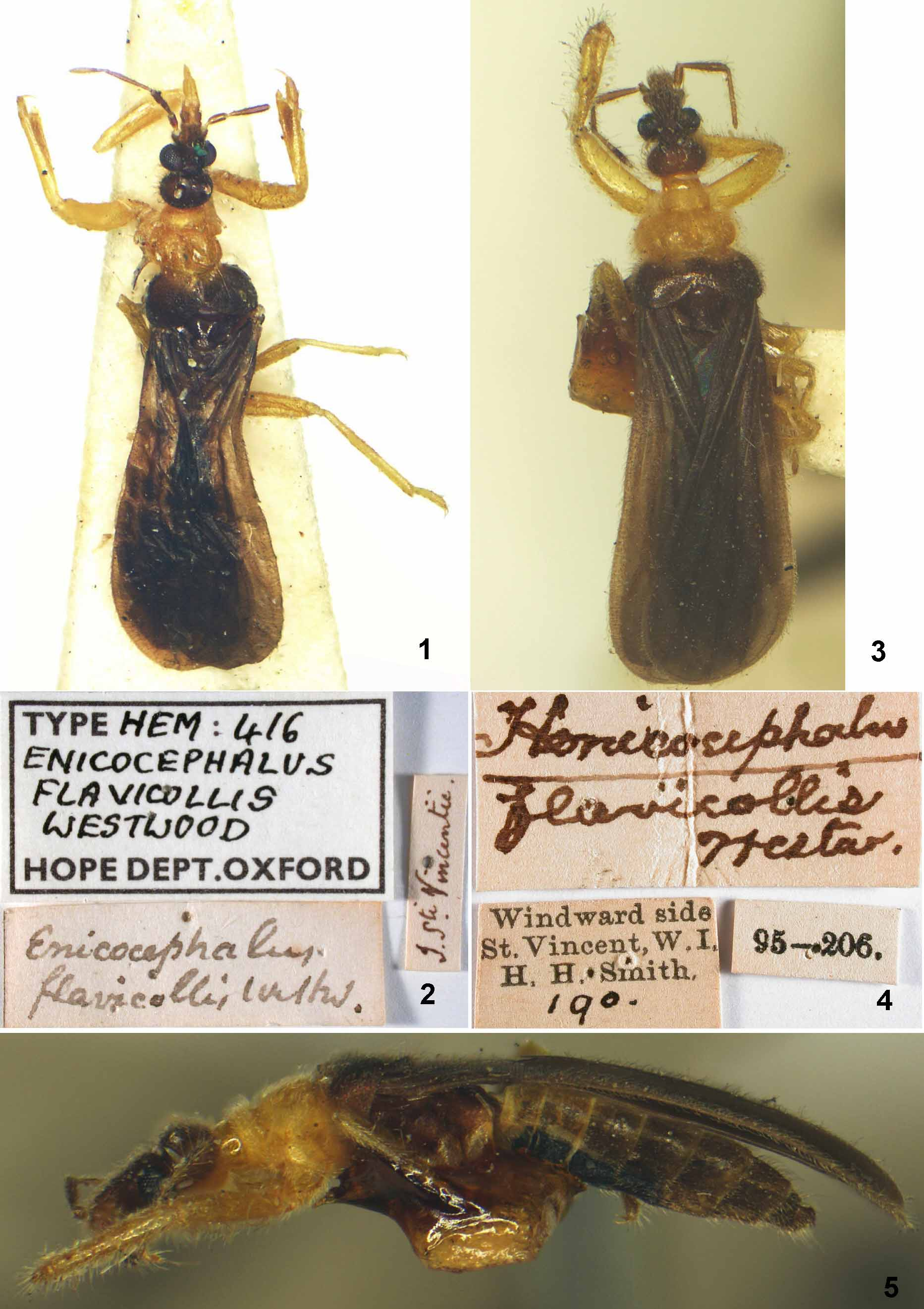
Material examined. Lectotype: male, first label: // I. S ti. Vincentii // [handwritten = Insula Sancti Vincentii (Latin)]; second label: // Enicocephalus / flavicollis Westw. // [handwritten]; third label: // Type Hem: 4/6 / Enicocephalus / flavicollis / Westwood / Hope Dept. Oxford // [partly printed and partly handwritten]. Specimen bears red label: // Enicocephalus flavicollis / Westwood, 1837 / LECTOTYPE ♂ / P. Štys & P. B aňař det. 2011 // [printed]. Specimen dry mounted, glued to triangle card, in very bad condition: midlobe of pronotum (and some sternal parts) strongly damaged, antennal segments III, IV, left mid- and hindtibiae and tarsi are missing, left hindfemur mounted separately ( Fig. 1 View FIGURES 1 – 5 ). Deposited in Hope’s collection, University Museum of Natural History, Oxford. Paralectotype: male, first label: // Windward side / St. Vincent, W.I. / H.H. Smith 190 // [printed; number ‘190’ handwritten]; second label: // 95-206. //; third label: // H enicocephalus [originally Enicocephalus - overwritten] / flavicollis Westw. // [handwritten]; specimen bears red label: // Enicocephalus flavicollis / Westwood, 1837 / PARALECTOTYPE ♂ / P. Št ys & P. B aňař det. 2011 // [printed]. Dry mounted specimen glued to a triangle, in good condition, right antennal segment 4 and left hind leg missing. Deposited in Department of Entomology, Natural History Museum, London.
Measurements (in mm): lectotype first ( paralectotype in parentheses, italics). Total body length —ca 3.9 (specimen damaged) ( 3.87). Head (without neck). Total length—0.62 ( 0.63); posterior lobe, length—0.24 ( 0.23), width—0.38 ( 0.37); distance of eye to apex of antennifer—0.13 ( 0.13); diatone (maximum width across eyes)— 0.39 ( 0.38); dorsal synthlipsis (minimum interocular distance)—0.18 ( 0.19); eye, length—0.17 ( 0.16); gena, length—0.03 ( 0.03), width (minimum)—0.19 ( 0.18). Labium. Total length—0.47 ( 0.46); segment I, length—0.06 ( 0.05); segment II, length—0.07 ( 0.07); segment III, length—0.20 ( 0.20); segment IV, length—0.13 ( 0.13). Antenna. Segment I, length—0.14 ( 0.13); segment II, length—0.29 ( 0.28), basal width—0.02 ( 0.02), distal width—0.05 ( 0.05); segment III, length—0.37 ( 0.37); segment IV, length—0.36 ( 0.36). Pronotum. Total length (median)—ca 0.58–0.62 (specimen damaged) ( 0.60); collum, length (median)—0.13 ( 0.13), width (maximum)— 0.29 ( 0.29); midlobe, length (median)—ca 0.25–0.29 (specimen damaged) ( 0.27), width (maximum)—0.54 ( 0.55); hindlobe, length (maximum)—0.40 ( 0.38), length (mediane)—0.19 ( 0.18), width (maximum)—0.80 ( 0.78). Foreleg. Femur, length—0.82 ( 0.81), width (maximum)—0.20 ( 0.19); tibia, length—0.74 ( 0.72), width (maximum)— 0.17 ( 0.17).
Coloration ( Figs. 1, 3, 5 View FIGURES 1 – 5 ). Bicolorous, piceous or yellow, the colours strongly contrasting except on abdomen. No red or scarlet pigments.
Head and thorax. Dorsum. Dark body parts piceous brown (dorsum of head, most of the posterior lobe of pronotum, mesoscutellum); neck of the head somewhat lighter; non-melanized parts unicolorous, contrastingly yellow (collum, midlobe of pronotum; linear, transverse, short, anterior strip of the hindlobe of pronotum). The yellow transverse strip of posterior lobe of pronotum with 1+1 submedial extensions shaped as segments of circle.
Lateral sides and venter of head and thorax. Anterior lobe of head (from apex to postocular constriction) piceous, posterior lobe yellow; prothorax yellow (the yellow area confluent with the anterior yellow strip on posterior lobe of pronotum), pterothorax (incl. all supracoxal lobes) piceous, pterosterna yellowish.
Antennae piceous brown; proximal part of segment IV non-contrastingly lighter. Labium: segments I and II brownish, III and IV yellow; legs (inclusive coxae) uniformly yellow.
Forewings piceous brown with blackish hue ( Figs. 1, 3 View FIGURES 1 – 5 ), veins concolorous except for the noncontrastingly yellowish marginal vein (costal margin cum ambient vein up to the level of cu-an). The light coloration of the marginal vein invisible in transmitted light owing to semitransparency of the wing; ventral side matching the dorsal one. Hindwings blackish-brown.
Abdomen. Dorsum not examined; venter: segments 1–3 yellowish brown, 3–7 piceous, 8 and 9 yellowish brown; posteromedial part of 2 and medial parts of 3–5 black.
Coloration of paralectotype London male matching the redescription of Oxford lectotype male (Wyhodzinsky& Schmidt 1991) except for the lectotype abdomen said to be whitish.
Microsculpture. Cuticle strongly lustrous and smooth. No setigerous tubercles. Posterior lobe of pronotum irregularly shallowly rugulose.
Ventral surface of fore trochanter with about 5 transverse rows of serrate microstructures. Ventral face of forefemur with a continuous percurrent longitudinal strips of minute black granules; these terminating nearly apically, leaving the area contacting basis of foretibia bare but provided with one large posterior dens; no microtrichiae or spicules present.
Venter of abdomen with minute transverse wrinkles.
Vestiture. Macrotrichia (further on ‘setae’) golden, rather soft, usually short and straight, rarely curved.
Head. Dorsal view. Anterior lobe of head covered by short and appressed, very dense short setae, obscuring its surface; longer curved setae occurring apically and in front of eyes only. Posterior lobe with sparse straight to curved, rather short setae. Lateral view. Dorsum with long, very dense, regularly curved, diagonal setae directed anteriad (excl. on preocular lobe), those on posterior lobe obscuring its outline. Venter with long, anteriorly directed, long straight or curved setae forming a conspicuous barb on the posterior lobe. Ventr al view. Very long and irregular macrotrichial cover all over.
Eyes with a few straight interommatidial setae in anterior half, with many curved setae in posterior half.
Antennae with short, dense, diagonal pubescence; segment 2 with few semi-erect hairs on its inner side, segment 3 on both sides and apically, segment 4 all over.
Labium. Segment 3 with moderately long and moderately dense diagonal straight macrotrichia interspersed with long, thin, erect to suberect trichobothrium-like setae along both sides (4+4) and clustering in the apical part.
Thorax. Dorsal view. Lateral outline: collum with moderately dense, straight short setae, midlobe with curved setae gradually longer caudad, hindlobe with very dense, curved, short setae.
Mesoscutellum with long curved dense setae.
Lateral view. Dorsum densely covered by extremely short, and very dense cover of straight to curved, diagonal, minute setae, only those on collum with a few erect setae. Prothoracic presupracoxale with a cluster of strikingly long hairs.
Forewings with short, curved hairs (mixture of golden and black ones) distributed in a manner modal for Enicocephalus (cf. Wygodzinsky & Schmidt 1991). Intervenial membrane bare.
Hindwings bare.
Legs. Anterior faces of all coxae densely pilose.
Foreleg. Dorsal face of femur with long diagonal setae, with interspersed few trichobothrium-like diagonal setae (recognizable only by their thinness); ventral face with long, thin, straight setae. Tibia covered all over by short, medium long, mostly appressed to diagonal straight setae (similar to those on dorsal face of femur), occurring on the anterior longitudinal tibial depression as well; dorsal and ventral faces additionally with numerous trichobothrium-like setae, all thin and all erect, rarely straight, mostly curved at the tip, nearly as long as local tibial diameter. The latter kind of setae occurring all over tarsus.
Mid - and hindtibiae with a long diagonal vestiture all over, setae longer than tibial diameters; the same true for mid - and hindtarsi. The setae of dorsal and ventral tibial faces thin, trichobothrium-like (?).
Abdomen. Margin without specialized setae, uniformly covered with uniformly curved setae becoming gradually longer posteriad, longest all along edge of segments 7 and particularly 8; the latter posteriorly with 3+3 very long setae, the longest exceeding apex of pygophore.
Venter. All segments (exc. pygophore) densely covered with very short and uniform diagonal pubescence with setae slightly dilated (not gradually pointed) at tips, a few longer setae present at the medial keel only.
Structure. Very small species, but rather robustly built.
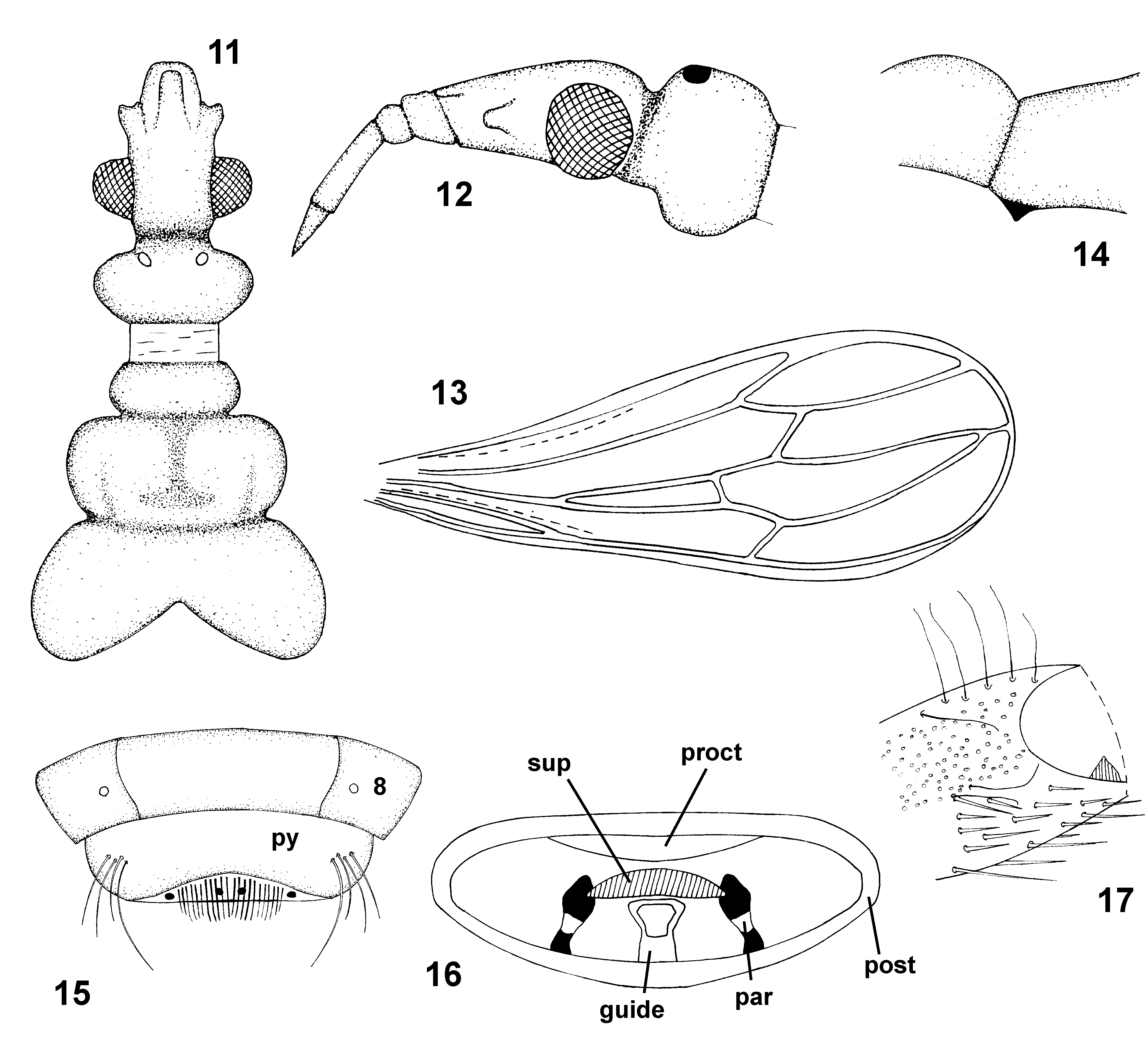
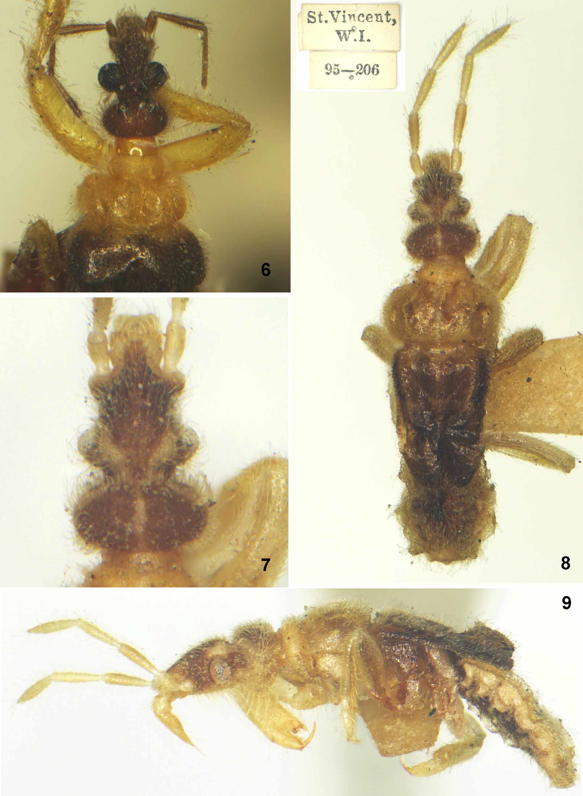
Head. Dorsal view ( Figs. 1–2 View FIGURES 1 – 5 , 6 View FIGURES 6 – 9 , 11 View FIGURES 11 – 17 ). Anterior lobe short, insertion of antennae far from apex of anteclypeus, midway beween eyes and anteclypeus, gena very short, ratio gena lenght to its width 0.17; ratio of gena length to length of eye 0.19. Postocular constriction deep and long (about as long as the gena) and sharply delimited. Postocular lobe strikingly transverse, laterally asymmetrically rounded, widest behind the middle, ratio of its length to maximum width 0.62, no structural indication of the median except for a slight interocellar concavity. Ocelli situated on low tubercles, interocellar distance shorter than distance eye–ocellus.
Head. Lateral view. Dorsal outline of the anteocular part of anterior lobe straight, slanting ventrad, strongly convex over the eye. Posterior lobe strongly convex, bulbous, its outline much exceeding that of the anterior lobe (more than shown by Wygodzinsky & Schmidt (1991, Fig. 80B)). Ventral outline of preocular part of anterior lobe straight, that of posterior lobe moderately convex.
Eyes large, strongly convex and outstanding, inner margins convex in dorsal view, ommatidia separately convex. Dorsal ocular index 2.00. Lateral view: dorsal margin far distant from dorsal margin of head, ventral margin slightly exceeding ventral margin of head. Ventral view: eyes strongly approaching each other.
Antennae short, first segment unusually thick, 2nd subclavate, strongly widening towards apex, segments III and IV stick-shaped, much thinner than segment II (but not flagelliform). Antennal formula (longest segment first) III-II-IV-I.
Labium. Segments I and II very short, their dorsal faces flat as well as that of segment III; the latter rather short, conical. Labial formula (longest segment first) III-IV-II-I.
Thorax. Pronotum in dorsal view. ( Figs. 1–2 View FIGURES 1 – 5 , 6 View FIGURES 6 – 9 , 11 View FIGURES 11 – 17 ). Collum long and laterally rounded, lacking lateroventral tubercles and any structurally indicated median, but with an unusual feature (natural or artifact?): posterior 3/4 with one medial and 1+1 sublateral subangular shallow impressions, contacting each other and reaching the short and sharp constriction between collum and midlobe.
Midlobe. Dorsal view. (Owing to the light colour and strong lustre of the area the description of surface sculpture is open to reinterpretation). Lateral margins strongly rounded, level of maximum width close to the anterior margin, posterior margin nearly straight, not interrupted. Ratio of midlobe maximum width to median length 3.05. Medial impression: antero-medial sector slightly depressed, with two impressed lines meeting in the centre and delimiting an anteromedial acutangular triangle with an apex pointing centrad; apex of the triangle sloping into a deep pit with a more shallow lateral linear extensions and rampart-like posterior margin coinciding with the posterior margin of the lobe itself (the impression resembling Oncylocotis -like inverse T-shaped impression.). The impression containing a linear, impressed median.
Sublateral impressions present as simple, shallow, depressions, not interrupting the posterior margin of the midlobe and situated anterolaterad to the median depression.
Posterior lobe. Dorsal view. Lateral margins regularly rounded inclusive indistinctly indicated posterolateral angles; medial part of posterior margin deeply angularly excised, lateral parts of this excision convex. No median structure. Ratio of hindlobe maximum width to its maximum length 2.05.
Pronotum in lateral view. Collum with no ventrolateral tubercles. Lateral sides of pronotum not clearly delimited from supracoxal lobes, no horizontal lateral ridge or sulcus present. ‘Proepimeral lobe’ short, rounded, ventrally not reaching the level of apex of posterior prosupracoxale.
Mesoscutellum with a convex surface, short, strongly rounded lateral margins, and unusually broad and long, apically rounded mucro separated from the disc by a transverse impression.
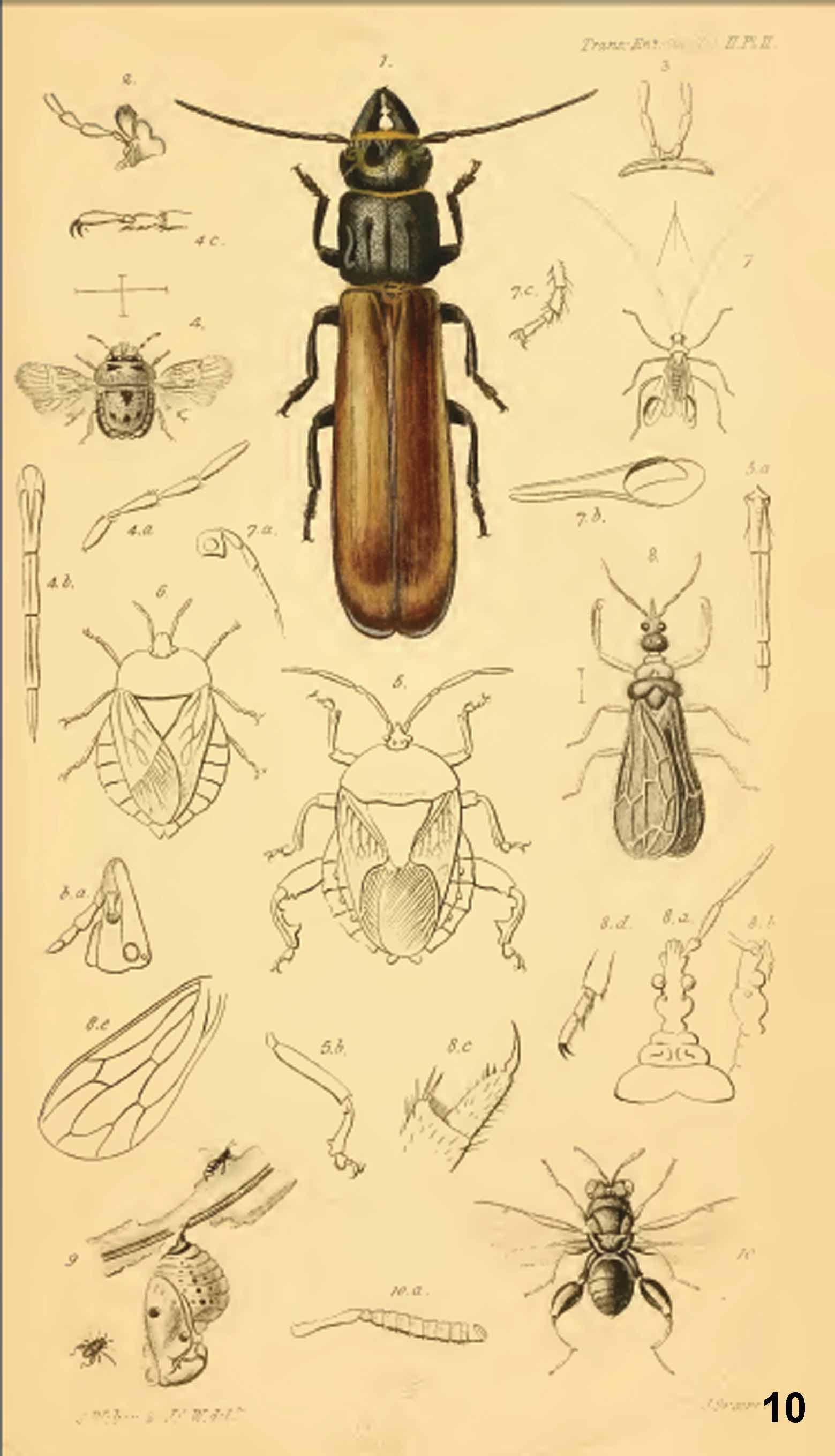
Wings macropterous, not caducous. Forewings much exceeding the abdomen, venation ( Fig. 13 View FIGURES 11 – 17 ) as illustrated by Wygodzinsky & Schmidt (1991, Fig. 80C), only the distal sector of AA + AP continuous, complete (checked on both the specimens).
Hindwings longer than abdomen
Legs. Foreleg. For microsculpture of trochanter and femur see above. Dorsal face of distal part of trochanter convex, slightly elevated over the neighbouring part of femur; ventral basis of femur slightly angularly produced and exceeding the surface of trochanter ventrad; ratio of femur length to maximum width 4.26. Tibia abruptly widening on dorsoventral plane in about a third of its length; ratio of tibia length to maximum width 4.24. Longitudinal (anterior) tibial depression starting at the points of tibial dilatation and running up to the cleaning comb. (Foretarsus pressed to the distal edge of tibia on both forelegs - consequently, apicitibial and tarsal armatures could not have been studied). Inner (anterior) claw large, posterior one represented by a short rounded hump only.
Mid - and hindtibiae with two apical bristle combs each; claws nearly isomorphic (the inner (anterior one) slightly longer.
Abdomen. Venter sclerotized, sulci between ventral laterotergites and sterna marked by deep impression and vaguely delimited laterosternal plates. Segments 3–7 with a sharp median keel, segment 8 without the keel, simply trapezoidal, posterolateral angles not protruding, embracing strikingly large, broadly transverse pygophore.
Pygophore ( Figs. 15–16 View FIGURES 11 – 17 ) (as seen in strictly posterior view and examined under 100 magnification; dorsum covered by abdominal segment 8 and wings) strongly transverse, posterior face oval, delimited by a rampart-like swollen margin of the posterior foramen of the pygophore. Guide slender and small, of a modal shape, with distinct shaft, head and frame, distal margin of guide head truncate. Supradistal plate ( Fig. 16 View FIGURES 11 – 17 ) seen as a sclerite dorsad and anterad to guide, parameres situated laterad to the former.
Diagnostic characters. Small species ( 3.9 mm), contrastingly bicolorous (lacking red pigment, yellow and brown - shared with E. boraceianus , E. guarani , E. schuhi (length 6.5–6.7 mm), and E. tupi only), legs uniformly yellow, collum and midlobe of pronotum uniformly yellow, the yellow part of pronotum involving also a short stripe on the disc of the hindlobe (unique!). Discal cell of forewing strikingly long and pointed, reaching nearly the wing margin (unique!). Male forelegs strikingly slender and genitalia small. Outer fore claw extremely reduced (shared with E. semirufus and E. usingeri only). Endemic to St. Vincent Island, sympatric occurrence of any other Enicocephalus species unknown.
No known copyright restrictions apply. See Agosti, D., Egloff, W., 2009. Taxonomic information exchange and copyright: the Plazi approach. BMC Research Notes 2009, 2:53 for further explanation.
|
Kingdom |
|
|
Phylum |
|
|
Class |
|
|
Order |
|
|
InfraOrder |
Enicocephalomorpha |
|
Family |
|
|
Genus |