
Troglohyphantes lanai Isaia and Pantini
|
publication ID |
https://doi.org/ 10.5281/zenodo.199535 |
|
DOI |
https://doi.org/10.5281/zenodo.6207534 |
|
persistent identifier |
https://treatment.plazi.org/id/03B57330-D71E-FFEB-FF40-FD5465FEFBC9 |
|
treatment provided by |
Plazi |
|
scientific name |
Troglohyphantes lanai Isaia and Pantini |
| status |
sp. nov. |
Troglohyphantes lanai Isaia and Pantini View in CoL new species
Figures 1–9 View FIGURES 1 – 3 View FIGURES 4 – 9
Types. Holotype male: Italy, Piemonte, Province of Vercelli: Borgosesia, Buco della Bondaccia Cave (2505Pi/VC), 690 m, 15/8/2008, E. Lana legit. Paratypes: same location as holotype 1 Ψ, 9/5/1992, E. Lana legit (CI); 1 Ψ, 2/6/1996, T. Pascutto legit; 1 Ψ, 6/4/1997, T. Pascutto legit (CI); 1 Ψ, 1 ɗ, 25/1/1998, T. Pascutto legit (CI); 1 Ψ, 26/1/2008, M. Isaia and E. Lana legerunt; 2 Ψ, 17/2/2008, E. Lana legit (CI); 1 ɗ, 13/ 4/2008, E. Lana legit (CI); 1 ɗ, 7/6/2008, M. Isaia and E. Lana legerunt (CI); 1 Ψ, 15/8/2008, E. Lana legit; 1 ɗ, 18/8/2008, E. Lana legit (CI).
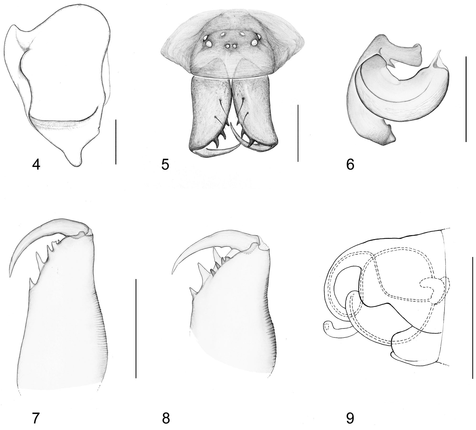
Diagnosis. Males of Troglohyphantes lanai are primarily distinguished from other species of Troglohyphantes by the shape of the lamella characteristica ( Figure 1 View FIGURES 1 – 3 ). The shape of the epigynum, ( Figures 2, 3 View FIGURES 1 – 3 and 9 View FIGURES 4 – 9 ) as well as the cymbium ( Figure 4 View FIGURES 4 – 9 ) are also diagnostic. The shape of the cymbium and embolus ( Figure 6 View FIGURES 4 – 9 ) of T. lanai is closest to that of T. bornensis and T. microcymbium , from which it is readily distinguishable by the shape of the lamella characteristica, the greater body size and the reduced eyes. Based on Pesarini’s complexes (2001), the new species is close to those included in the microcymbium complex.
Description. Male holotype: prosoma 1.38 long, 1.23 wide, light-yellowish. Anterior part of prosoma faintly darker. Thoracic region rounded, dorsally with a narrow ridge, marked with a dark longitudinal streak. Cephalic region slightly elevated, interspersed with black bristles starting from the eye region and continuing in three longitudinal rows that converge to the anterior end of fovea. Clypeus slightly indented under the eyes, then convex. Eyes reduced, without pigment ( Figure 5 View FIGURES 4 – 9 ). AME smallest. PLE very slightly bigger than PME, ALE slightly larger than PLE. ALE and PLE nearly contiguous (distance = 0.014). PLE-PME distance = 0.110, ALE-AME distance = 0.138, PME-PME distance = 0.128, AME-AME distance = 0.013. Eye diameters AME 0.027, PME 0.038, ALE 0.046, PLE 0.050. Sternum heart-shaped, yellowish with flimsy darkened anterior edges. Chelicerae light brownish, with lateral stridulatory ridges and armed with three anterior teeth. Posterior margin with a small lamina and a small proximal tooth ( Figure 7 View FIGURES 4 – 9 ). Legs medium-long (femur twice as long as the prosoma), uniformly light yellowish. Leg I: femur 2.87, patella 0.43, tibia 3.13, metatarsus 2.70, tarsus1.56 TLL 10.69; leg II: Fe 2.60, patella 0.50, tibia 3.00, metatarsus 2.66, tarsus 1.33, TLL 10.09; leg III: femur 2.33, patella 0.43, tibia 2.33, metatarsus 2.16, tarsus 1.00, TLL 11.01; leg IV: femur 3.00, patella 0.43, tibia 2.97, metatarsus 2.66, tarsus 1.33, TLL 10.03. Abdomen 1.93 long, 1.10 wide; whitish-grey with dark setae. Palp ( Figure 1 View FIGURES 1 – 3 ): femur 0.83, patella 0.36. Cymbium faintly convex, subtriangular when seen from above, ending proximally in a single tooth-like apophysis, rounded at the proximal border ( Figure 4 View FIGURES 4 – 9 ). Posterior part of paracymbium sub-triangular, apical part dimly longer than the latter and distally bent nearly at a right angle, gradually narrowed anteriorly. Lamella characteristica branched in two. The upper branch ending in a small and narrow lobe, twisted and rounded at top. Lower branch of the lamella directed anteriorly, nearly orthogonal to the upper branch and enlarged at the end in the shape of a right-angled trapezium. Suprategular apophysis directed upwards, with a sharp end. Tip of the embolus spiky, tubular ( Figure 6 View FIGURES 4 – 9 ). Spination: femur I with one dorsal, two prolateral spines; femur II with one dorsal spine and one small prolateral spine. Femur III and IV with one dorsal spine. Patella I–IV with one dorsal spine. Tibia I and II with two dorsal, four prolateral (apical included), three retrolateral (apical included) spines; tibia III and IV with one dorsal, three prolateral (apical included) and two retrolateral spines (apical included). Metatarsus I– IV with one dorsal and one prolateral spine. Metatarsus ventrally with several small spines. Position of TmI: 0.2. Trichobothrium on Mt IV absent.
Female (paratype from same locality as holotype, 9/05/1992, E. Lana legit): prosoma 1.53 long, 1.33 wide, convex, smooth and slightly darker than male. Cephalic region slightly elevated. Carapace, ocular area, clypeus, and sternum similar to male in all features except cephalic bristles, being smaller. Anterior margin of the chelicerae armed with three teeth, posterior margin distally with a small lamina and three small teeth ( Fig. 8 View FIGURES 4 – 9 ). PLE-PME distance = 0.100, ALE-AME distance = 0.110, PME-PME distance = 0.110, AME-AME distance = 0.020, ALE-PLE distance = 0.015. Eye diameters: AME 0.030, PME 0.046, ALE 0.061, PLE 0.046. Abdomen 2.00 long, 1.33 wide, whitish/grey with dark hairs.. Leg I: femur 2.90, patella 0.56, tibia 3.13, metatarsus 2.61, tarsus 1.50, TLL 10.07; leg II: Fe 3.23, patella 0.56, tibia 3.31, metatarsus 2.76, tarsus 1.56, TLL 10.82; leg III: femur 2.63, patella 0.50, tibia 2.42, metatarsus 2.16, tarsus 1.00, TLL 8.71; leg IV: femur 3.13, patella 0.55, tibia 3.13, metatarsus 2.66, tarsus 1.37, TLL 10.84. Female palp: femur 0.56, patella 0.13, tibia 0.30, tarsus 0.83, total palp length 1.82. Spination: Pedipalpal patella armed with a dorsal spine, tibia with three long spines (one dorsal and two prolateral) and metatarsus armed with nine smaller spines of which two dorsal proximal, three prolateral, 2 retrolateral and two ventral apical. Femur I with two dorsal, two prolateral spines; femur II with one dorsal spine and two prolateral spine. Femur III and IV with one dorsal spine. Patella I–IV with one dorsal spine. Tibia I and II with two dorsal, four prolateral (apical included) and three retrolateral (apical included) spines; tibia III and IV with one dorsal, three prolateral (apical included) and two retrolateral spines (apical included). Metatarsus I–IV with one dorsal and one prolateral spine. Position of TmI: 0.2. Trichobothrium on Mt IV absent. Epigynum strongly protruding. Scape short, wider than long, U-shaped with two lateral incisions, arched in lateral view. Internal genitalia as in Figure 9 View FIGURES 4 – 9 .
Etymology. The species is dedicated to our friend Enrico Lana, Piedmontese biospeleologist, who lead us in the Fenera caves and collected most of the type material.
Distribution. The species is confined to several caves of Monte Fenera, section of Pennine Alps, sector of North-western Alps.
Biospeleological notes. Specimens of T. lanai have been found among stony debris in the caves of Buco della Bondaccia (2505Pi/VC; UTM 32T 4465205062417); Grotta delle Arenarie (2509Pi/VC; UTM 32T 4467265062472); Pozzo di San Quirico (2567Pi/VC; UTM 32T 4467135061515); Buco delle Radici (2540Pi/ VC; UTM 32T 4473555062438); Bocc d’la Mocia (2541Pi/VC; UTM 32T 4473495062452); Ciota Ciara (2507Pi/VC; UTM 32T 4464745062356). All caves are included in the cave complex of Monte Fenera (limestone with intercalated sandstone), in the municipalities of Borgosesia and Valduggia (about 140 km NE of Torino). All caves have openings into beech woods, with a prevalent northerly aspect to the cave opening. The temperature of Buco della Bondaccia (nearly 500 m in length) and Grotta delle Arenarie (extending for over 3000 m) are constantly about 9°C. Pozzo di San Quirico cave extends for 120 m, Buco delle Radici cave for 38 m, Bocc d’la Mocia for 11 m and Ciota Ciara cave for 202 m. The caves and their fauna have been previously studied by several researchers ( Jeannel, 1934; Martinotti 1968; Brignoli 1972; Casale 1988; Arnò & Lana 2005) and several interesting endemic species have been recognised, including Bathysciola adelinae Jeannel ( Coleoptera , Cholevidae ); Alpioniscus feneriensis (Parona) ( Isopoda , Trichoniscidae ), Trechus lepontinus Ganglbauer ( Coleoptera , Carabidae ) and Sphodropsis ghilianii (Schaum) ( Coleoptera , Carabidae ).
Additional specimens studied: Other material: same location as holotype, 1 Ψ, [no date], T. Pascutto legit; 1 ɗ, [no date], T. Pascutto legit (CI); Province of Vercelli: Borgosesia, Pozzo di San Quirico Cave (2567 Pi/VC), 640 m 3 Ψ, 1 ɗ, 6/6/2009, E. Lana legit (CI); 1 Ψ, 1 ɗ 17/6/2009, E. Lana legit (CI); Borgosesia, Ciota Ciara (2507 Pi/VC) ramo dei Pipistrelli/Torre, 675 m, 1Ψ 28/2/2010, E. Lana legit; Valduggia, Grotta delle Arenarie Cave (2509 Pi/VC), 780 m, 2 ɗ, 21/1/2009, E. Lana legit (CI); 3Ψ, 1 ɗ, 7/6/2009,M. Isaia and E. Lana legerunt (CI); 2ɗ, 3/1/2010, E. Lana legit. Valduggia, Buco delle Radici Cave (2540 Pi/VC), 804 m, 2 Ψ, 1 ɗ 17/6/2009, E. Lana legit (CI); Valduggia, Bocc d’la Mocia Cave (2541 Pi/VC), 805 m, 3 Ψ, 1 ɗ 17/6/ 2009, E. Lana legit (CI).

No known copyright restrictions apply. See Agosti, D., Egloff, W., 2009. Taxonomic information exchange and copyright: the Plazi approach. BMC Research Notes 2009, 2:53 for further explanation.