
Doris immonda, RISBEC, 1928
|
publication ID |
https://doi.org/10.1046/j.1096-3642.2002.00039.x |
|
DOI |
https://doi.org/10.5281/zenodo.5490894 |
|
persistent identifier |
https://treatment.plazi.org/id/03B5879A-7547-6C52-90F8-FB282582AE26 |
|
treatment provided by |
Carolina |
|
scientific name |
Doris immonda |
| status |
|
DORIS IMMONDA RISBEC, 1928 View in CoL ( FIGS 4B View Figure 4 , 7 View Figure 7 , 8 View Figure 8 )
Platydoris immonda Risbec, 1928: 84 , pl. 1, fig. 4, text fig. 12.
Type material
SYNTYPE: New Caledonia, date unknown, one specimen, leg. J. Risbec ( MNHN) .
Additional material
Tengan Pier , 14 km west of Ikei-Shima, Okinawa, Ryukyu Islands, Japan, 12 m depth, 20 March 1993, one specimen, 21 mm long, leg. T . M. Gosliner ( CASIZ 089023 ) .
External morphology
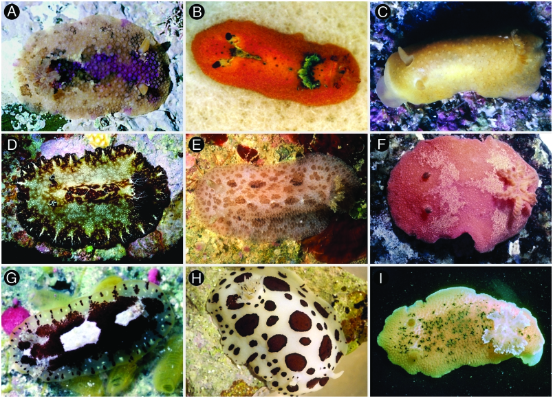
The background colour of the living animals is yelloworange to pale brown. There is an opaque white or brown inverted ‘Y’ or hourglass pattern extending mid-dorsally from between the rhinophores to just in front of the gill ( Fig. 4B View Figure 4 ). In some specimens this pattern can be interrupted or almost absent. Some of the dorsal tubercles, and those situated on the dorsal hourglass pattern, are dark purple-brown. The rhinophores have a purple club and a white base. The branchial leaves are yellow-orange with some of the apices dark brown. The whole dorsum is covered with rounded, slightly conical tubercles, all of them similar in size ( Fig. 7D View Figure 7 ). The largest tubercles are those situated in the central region of the body. The rhinophoral sheaths have several slightly stalked tubercles, larger than those surrounding the sheath, but not larger than the largest tubercles on the dorsum. The tubercles surrounding the branchial sheath are similar to the rest of the dorsal tubercles. There are five tripinnate branchial leaves, forming a circle. The anal papilla is small, situated in the centre of the branchial circle of leaves. The rhinophores are elongate, having eight lamellae in a 21-mm preserved length specimen.
Ventrally there are no oral tentacles, but two blunt prolongations on each side of the mouth opening ( Fig. 8E View Figure 8 ). The anterior border of the foot is grooved but not notched.
Anatomy
The posterior end of the glandular portion of the oral tube has six strong retractor muscles ( Fig. 8C View Figure 8 ) which attach to the body wall. The oval, muscular buccal bulb has two additional muscles attached. Two short salivary glands connect with the buccal bulb at each side of the oesophageal junction. The buccal bulb is several times longer than the glandular portion of the oral tube. The labial cuticle is smooth. The radular formula is 34 ¥ 43.0. 43 in a 21-mm long specimen. Rachidian teeth are absent. The lateral teeth are narrow and elongate, having a single cusp and lacking denticles ( Fig. 7A View Figure 7 ). The teeth from the middle portion of the half-row are larger than those closer to the medial portion of the radula ( Fig. 7B View Figure 7 ). The mid-lateral teeth near to the outer edge bear 2–3 large and blunt denticles on the main cusp. The 2–4 outermost teeth are smaller and have a number of thin denticles ( Fig. 7C View Figure 7 ). The oesophagus is short and connects directly to the stomach.
The ampulla is long and convoluted, and branches into a short oviduct and the prostate ( Fig. 8B View Figure 8 ). The oviduct enters the female gland mass near to its centre. The prostate is tubular, long, folded and granular. It connects with a short duct that narrows and expands again into the ejaculatory portion of the deferent duct. The muscular deferent duct opens into a common atrium with the vagina. The vagina is long. Near to its proximal end it joins the bursa copulatrix. From the bursa copulatrix leads another duct that connects to the uterine duct and the seminal receptacle. The bursa copulatrix is oval in shape, about three times larger than the seminal receptacle.
In the central nervous system ( Fig. 8D View Figure 8 ) the cerebral and pleural ganglia are fused and distinct from the pedal ganglia. There are three cerebral nerves leading from each cerebral ganglion and two pleural nerves leading from each pleural ganglion. There is no separate abdominal ganglion on the right side of the visceral loop. The buccal ganglia are near to the rest of the central nervous system, joined to the cerebral ganglia by two relatively short nerves. Gastro-oesophageal, rhinophoral and optical ganglia are present. The pedal ganglia are clearly separated, having three nerves leading from each one. The pedal and parapedal commissures are enveloped together with the visceral loop.
The circulatory system ( Fig. 8A View Figure 8 ) consists of a large heart and two blood glands situated in front of and behind the central nervous system.
Remarks
Pease (1860) described Doris nucleola based on specimens collected from Hawaii as an orange species, dusky along the dorsal region and shaded with purple on each side of the branchiae. Pruvot-Fol (1947) revised the original description of this species and regarded it as nonidentifiable.
Kay & Young (1969) redescribed Doris nucleola also from Hawaiian material and introduced the new genus Doriorbis to include this species. Their animals were described as having a brown or grey-blue background colour with a T or Y shaped yellow pattern extending mid-dorsally from the rhinophores. They synonymized Doris papillosa Pease, 1860 and Doris tincta Pease, 1864 with Doris nucleola .
Brodie & Willan (1993) studied some specimens from Australia and Norfolk Island, which they assigned to Doris nucleola . At the same time they regarded Doriorbis Kay & Young, 1969 as a synonym of Siraius Er. Marcus, 1955 and added Doris carinata Alder & Hancock, 1864 , Doris carina Abraham, 1877 , Platydoris immonda Risbec, 1928 to the synonymy of this species. The Australian specimens are dull orange-yellow with a pale hourglass-shaped patch extending mid-dorsally from between the rhinophores to just in front of of the gill.
More recently Rudman (2000) argued that Pease (1860) did not mention any dorsal markings between the rhinophores and the gill. He also posted a copy of Garret’s illustration of Pease’s (1860) original specimen published by Bergh (1881) in which there are no traces of the hourglass pattern between the gills and the rhinophores. Therefore he considered that Pease’s Doris nucleola is a different species, and that the first name available and recognizable for the species studied by Kay & Young (1969) and Brodie & Willan (1993) is Platydoris immonda Risbec, 1928 . A close examination of the drawing by Garret reveals that Doris nucleola has well-developed oral tentacles that are absent in Doris immonda and the material examined by Kay & Young (1969) and Brodie & Willan (1993), and it is very likely that Doris nucleola belongs to a different genus. Rudman (2000) also commented that Doris carinata Alder & Hancock, 1864 is a different species because of the higher body profile and larger number of branchial leaves compared to those of Doris immonda . A re-examination of Alder & Hancock’s (1864) paper shows not only that, but the rhinophores are described as brownish, whereas they are whitish or cream with the club black or violet in Doris immonda . The identity of Doris nucleola and Doris carina remains unknown.


| MNHN |
Museum National d'Histoire Naturelle |
| T |
Tavera, Department of Geology and Geophysics |
No known copyright restrictions apply. See Agosti, D., Egloff, W., 2009. Taxonomic information exchange and copyright: the Plazi approach. BMC Research Notes 2009, 2:53 for further explanation.
|
Kingdom |
|
|
Phylum |
|
|
Class |
|
|
Order |
|
|
Family |
|
|
Genus |
Doris immonda
| Valdés, Ángel 2002 |
Platydoris immonda
| Risbec J 1928: 84 |