
Doris pseudoargus, RAPP, 1827
|
publication ID |
https://doi.org/ 10.1046/j.1096-3642.2002.00039.x |
|
persistent identifier |
https://treatment.plazi.org/id/03B5879A-755C-6C51-930C-FBD424BCAC48 |
|
treatment provided by |
Carolina |
|
scientific name |
Doris pseudoargus |
| status |
|
DORIS PSEUDOARGUS RAPP, 1827 View in CoL ( FIGS 4A View Figure 4 , 5 View Figure 5 , 6 View Figure 6 )
Doris pseudoargus Rapp, 1827: 519 View in CoL .
Doris flavipes Leuckart, 1828: 14 View in CoL .
Doris leuckartii Delle Chiaje, 1841: 19 View in CoL , pl. 40, fig. 3. Doris schembrii Verany, 1846: 21–22 View in CoL .
Type material
Doris pseudoargus Rapp , the type material, collected from Le Havre, France, is untraceable. NEOTYPE (here designated): Locmariaquer, France, 13 April 1972, one specimen, 22 mm preserved length, leg. P. Bouchet ( MNHN). Doris flavipes Leuckart , the type material collected from the Mediterranean Sea is untraceable. Doris leuckartii Delle Chiaje , the type material collected from Nice, France, is untraceable. Doris schembrii Verany , SYNTYPES: Gulf of Geneva, Italy, two specimens ( MNHN). The type material of Doris britannica Leach could not be located at BMNH and is probably lost.
Additional material
Las Llanas Beach, Muros de Nalón , Asturias, Spain , 16 August 1987, one specimen, 17 mm preserved length, leg. A. Valdés ( CASIZ 121105 ). Naples , Italy
1902–03, one specimen, 33 mm preserved length, leg. F. M. MacFarland ( CASIZ 081871 ) .
External morphology
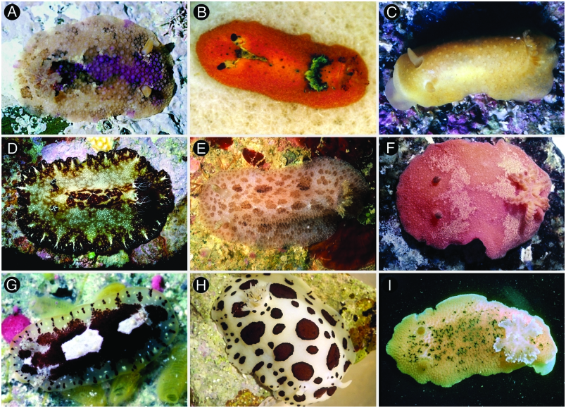
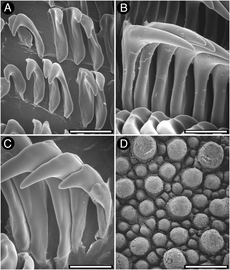
The general colour of the living animals varies from yellowish to pale brown, with pale purple, whitish, green, dark brown or reddish irregular patches on the dorsum ( Fig. 4A View Figure 4 ). In some specimens there are only dark brown patches. The rhinophores and the gill are yellowish to pale brown. The whole dorsum is covered with rounded and simple tubercles, all of them similar in size ( Fig. 5D View Figure 5 ). The largest tubercles are those situated in the central region of the body. The rhinophoral and branchial sheaths have several tubercles which are slightly stalked but otherwise similar to the rest of the dorsal tubercles. There are 8–9 tripinnate branchial leaves, forming a circle. The anal papilla is prominent, situated in the centre of the branchial circle of leaves. The rhinophores are elongate, having 14 lamellae in a 17-mm preserved length specimen.
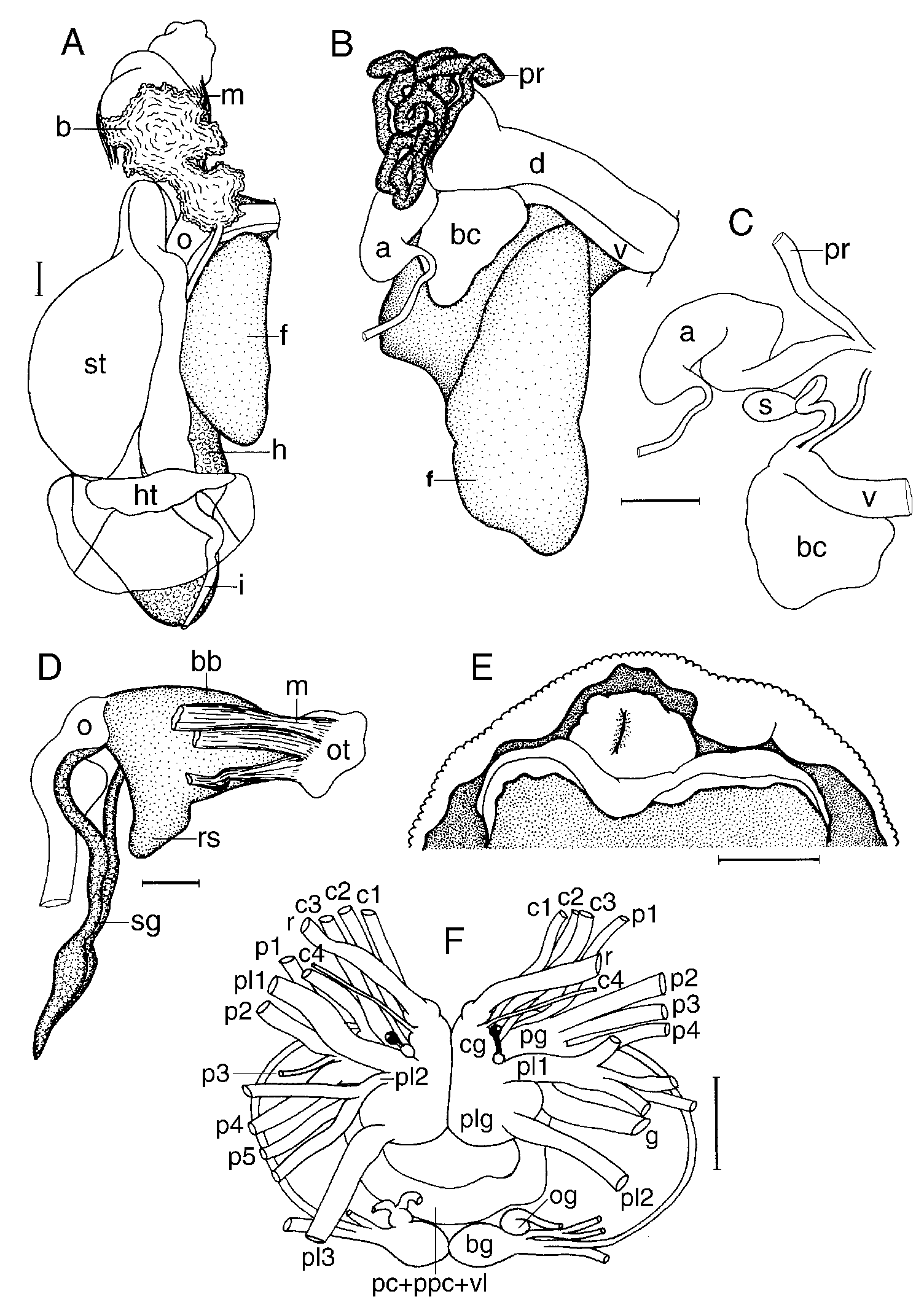
Ventrally, there are no oral tentacles, but two blunt prolongations on each side of the mouth opening ( Fig. 6E View Figure 6 ). The anterior border of the foot is grooved but not notched.
Anatomy
The posterior end of the glandular portion of the oral tube has six strong retractor muscles ( Fig. 6D View Figure 6 ) which attach to the body wall. Two long salivary glands connect with the buccal bulb at each side of the oesophageal junction. The buccal bulb is several times longer than the glandular portion of the oral tube. The labial cuticle is smooth. The radular formula is 41 ¥ 73.0. 73 in a 33-mm long specimen. Rachidian teeth are absent. The lateral teeth are narrow and elongate, having a single cusp and lacking denticles ( Fig. 5A View Figure 5 ). The teeth from the middle portion of the half-row are larger than those closer to the medial portion of the radula ( Fig. 5B View Figure 5 ). The outermost teeth are smaller and have a number of thin denticles ( Fig. 5C View Figure 5 ). The oesophagus is short and connects directly to the stomach ( Fig. 5A View Figure 5 ).
The ampulla is convoluted and branches into a short oviduct and the prostate ( Fig. 6C View Figure 6 ). The oviduct enters the female gland mass near to its centre. The prostate is tubular, very long, folded and granular ( Fig. 6B View Figure 6 ). It connects with a long duct that narrows and expands again into the huge ejaculatory portion of the deferent duct. The muscular deferent duct opens into a short common atrium with the vagina. The vagina is long and wide. Near to its proximal end it joins the duct connecting the bursa copulatrix and the seminal receptacle. The uterine duct also leads from this duct. The bursa copulatrix is irregular in shape, about 10 times larger than the seminal receptacle ( Fig. 6C View Figure 6 ).
In the central nervous system ( Fig. 6F View Figure 6 ) the cerebral and pleural ganglia are fused and distinct from the pedal ganglia. There are four cerebral nerves leading from each cerebral ganglion, and three pleural nerves leading from the left pleural ganglion and two from the right one. There is no separate abdominal ganglion on the right side of the visceral loop. The buccal ganglia are near to the rest of the central nervous system, joined to the cerebral ganglia by two relatively short nerves. Gastro-oesophageal, rhinophoral and optical ganglia are present. The pedal ganglia are clearly separated, having five nerves leading from the left ganglion and four from the right one. The pedal and parapedal commissures are enveloped together with the visceral loop.
The circulatory system ( Fig. 6A View Figure 6 ) consists of a large heart and a single large blood gland situated over the central nervous system.
Remarks
Doris tuberculata Müller, 1778 View in CoL was described on the basis of an undetermined number of specimens collected from Norway. Müller (1778) described the animals as golden, patelliform, with the dorsum covered with numerous hair-like yellowish tubercles. The description of the animals clearly represents a species of phanerobranch dorid, probably a member of the genera Acanthodoris J.E. Gray, 1850 View in CoL , Adalaria Bergh, 1878 View in CoL or Onchidoris Blainville, 1816 View in CoL .
Years later, Cuvier (1804) reported Doris tuberculata Müller, 1778 View in CoL from the Atlantic coast of France based on two newly collected specimens, but indicating that his material was clearly different from Müller’s (1778). The animals described by Cuvier are large cryptobranch dorids with the dorsum covered with rounded tubercles. Rapp (1827) described Doris pseudoargus View in CoL from Le Havre, France, with the same characteristics of the specimens studied by Cuvier (1804): ‘ash colour with dull reddish spots’, and therefore this is the first valid introduction of a name for this species.
Johnston (1838) introduced the names D. britannica and D. montagui , without a description and in the synonymy of D. Tuberculata . Therefore they are nomina nuda and if they have not been used as valid before 1961 they are also not available ( ICZN, 1999).
In the following years most authors referred to this species as Doris tuberculata , but with authorship of Cuvier. Examples include Delle Chiaje (1841), Bergh (1878b), Eliot (1910), Vayssière (1913) O’Donoghue (1929), Pruvot-Fol (1935), Odhner (1939). The scientific influence of Cuvier’s papers probably explains why subsequently many authors applied the name Doris tuberculata to this cryptobranch dorid species.
The usage of the name Doris tuberculata for this species was challenged by the British School. Early on, Iredale & O’Donoghue (1923) for some unexplained reason decided that the animals named Doris tuberculata by Cuvier are a different species from specimens identified as such by Alder & Hancock and Eliot; they used the unavailable name Doris britannica , combined with the genus name Archidoris , for the latter. On the other hand, Pruvot-Fol (1931) argued that all these animals belonged to the same species - Doris tuberculata with authorship of Cuvier the valid name. The name Doris britannica very rarely appears in the literature. Thompson (1966) reintroduced the usage of the name Doris pseudoargus , also combined with Archidoris , but without a justification.
Both Doris pseudoargus View in CoL and Doris tuberculata View in CoL have been equally used in modern literature, usually combined with the genus name Archidoris View in CoL . Examples of the former in taxonomic papers include Schmekel & Portmann (1982), Thompson & Brown (1984), Cattaneo-Vietti et al. (1990), Picton & Morrow (1994); examples of the latter include Ros (1975), Barletta (1981), Swennen & Dekker (1987), Sabelli, Giannuzzi- Savelli & Bedulli (1990). In addition, most papers on physiology, ecology or histology of this species have used the former ( Thompson, 1966; Rose, 1971; Potts, 1983; Jonas, 1986), whereas biochemistry papers have used the latter ( Cimino et al. 1993). In no cases did authors specify their reasons for using one or the other name, which increased the general confusion. Because both names are currently in use, the maintenance of the usage of the valid name for this species, Doris pseudoargus View in CoL , would certainly not cause a larger disruption than the validation of the name Doris tuberculata View in CoL .
Doris pseudoargus View in CoL is a well-known species that ranges from Nordkapp ( Norway), Iceland and the Faroes to the Mediterranean Sea ( Thompson & Brown, 1984). The name D. tuberculata View in CoL has been used for specimens that occur beyond the geographical range of this species. Savigny (1817) reported this species from the Red Sea, Bergh (1894) from the North Pacific and Lemche (1929) from the Gulf of Mexico. These three records are probably misidentifications (see Pruvot-Fol, 1935 and Thompson & Brown, 1984, who have also listed several other synonyms for this species discussed here).
Doris schembrii Verany, 1846 View in CoL was originally described with the same external features of A. pseudoargus View in CoL (see Verany, 1846), and the re-examination of its type material confirmed that these names are synonyms. Also, the original descriptions of Doris flavipes View in CoL (see Leuckart, 1828) and Doris leuckartii View in CoL (see Delle Chiaje, 1841) clearly show that they should be regarded as junior synonyms of A. pseudoargus View in CoL .
Doris flammea Alder & Hancock, 1844 and Doris mera Alder & Hancock, 1844 View in CoL have been regarded as synonyms of D. pseudoargus View in CoL (see Thompson & Brown, 1984). However, the original description of these species ( Alder & Hancock, 1845 -55) shows that they are externally very different from D. pseudoargus View in CoL . Doris flammea is a bright orange-scarlet species, occasionally blotched with purple. The dorsum is covered with short, obtuse, spiculose tubercles. The rhinophores are large, tapering, orange with 10 or 11 scarlet lamellae. There are nine scarlet branchial leaves. This description resembles Rostanga rubra Risso, 1818 View in CoL , but whether these two names are synonyms requires further investigation. Doris mera View in CoL was described as a white species, ‘rather broad and elevated on the back’. This is very different from D. pseudoargus View in CoL , which is a brownish species. Also, the dorsal tubercles of D. mera View in CoL were described as being moderately sized, unequal and round. This is very similar to Aldisa zetlandica ( Alder & Hancock, 1854) View in CoL , for which D. mera View in CoL could be a synonym.
| MNHN |
Museum National d'Histoire Naturelle |
No known copyright restrictions apply. See Agosti, D., Egloff, W., 2009. Taxonomic information exchange and copyright: the Plazi approach. BMC Research Notes 2009, 2:53 for further explanation.
|
Kingdom |
|
|
Phylum |
|
|
Class |
|
|
Order |
|
|
Family |
|
|
Genus |
Doris pseudoargus
| Valdés, Ángel 2002 |
Doris leuckartii
| Verany DB 1846: 22 |
| Delle Chiaje S 1841: 19 |
Doris flavipes
| Leuckart FS 1828: 14 |
Doris pseudoargus
| Rapp WL 1827: 519 |