
Doris verrucosa, LINNAEUS, 1758
|
publication ID |
https://doi.org/ 10.1046/j.1096-3642.2002.00039.x |
|
persistent identifier |
https://treatment.plazi.org/id/03B5879A-755F-6C4A-90F6-FAAE20CCABCE |
|
treatment provided by |
Carolina |
|
scientific name |
Doris verrucosa |
| status |
|
DORIS VERRUCOSA LINNAEUS, 1758 View in CoL ( FIGS 2 View Figure 2 , 3 View Figure 3 )
Doris verrucosa Linnaeus, 1758: 653 View in CoL View Cited Treatment .
Doris derelicta Fischer, 1867: 7–8 View in CoL .
Doris biscayensis Fischer, 1872: 6–8 View in CoL .
Staurodoris januari Bergh, 1878a: 583–585 , pl. 63, fig. 24, pl. 64, figs 8-12.
Staurodoris verrucosa var. mollis Eliot, 1906a: 338– 339 View in CoL .
Staurodoris bobretzkii Gadzikiewicz, 1907: 509–510 View in CoL .
Type material
Doris verrucosa Linnaeus View in CoL , NEOTYPE (designated by Bouchet & Valdés, 2000 and validated by Opinion 1980 - ICZN, 2001): Castropol, Asturias, Spain, leg. J. Cigarría ( MNHN). Doris derelicta Fischer View in CoL , NEOTYPE (designated by Bouchet & Valdés, 2000): Castropol , Asturias, Spain , leg. J. Cigarría ( MNHN). The type material of Staurodoris januari Bergh could not be located at ZMUC and is presumed lost; the original type locality is near Rio de Janeiro, Brazil .
Additional material
Naples , Italy 1898, three specimens, 28–33 mm preserved length, leg. F. M. MacFarland ( CASIZ 082119 ) .
External morphology
The external morphology of this species has been described and illustrated by many authors. Three recent examples can be found in the papers by Schmekel (1968), Ortea, Pérez & Llera (1982) and Thompson & Brown (1984).
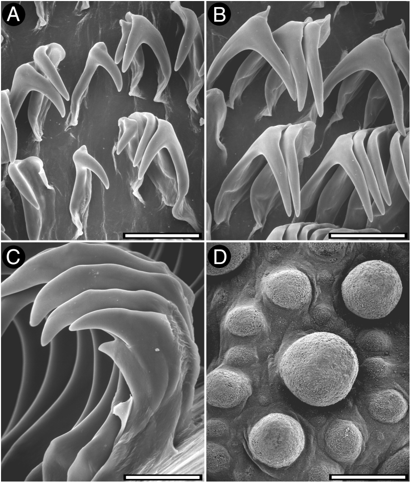
The general colour of the living animals is uniformly yellow to yellowish-grey. The whole dorsum is covered with hemispherical tubercles varying in size ( Fig. 2D View Figure 2 ). The largest tubercles are situated in the central region of the body. The rhinophoral sheath has one prominent, stalked tubercle on each side. The branchial sheath has 8–12 stalked tubercles all around. There are 13–18 unipinnate branchial leaves, forming a circle. The anal papilla is prominent, situated in the centre of the branchial circle of leaves. The rhinophores are elongate, having 11 lamellae in a 28-mm preserved length specimen.
Ventrally there are no oral tentacles, but two blunt prolongations on each side of the mouth opening ( Fig. 3F View Figure 3 ). The anterior border of the foot is grooved but not notched.
Anatomy
The posterior end of the glandular portion of the oral tube has six strong retractor muscles ( Fig. 3D View Figure 3 ) which attach to the body wall. The oval, muscular buccal bulb has two additional muscles attached; two long salivary glands connect with it at each side of the oesophageal junction. The buccal bulb is as long as the glandular portion of the oral tube. The labial cuticle is smooth. The radular formula is 38 ¥ (50.0.50) in a 33- mm long specimen. Rachidian teeth are absent. The lateral teeth are narrow and elongate, having a single cusp and lacking denticles ( Fig. 2A View Figure 2 ). The teeth from the middle portion of the half-row are larger than those closer to the medial portion of the radula ( Fig. 2B View Figure 2 ). The outermost teeth are smaller and also lack denticles ( Fig. 2C View Figure 2 ). The oesophagus is short, convoluted and connects directly to the stomach ( Fig. 3A View Figure 3 ). The ampulla is very large and branches into a short oviduct and the prostate ( Fig. 3C View Figure 3 ). The oviduct enters the female gland mass near to its centre. The prostate is tubular, folded and granular ( Fig. 3B View Figure 3 ). It connects with a long duct that narrows and expands again into the long ejaculatory portion of the deferent duct. The muscular deferent duct opens into a common atrium with the vagina. The vagina is long and undulate. Near to its proximal end it joins the duct connecting the bursa copulatrix and the seminal receptacle. The uterine duct also leads from this duct. The bursa copulatrix is irregular in shape, about twice as large as the seminal receptacle ( Fig. 3C View Figure 3 ).
In the central nervous system ( Fig. 3E View Figure 3 ) the cerebral and pleural ganglia are more or less fused and distinct from the pedal ganglia. There are four cerebral nerves leading from the right cerebral ganglion and five from the left one, and four pleural nerves leading from each pleural ganglion. There is no separate abdominal ganglion on the right side of the visceral loop. The buccal ganglia are near to the rest of the central nervous system, joined to the cerebral ganglia by two relatively short nerves. Gastro-oesophageal, rhinophoral and optical ganglia are present. The pedal ganglia are clearly separated, having three nerves leading from each one. The pedal and parapedal commissures are enveloped together, and also partially enveloped with the visceral loop.
The circulatory system ( Fig. 3A View Figure 3 ) consists of a large heart and a single large blood gland situated over the central nervous system.
Remarks
Doris verrucosa View in CoL , in the sense of the neotype proposed by Bouchet & Valdés (2000) and other many authors (e.g. Schmekel, 1968; Ortea et al., 1982; Thompson & Brown, 1984), is a well-known species distributed through the Atlantic and Mediterranean coasts of Europe down to the Canary Islands. Records from the Atlantic coast of the Americas probably belong to this species ( Marcus, 1955; Franz, 1970). Indeed, Doris januari Bergh, 1878 , originally described from Brazil, is very likely a synonym ( Thompson & Brown, 1984). Gosliner’s (1987) reference to South Africa probably represents a distinct species.
Fischer (1867), recognized that the specific name Doris verrucosa Linnaeus View in CoL originally refers to a species from the Indian Ocean and cannot be used for a European species. For the latter he introduced the name Doris derelicta View in CoL . Bouchet & Valdés (2000) proposed designating the same specimen as the neotype of Doris verrucosa Linnaeus View in CoL and Doris derelicta P. Fischer View in CoL , so these two names would become objective synonyms. They also proposed that Doris derelicta P. Fischer View in CoL should be placed in the Official List of Rejected and Invalid Specific Names in Zoology. These proposals were endorsed by the ruling of the ICZN in Opinion 1980 ( ICZN, 2001).
Doris biscayensis View in CoL was described by Fischer (1872) with the same characteristics of Doris verrucosa View in CoL . The uniform pale yellow colour, the presence of two tubercles in the rhinophoral sheath (one on each side), the presence of 13 unipinnate branchial leaves arranged in a circle, and the absence of oral tentacles, are the main diagnostic features of this species. Doris verrucosa View in CoL is the only species from the Atlantic coast of Europe that has this combination of external characteristics. The variety mollis of Staurodoris verrucosa View in CoL described by Eliot (1906a), is also identical to Doris verrucosa View in CoL and is here regarded as a synonym. Gadzikiewicz (1907) described Staurodoris bobretzkii View in CoL on the basis of several specimens collected from the Black Sea, characterized by having a bright orange body covered by large tubercles spotted in black. The eight branchial leaves have the same colour as the body and vary in size, the anterior ones being much longer than the posterior ones. The gill and rhinophoral sheaths are surrounded by tubercles similar to the dorsal tubercles. The tubercles around the gill sheath are much larger than the ones around the rhinophoral sheaths. This description fits with the characteristics of D. verrucosa View in CoL described above, and both names are regarded as synonyms. The three names discussed in this paragraph have been already considered by Thompson & Brown (1984) as synonyms of Doris verrucosa View in CoL .
Thompson & Brown (1984) also included Doris seposita P. Fischer, 1872 and Doris eubalia P. Fischer, 1872 View in CoL in the synonymy of Doris verrucosa View in CoL . However, these two species are easily differentiated from D. verrucosa View in CoL on the basis of their external morphology. Doris eubalia View in CoL is characterized by the presence of large, dark tubercles surrounded by a purple area ( Fischer, 1872). This and other features of this species are very similar to those of Doris sticta Iredale & O’Donoghue, 1923 View in CoL , and both names are probably synonyms. Doris seposita is an uncertain species. According to Fischer (1872) it is different from Doris biscayensis View in CoL (= Doris verrucosa View in CoL ) in having a different rhinophoral morphology, a small number of branchial leaves, the dorsal tubercles more compacted and a darker colour. It is difficult, however, a definitive identification of this species based on the original description, and anatomical studies would be necessary. Unfortunately, the type material of Doris seposita could not be located in MNHN, and is presumed lost.

No known copyright restrictions apply. See Agosti, D., Egloff, W., 2009. Taxonomic information exchange and copyright: the Plazi approach. BMC Research Notes 2009, 2:53 for further explanation.
|
Kingdom |
|
|
Phylum |
|
|
Class |
|
|
Order |
|
|
Family |
|
|
Genus |
Doris verrucosa
| Valdés, Ángel 2002 |
Staurodoris bobretzkii
| Gadzikiewicz W 1907: 510 |
Staurodoris verrucosa var. mollis
| Eliot CN 1906: 339 |
Staurodoris januari
| Bergh R 1878: 585 |
Doris biscayensis
| Fischer P 1872: 8 |
Doris derelicta Fischer, 1867: 7–8
| Fischer P 1867: 8 |