
Urotricha antarctica, Wilbert & Song, 2008
|
publication ID |
https://doi.org/10.1080/00222930701877540 |
|
persistent identifier |
https://treatment.plazi.org/id/03B587B3-FF84-C447-FE7D-63F9FB75FD56 |
|
treatment provided by |
Felipe |
|
scientific name |
Urotricha antarctica |
| status |
|
Order PROSTOMATIDA Schewiakoff, 1896 View in CoL View at ENA
Urotricha antarctica nov. spec.
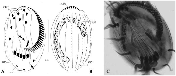
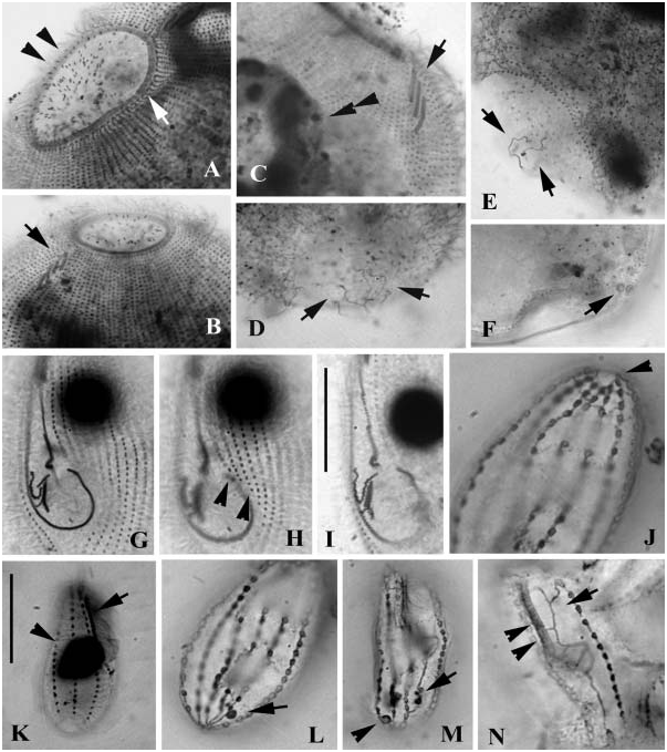
( Figures 1A–C View Figure 1 , 8A–F View Figure 8 )
Diagnosis
Large marine Urotricha with oval body shape and a large cilia-free caudal region; 90–150×80–120 Mm after protargol impregnation; macronucleus long and sausagelike; about 100 somatic kineties; ca. six caudal cilia; circumoral kinety consisting of about 70 dikinetids; three genus-typical dorsal brushes; single large contractile vacuole caudally positioned.
Type locality
Rockpools and the littoral zone on the sea coast near Bellinghausen Station, King George Island (62 ° 139S, 58 ° 589W).
Description
We were unable to make a live observation of this ciliate, so it is identified according to silver-impregnated specimens.
Shape generally symmetrical and broadly oval, cytoplasm colourless. Both cell ends slightly truncated, cytostome wide and prominent; numerous extrusomes spindle-shaped, about 2 Mm in length, entirely contained within the cytopharynx ( Figures 1B View Figure 1 ; 7A View Figure 7 , arrowheads). Cilia-free area in rear pole conspicuously large ( Figures 1C View Figure 1 , 7E View Figure 7 ). Single contractile vacuole, caudally positioned (CV, Figure 1A View Figure 1 ), 2– 3 excretory pores found in posterior cell end ( Figure 1C View Figure 1 , arrow). Cilia about 7 Mm long, with 5–7 caudal cilia arranged more or less in a circle around posterior pole ( Figure 1C View Figure 1 , double-arrowheads). Single sausage-shaped macronucleus uniquely long and always highly twisted (Ma, Figure 1A View Figure 1 ), micronuclei not detected. About 100 somatic kineties not extending to the rear pole, which are more densely ciliated in anterior portion than in posterior and form a closed circle of single kinetosomes ( Figure 1C View Figure 1 , arrowheads) around the rear cilia-free area. At anterior end, circumoral kinety (CK) consisting of about 70 pairs of basal bodies, which surrounds the prominent cytostome ( Figures 1B View Figure 1 ; 7A View Figure 7 , arrow); three (seldom four) dorsal brushes with two-rowed structure, closely packed and positioned near the CK ( Figure 1B View Figure 1 , arrow). Silverline system genus-typical (not illustrated).
Remarks
Considering the combination of several criteria, e.g. the large size, ca. 100 somatic kineties, several caudal cilia, long sausage-shaped macronucleus and the marine (Antarctic) habitat, this new species can be clearly identified ( Kahl 1930). Urotricha baltica Czapik and Jordan, 1976 living in the interstitial of the Baltic is completely different in body size (65–80 Mm), number of kineties (32–36) and infraciliature: there is no circumoral and no postoral circle of kinetosomes ( Figures 1B; 1C View Figure 1 ).
Order CYRTOPHORIDA Fauré-Fremiet in Corliss, 1956 Chlamydonella apoprostomata nov. spec.
( Figures 1D–F View Figure 1 , 9A–G View Figure 9 )
Diagnosis
Large marine Chlamydonella with conspicuous snout-like projection on anterior left; 80–140×50–80 Mm in fixed specimens; perioral kineties often slightly fragmented; totally about 30 densely-ciliated somatic kineties, including consistently seven preoral ones, of which approximately six on the left side are shortened gradually posteriad; about 23 argentophilic granules around cytostome; single oval to globular macronucleus and one micronucleus.
Type locality
In benthos samples from rockpools and the littoral zone on the sea coast near Bellinghausen Station, King George Island (62 ° 139S, 58 ° 589W).
Description
This new species was also described from mounted slides, hence no live information can be supplied. According to the protargol-impregnated specimens, body size and shape highly variable, from oval to slender with narrowed posterior portion, but generally broadly oval ( Figures 1D, F View Figure 1 ). Cells often full of large diatoms ( Figure 1F View Figure 1 ). No contractile vacuoles detected.
Twenty-eight to 35 somatic kineties densely ciliated. Consistently seven preoral kineties arched to left margin of cell, among which four are ‘‘interrupted’’ by the oral apparatus (perioral kineties) and only three rows extend posteriorly to the rear end of cell ( Figure 1D View Figure 1 ). One apical fragment (AF) with about 20 densely arranged kinetosomes ( Figure 9D View Figure 9 , arrow). Perioral kineties genus-typical, Y-shaped and extending from left to right margin of cell, but often slightly fragmented ( Figure 1E View Figure 1 ); all structures consisting of densely arranged dikinetids. On ventral side, postoral kineties, except three preoral ones (PrK), terminating anteriorly at about buccal level and forming a prominent buccal area ( Figures 1D View Figure 1 , 9A–C View Figure 9 ). About six on left posteriorly shortened gradually leftwards ( Figure 9F View Figure 9 , arrowheads). Always one terminal (TF) and one equatorial (EF) fragment ( Figure 1D View Figure 1 ) on left and right margins. Macronucleus (Ma) globular, about 20–30 Mm across, only one micronucleus recognized ( Figure 1D View Figure 1 , double-arrowheads).
Remarks
This new species differs from the closely-related congener, Chlamydonella prostomata Song and Wilbert, 2000 in its consistently higher number of preoral kineties (seven versus four) and more somatic kineties (28–35 versus 21–29), as well as in the globular shape of the macronucleus (versus elongate in C. prostomata ). Agatha et al. (1993) described an isolate from the Antarctic as Chlamydonella sp. as well. As mentioned by Song and Wilbert (2000), it could be a population of C. prostomata .
Hartmannula angustipilosa Deroux and Dragesco, 1968 View in CoL
( Figure 9J View Figure 9 )
Based on many well-impregnated specimens, identification should be quite certain. It is basically similar to the 2002 population described by the present authors ( Wilbert and Song 2005), hence only some points will be documented here.
Description
Body size about 70×40 Mm after protargol impregnation. Oral ciliature genus typical, consistently comprising three obliquely positioned dikinetids. About 8–9 preoral kineties curving towards the left five of which consistently terminate at the leftmost margin. Anterior ends of ca. 25 kineties extend post-orally or around cytostome, forming a conspicuous cilia-free buccal area. Approximately 25 kineties on left side evenly shortened posteriad ( Figure 9J View Figure 9 , arrowheads). Macronucleus and podite similar to that of the 2002 population ( Wilbert and Song 2005). In addition to these normal kineties, one equatorial right kinety resembles a fragment, with about 15 basal bodies found in mid-body.
Remarks
Deroux and Dragesco (1968) described three ‘‘populations’’ under the name Hartmannula angustipilosa , but these are remarkably different in size and numbers of somatic kineties, which indicates that they might be different species. We therefore suggest that this species name be limited to the largest form. The Antarctic form resembles the largest morphotype in both body shape and size, as well as the number of kineties.
Order PLEUROSTOMATIDA Schewiakoff, 1896 Litonotus sp.
( Figures 9H, I, K View Figure 9 )
We identified this organism from protargol-impregnated slides. Since no living observation was carried out—that is, no information regarding position/number of the contractile vacuoles or the extrusome characters is available—this organism must be treated as an unknown form here. Considering the cell size, the habitat, and the general appearance of the infraciliature and nuclear apparatus, the present organism possibly represents an undescribed form, but further live information is required.
Description
Cells after protargol impregnation are generally form-constant, about 200 Mm in length, with both anterior and posterior ends conspicuously narrowed. Two globular macronuclear nodules are present in the mid-body, arranged close together ( Figure 9K View Figure 9 , arrowheads). No micronucleus are detected. Left cell side always with several longitudinal ridges ( Figure 9K View Figure 9 , arrow). Infraciliature typical of genus. On the right side, ca. 12 densely ciliated somatic kineties which are shortened near the slitlike cytostome (arrowheads in Figure 9I View Figure 9 ), whereas on left side probably 10 (? not clearly detected) loosely ciliated kineties (including perioral kinety), all of which seem to extend along the whole cell length. Dorsal brush composed of over 30 basalbody pairs and extending posteriorly to about half the cell length ( Figure 9H View Figure 9 , arrow).


No known copyright restrictions apply. See Agosti, D., Egloff, W., 2009. Taxonomic information exchange and copyright: the Plazi approach. BMC Research Notes 2009, 2:53 for further explanation.
|
Kingdom |
|
|
Phylum |
|
|
Class |
|
|
Order |
|
|
Family |
|
|
Genus |
Urotricha antarctica
| Wilbert, Norbert & Song, Weibo 2008 |
Hartmannula angustipilosa
| Deroux and Dragesco 1968 |