
Naisdoris vitiligata, Paz-Sedano & Cobb & Gosliner & Pola, 2024
|
publication ID |
https://doi.org/ 10.11646/zootaxa.5443.4.3 |
|
publication LSID |
lsid:zoobank.org:pub:F4D19D80-3772-4F85-ACB2-6140D2F3BABB |
|
DOI |
https://doi.org/10.5281/zenodo.11074268 |
|
persistent identifier |
https://treatment.plazi.org/id/03B587E3-FFDC-EE01-00F9-4C83FD15FD2F |
|
treatment provided by |
Plazi |
|
scientific name |
Naisdoris vitiligata |
| status |
sp. nov. |
Naisdoris vitiligata sp. nov.
( Figs. 7E‒F View FIGURE 7 , 6G‒H View FIGURE 6 , 8H‒J View FIGURE 8 )
Zoobank: urn:lsid:zoobank.org:act:B31C27EF-752A-4A97-ABEB-5F77074C902B
Okenia liklik — Anderson (2016)
Okenia cf. liklik — Behrens (2019)
Naisdoris sp. F — Paz-Sedano et al. (2024)
Type material. Holotype. ZCR.MOL.15410, Hantu Island , Singapore, 15 m depth, 28 January 2018, col. by C. H. Toh, 96% EtOH, dissected ( SEM: radula, labial cuticle, penis) . Paratypes. ZCR.MOL.15411, Hantu Island , Singapore, 15 m depth, 28 January 2018, col. by C. H. Toh, 96% EtOH, dissected ( SEM: labial cuticle, penis) ; CASIZ 186126 , Ladder dive site, Caban Island, Maricaban Island, Batangas Province, the Philippines, 13.6842 ºN 120.84119 ºE, 10 May 2011, col. by GoogleMaps T. Gosliner.
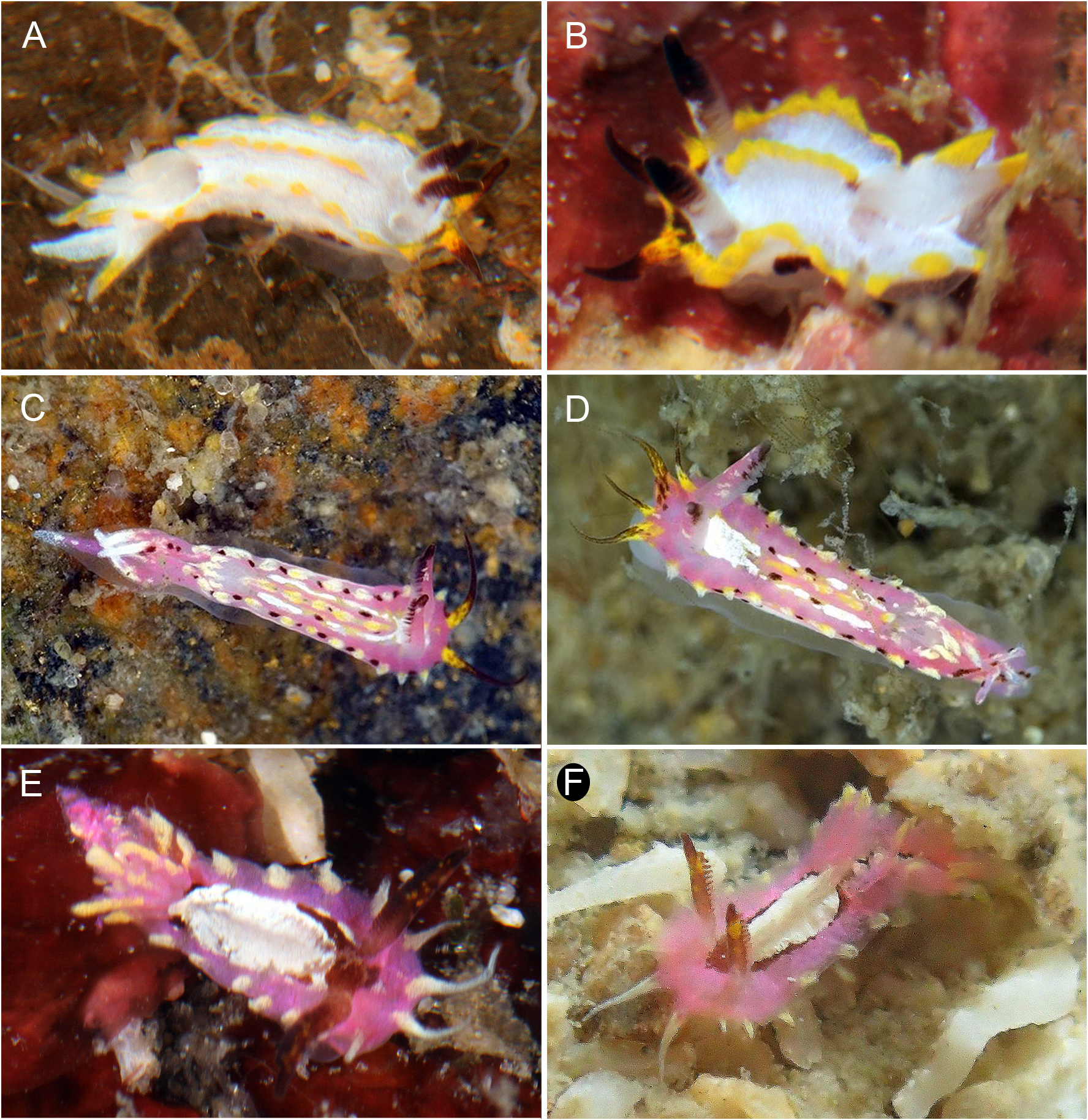
External morphology ( Figs. 7E‒F View FIGURE 7 ). Preserved specimens 2–3 mm in length. Body elongated, notal border well developed, markedly serrated due to presence of internal spicules. Ends of large spicules resemble small papillae from outside of specimens. Middorsal crest also supported by spicules. Two elongated, conical, and thin papillae located in front of rhinophores, one on each side. Tips of anterior papillae thinner than bases. Posterior part of mantle with a small extension that seems a short posterior papilla on each side of body mantle. Anterior papillae slightly larger than posterior ones. Rhinophores non-retractile, elongated, and slender, bearing 12–13 lamellae oriented to posterior part. Rhinophoral sheath absent. Four thin, simple gill branches form a semicircle around anus, two anteriormost sharing common stalk. One tentacular oral tentacle on each side of mouth, short and muscular, somehow triangular shape. Reproductive opening located on first third of right lateral side of body. Mantle covered by spicules.
Color pattern ( Figs. 7E‒F View FIGURE 7 ). Body pink. Large white patch in the dorsal part of the body, may be surrounded by a thin reddish-brown line. Mantle margin with light yellow patches, matching with spikes that make it serrated. Anteriormost papillae translucent yellow at tip, light yellow at base. Posteriormost papillae and gill branches with yellow tips and pink base. Rhinophores same color as dorsal reddish-brown line, may have a bright yellow band. Most external lamellae with yellow tones.
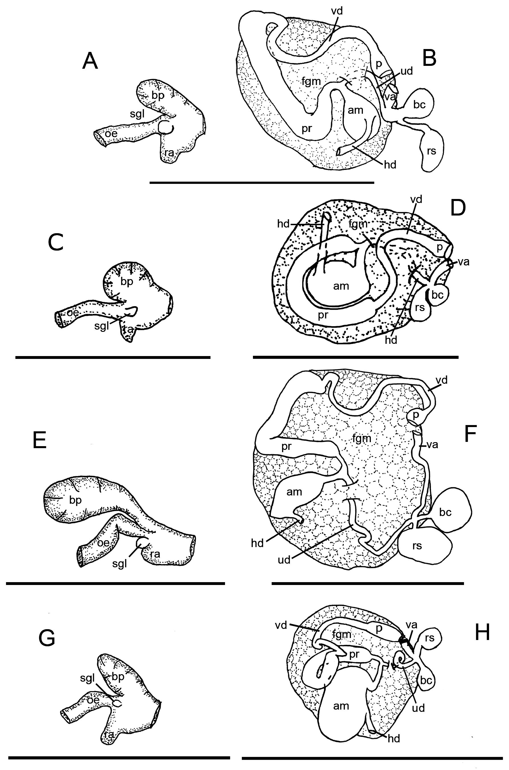
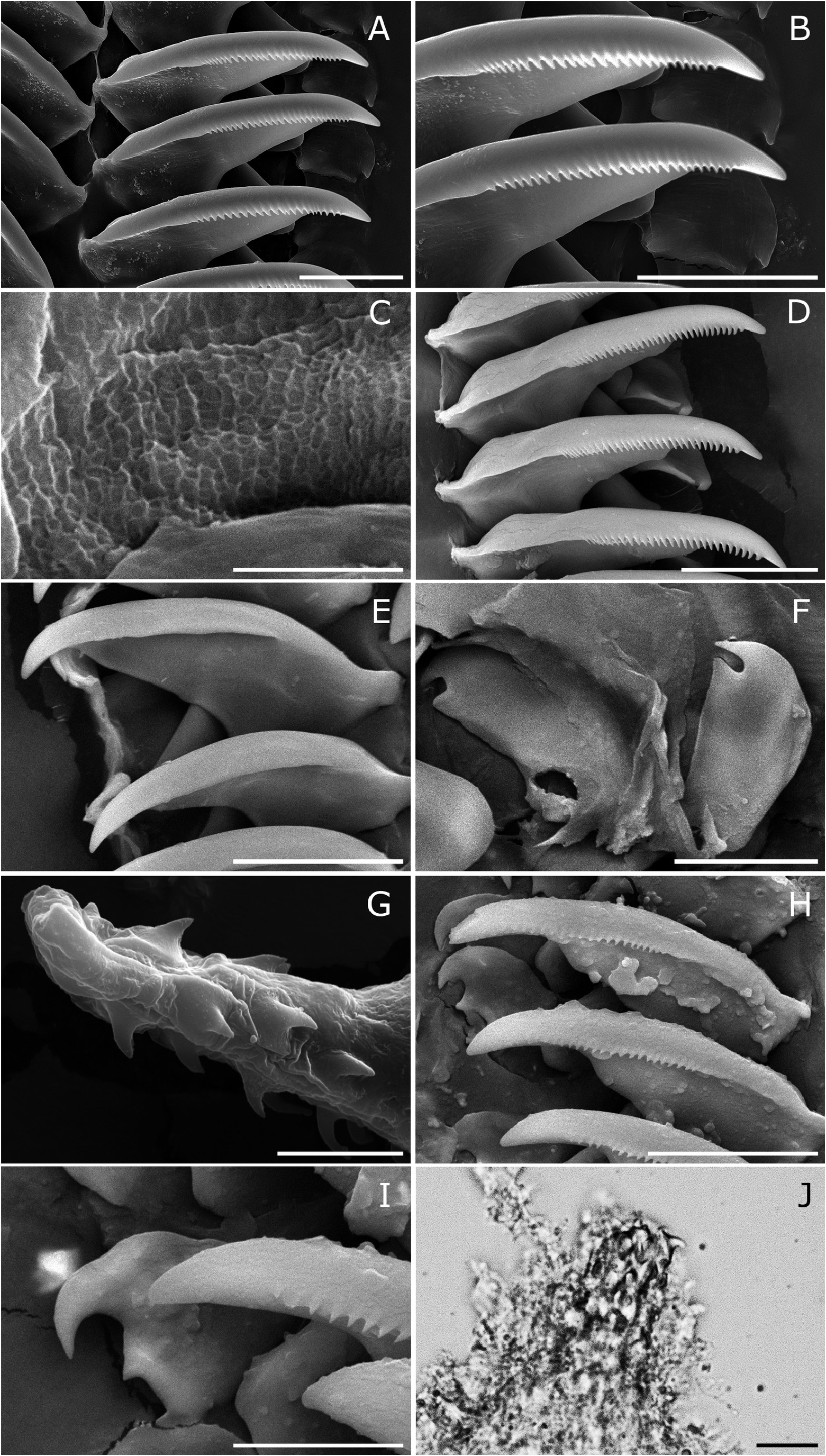
Foregut anatomy ( Figs. 6G View FIGURE 6 , 8H‒I View FIGURE 8 ). Buccal bulb thick and muscular ( Fig. 6G View FIGURE 6 ). Elongated, wide, dorsal buccal pump expanding posteriorly. Small radular sac located ventrally, expanding backwards. Esophagus begins from buccal bulb behind buccal pump. Salivary glands very small, rounded, located at junction of esophagus with buccal bulb. Nervous system covers this junction.Esophagus continues posteriorly and inserts into digestive-hermaphroditic gland. Labial cuticle surrounds lips and expands within buccal pump. Radular formula 14 × 1.1.0.1.1. Inner lateral tooth with single large and thin cusp, and wide, rectangular base ( Fig. 8H View FIGURE 8 ). Cusp large and pointed, masticatory margin with 12–21 very small, pointed denticles ( Fig. 8H View FIGURE 8 ). Denticles located at middle part of masticatory margin larger than lateral ones. Outer base ends in somewhat prominent wing. Outer lateral tooth much smaller, rectangular with two thin hooks, upper hook large and lower one very small ( Fig. 8I View FIGURE 8 ).
Reproductive system ( Figs. 6H View FIGURE 6 , 8J View FIGURE 8 ). Reproductive system located at anterior-third of body. Thin hermaphroditic duct begins at ovotestis, located inside digestive-hermaphroditic gland. Hermaphroditic duct expands into big and oval ampulla. Postampullary duct emerges from ampulla and divides into two different, thin ducts. Short, oviduct enters inside female gland mass. Second duct connects with first portion of prostate. Prostate sausage-shaped. Prostate folds, narrows and become a very thin vas deferens. Prostate and vas deferens have similar length. Vas deferens continues and expands to ejaculatory duct. Penis with small, hooked penial spines ( Fig. 8J View FIGURE 8 ). Vagina small, slightly wider than vas deferens. Vagina connects with rounded bursa copulatrix. From middle of vagina arises a duct which connects with rounded receptaculum seminis. At same point of division of ducts arises thin uterine duct that enters female gland mass. Receptaculum seminis similar in size to bursa copulatrix.
Etymology. “vitiligata ” refers to the distinctive white patch on the dorsum of the species, in Latin vitiligo refers to the lack of pigmentation in an area of the skin.
Distribution. Indonesia ( Behrens 2019), the Philippines ( Anderson 2016; present study) and Singapore (present study)
Natural history. This species is found on flat rubble plains in about 30 m of water.
Remarks. The species N. vitiligata sp. nov. resembles N. liklik and N. labalsaensis sp. nov. by its pink background coloration but differs in the color pattern of the mid-dorsal part of the body ( Gosliner 2004). Naisdoris vitiligata sp. nov. is distinguished by its large white patch in the middle of dorsum while N. labalsaensis sp. nov. has a pink mid-dorsal line with yellow and dark brown spots. The rhinophores are dark in N. vitiligata sp. nov., pink with brown tips in N. labalsaensis sp. nov. and pink with orangish tips in N. liklik ( Gosliner 2004) . Naisdoris vitiligata sp. nov. has serrated margin and lacks the dorsal papilla that are present in N. liklik ( Gosliner 2004) . Internally, in N. vitiligata sp. nov. the cusp located at the external part of the outer lateral tooth has a hooked point, larger and more evident than in N. liklik and N. labalsaensis sp. nov. ( Gosliner 2004). Moreover, N. vitiligata sp. nov. differs from N. labalsaensis sp. nov. by the length of the vagina, uterine duct, and buccal pump, which are much shorter in N. vitiligata sp. nov. than in N. labalsaensis sp. nov. The ducts that connect with the bursa copulatrix, the receptaculum seminis and the uterine duct arises from the same point in N. vitiligata sp. nov. while in N. liklik the uterine duct arises from the base of the vagina ( Gosliner 2004).
| T |
Tavera, Department of Geology and Geophysics |
No known copyright restrictions apply. See Agosti, D., Egloff, W., 2009. Taxonomic information exchange and copyright: the Plazi approach. BMC Research Notes 2009, 2:53 for further explanation.
|
Kingdom |
|
|
Phylum |
|
|
Class |
|
|
Order |
|
|
Family |
|
|
Genus |