
Caligus dasyaticus Rangnekar, 1957
|
publication ID |
https://doi.org/10.11646/zootaxa.4398.1.1 |
|
publication LSID |
lsid:zoobank.org:pub:79E3EB78-D1C3-45CF-AB13-F8E61C936252 |
|
DOI |
https://doi.org/10.5281/zenodo.5952162 |
|
persistent identifier |
https://treatment.plazi.org/id/03B587F2-AA54-4D11-B6F8-FF7C3AD2F87E |
|
treatment provided by |
Plazi |
|
scientific name |
Caligus dasyaticus Rangnekar, 1957 |
| status |
|
Caligus dasyaticus Rangnekar, 1957
( Fig. 26 View FIGURE 26 )
Material examined. 1♀ from Neotrygon kuhlii (Müller & Henle, 1841) (TC17336) 20 January 2016, QM Reg. No. W53064.
Site on host. Lower body surface.
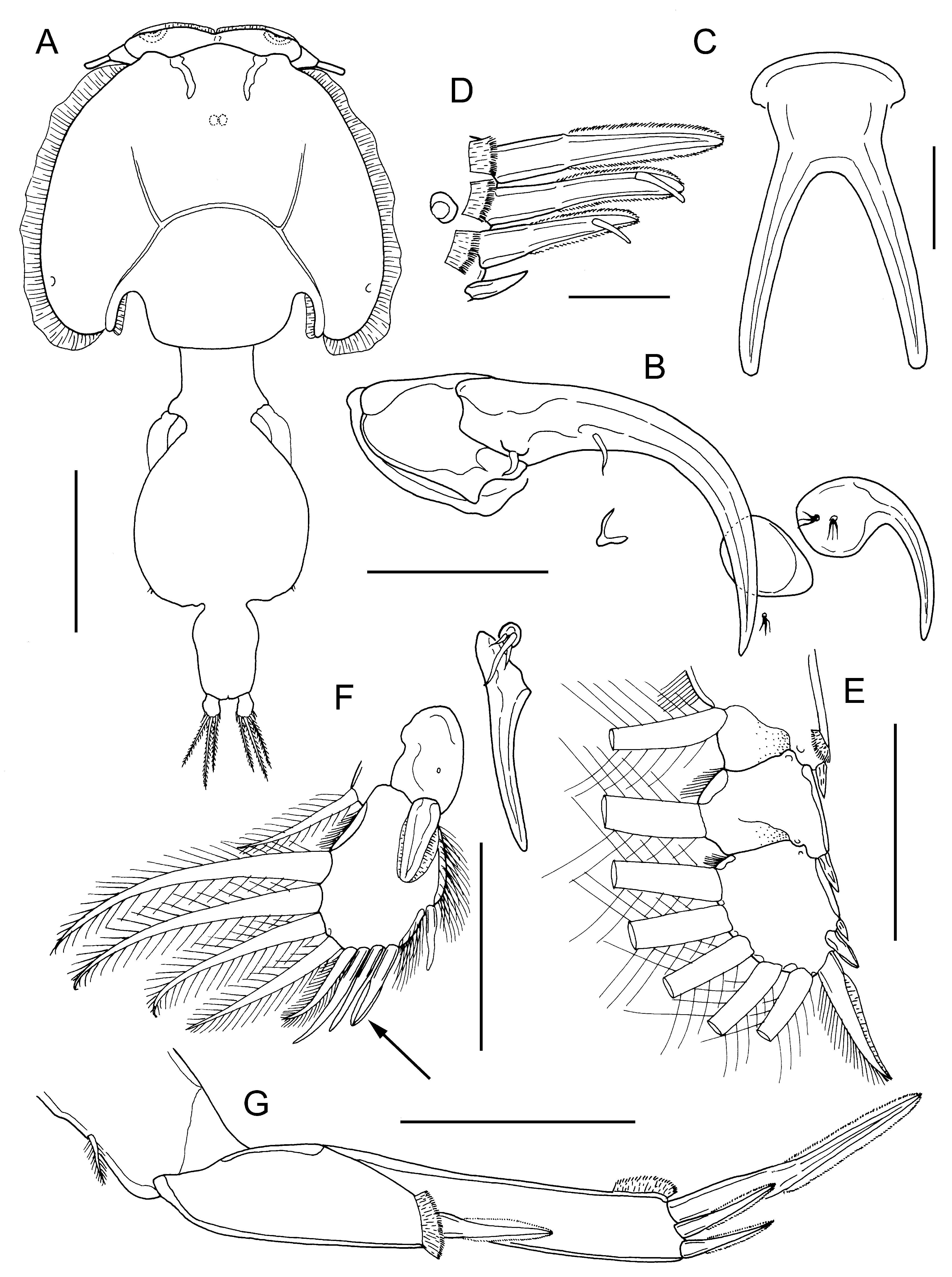
Differential diagnosis. Cephalothorax dorsoventrally flattened with well-developed marginal membranes along lateral zones; frontal plates with very small lunules. Genital complex just longer than wide (1.02 to 1.10 times); abdomen indistinctly 2-segmented, about 1.8 times longer than wide ( Fig. 26A View FIGURE 26 ); genital complex about 1.7 times longer than abdomen. Antenna without posterior process on proximal segment ( Fig. 26B View FIGURE 26 ). Post-antennal process strongly recurved, tine without marginal flange; associated papillae multisensillate ( Fig. 26B View FIGURE 26 ). Posterior process of maxillule long, slender and slightly curved outwards ( Fig. 26B View FIGURE 26 ). Maxilliped of female slender with smooth myxal margin. Sternal furca with long, slender divergent tines ( Fig. 26C View FIGURE 26 ). Distal exopodal segment of leg 1 with 3 plumose setae on posterior margin; distal spine 1 longer than other spines, ornamented with finely striated membrane bilaterally; spines 2 and 3 each with accessory process and striated marginal membrane around tip; seta 4 naked, less than half length of spines 2 and 3 ( Fig. 26D View FIGURE 26 ). Leg 2 with marginal setules on endopodal segments 2 and 3; outer spines on exopodal segments 1 and 2 small (each shorter than next segment) and aligned parallel with longitudinal axis of ramus ( Fig. 26E View FIGURE 26 ). Leg 3 apron lacking distinctive ornamentation; exopod 2-segmented ( Fig. 26F View FIGURE 26 ) with second and third segments fused to form compound distal segment; first segment bearing straight outer spine with marginal flanges and inner plumose seta; compound distal segment with 4 outer spines and 5 plumose setae; proximal outer spine derived from third segment (arrowed in Fig. 26F View FIGURE 26 ) ornamented with marginal membrane distally. Leg 4 3-segmented ( Fig. 26G View FIGURE 26 ); first exopodal segment with outer spine and large pecten; distal exopodal segment with 3 spines around apex, long spine more than twice as long as 2 short spines; segment ornamented with marginal membrane distally. Body length of adult female 4.23 mm.
Remarks. This species was originally described by Rangnekar (1957) based on two females from the body surface of a dasyatid sting ray named as “ Dasyaticus [sic!] uarnac ” by Rangnekar and caught off Bombay, India. The identity of the host should be considered as uncertain given the changes in understanding of species level taxonomy of chondrichthyan fishes in general and of the Himantura uarnak (Gmelin, 1789) species group in particular (see Naylor et al., 2012). The body length of Rangnekar’s single intact female was 3.80 mm ( Rangnekar, 1957). Shiino (1960) subsequently reported this species from Dasyatis akajei (Müller & Henle, 1841) taken off Hamazima, Japan and provided the first description of the male. The body length of the largest female recorded by Shiino was 6.00 mm and that of the male was 2.89 mm ( Shiino, 1960). Kabata (1966) was the first to report C. dasyaticus from Australian waters: based on two males collected from Neotrygon kuhlii (as Amphotistius kuhlii ) caught in Moreton Bay. With a body length of 2.23 mm, Kabata’s male was smaller than that of Shiino, but so is the female from Moreton Bay (at 4.23 mm).
As already highlighted by Ho et al. (2007), the taxonomy of C. dasyaticus became confused after Pillai (1968) published a detailed description of a different species of Caligus as “ C. dasyaticus Rangnekar ” from a specimen of the sawfish Pristis examined at Trivandrum, India. The female described by Pillai (1968) had a body length of 7.0 mm and the male was 3.3 mm in length. In his 1985 monograph, Pillai (1985) re-used the figures from his 1968 description. Ho et al. (2007) redescribed female C. dasyaticus from a single specimen with a body length of 5.62 mm, collected from the body surface of Dasyatis navarrae (Steindachner) landed in Taiwan. Ho et al. (2007) stated that “Pillai’s (1968) “ C. dasyaticus ” very likely represents a different species”. A new species, Caligus elasmobranchi sp. nov., is established below, to accommodate the species described by Pillai (1968) from Pristis sp.
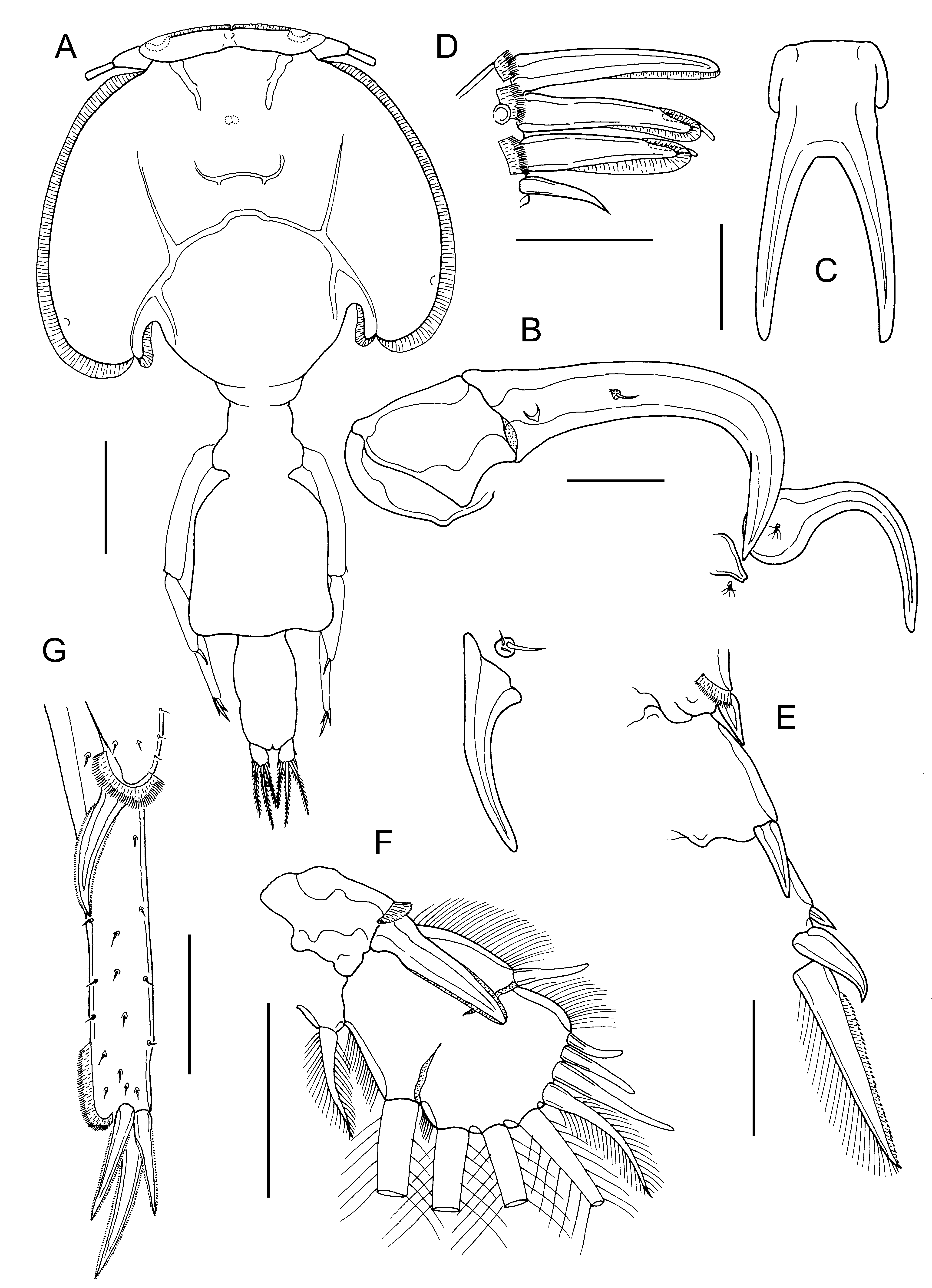
Both species are present in Moreton Bay: typical C. dasyaticus as described by Rangnekar (1957) was only found on Neotrygon kuhlii (Kabata, 1966; present account) while C. elasmobranchi sp. nov. was found on five different elasmobranch hosts. The best character for distinguishing between females of these two widely distributed species is leg 4: in C. dasyaticus leg 4 is shorter than the genital complex and bears 1 long and 2 short spines at its apex (cf. Fig. 26G View FIGURE 26 ), whereas in C. elasmobranchi sp. nov. the leg is much longer than the genital complex, extending beyond the middle of the abdomen, and bears 3 short to medium length apical spines (cf. Fig. 27G View FIGURE 27 ). Another significant difference is the size of the outer margin spine on exopodal segment 1 of leg 3: in C. dasyaticus it is short and does not reach the articulation between exopodal segments 2 and 3, whereas in C. elasmobranchi sp. nov. it is longer, extending well beyond this articulation.
No known copyright restrictions apply. See Agosti, D., Egloff, W., 2009. Taxonomic information exchange and copyright: the Plazi approach. BMC Research Notes 2009, 2:53 for further explanation.
|
Kingdom |
|
|
Phylum |
|
|
Class |
|
|
Order |
|
|
Family |
|
|
Genus |