
Callanthias platei, Steindachner, 1898
|
publication ID |
https://doi.org/ 10.11646/zootaxa.4751.1.8 |
|
publication LSID |
lsid:zoobank.org:pub:B79BA86F-4F61-4016-8AEE-0AE9110BA253 |
|
DOI |
https://doi.org/10.5281/zenodo.4323840 |
|
persistent identifier |
https://treatment.plazi.org/id/03B59B5D-FFB4-FFEC-FF26-FE58FDBDF84E |
|
treatment provided by |
Felipe |
|
scientific name |
Callanthias platei |
| status |
|
total of five larvae of Callanthias platei were obtained, ranging from 3.0 mm NL to 6.1 mm SL ( Table 2). Standardised abundance was low, varying from 0.30 to 3.27 individuals per 1000 m-3 ( Table 1).
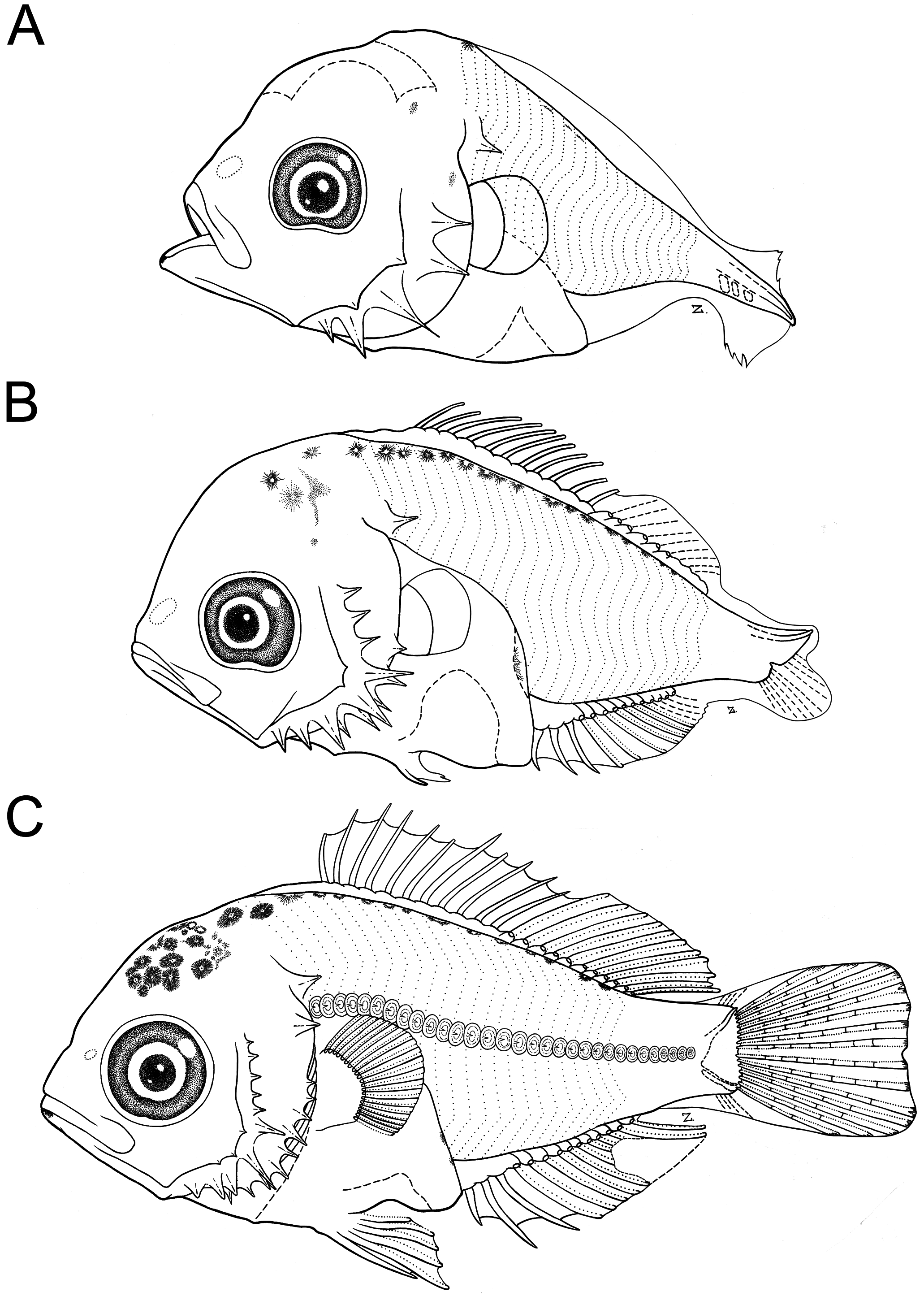
We describe three larvae that represent preflexion, flexion, and postflexion stages. The body of the larvae ( Fig. 1 View FIGURE 1 ) is deep at all stages (BD 48-54%), with a large head (HL 40-50%), and a long (PAL 60-69%) and a coiled gut.
Pigmentation
The preflexion larva has a few internal melanophores on the neurocranium and along the base of the dorsal fin. A conspicuous melanophore marks the symphysis of the lower jaw, and is present in all specimens. At flexion, there are external stellated melanophores in the dorsal area of the neurocranium, along the base of the dorsal fin, and an internal dendritic melanophore in the posterior margin of the gut, near the anus. In the postflexion stage, the number of melanophores in the dorsal margin of the neurocranium has increased, and a few long, stellated melanophores are present in the base of the anal fin.
Osteological development
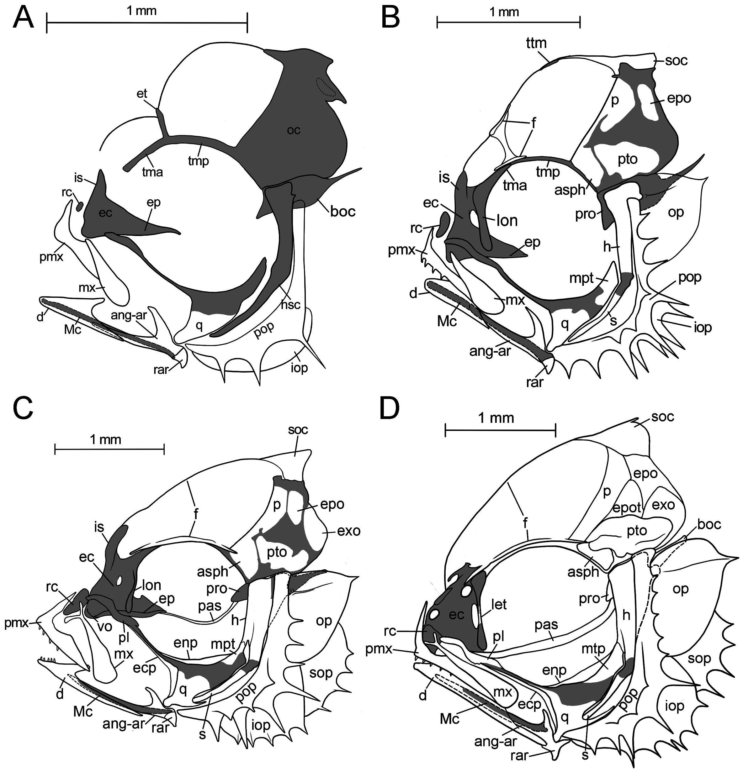
Neurocranium: The occipital region, the posterior portion of the neurocranium, is closed dorsally already in our smallest specimens ( Fig. 2A, B View FIGURE 2 ). It forms a small crest that is later covered by the dome-shaped supraoccipital ( Fig. 2C, D View FIGURE 2 ). The basioccipital has a caudad oriented projection ( Fig. 2A View FIGURE 2 ) that approaches the first vertebra in larger specimens ( Fig. 2D View FIGURE 2 ), and, since dissections are not possible, we are not able to interpret or relate this structure to anything else. The taenia marginalis (anterior and posterior) is connected via an epiphysal bridge to its counterpart ( Fig. 2A View FIGURE 2 ). Anteriorly, it fuses to the lamina orbitonasalis and is covered by the frontal ( Fig. 2B View FIGURE 2 ) laterally. The frontal appears as a small ossification lateral to the taenia marginalis, above the orbit ( Fig. 2B View FIGURE 2 ). It spreads medially to meet its counterpart in the midline in the larger stages ( Fig. 2B, D View FIGURE 2 ). The parietal starts as a small and narrow ossification posterior to the frontal and in the largest stage it covers the dorsal part of the neurocranium between the frontal anteriorly, the supraoccipital and epioccipital posteriorly, and the pterotic and autosphenotic laterally ( Fig. 2B, D View FIGURE 2 ). The prootic and the pterotic form the facet for the articulation with the dorsal head of hyomandibula ( Fig. 2D View FIGURE 2 ). The ethmoid region is already well developed with a large internasal septum in the smallest specimen ( Fig. 2A View FIGURE 2 ). The connection between the ethmoid plate and the otic capsule via the trabeculae is already absorbed ( Fig. 2A View FIGURE 2 ). A welldefined lamina orbitonasalis bears a small lateral ethmoid. The posterior edge of the ethmoid cartilage articulates with the pars autopalatina of the palatoquadrate ( Fig. 2B, C View FIGURE 2 ).
Jaws, suspensorium and dorsal hyoid arch: All elements of the upper jaw are already present in the smallest stage available to us ( Fig. 2A View FIGURE 2 ). The premaxilla has a well-developed ascending process that articulates in older stages with a large rostral cartilage ( Fig. 2B, D View FIGURE 2 ). Initially, the maxilla is a simple leaf-shaped element that develops its complex anterior end for the articulation with the premaxilla and the autopalatine in the early postflexion stages ( Fig. 2C, D View FIGURE 2 ). The quadrate is the first element to ossify in the palatoquadrate ( Fig. 2A View FIGURE 2 ), that develops the posteroventral process in a later stage ( Fig. 2C, D View FIGURE 2 ). Following the ossification of the quadrate in sequence is the metapterygoid ( Fig. 2B View FIGURE 2 ) and then the autopalatine ( Fig. 2C, D View FIGURE 2 ). The metapterygoid develops a lamina of membrane bone which contacts a membranous ossification of the hyomandibula ( Fig. 2C View FIGURE 2 ). The autopalatine articulates with the lateral ethmoid of the neurocranium early on, and the maxilla in later stages ( Fig. 2B, D View FIGURE 2 ). In the flexion stage larvae, the hyomandibula and the symplectic are present as separate ossifications of the hyosymplectic cartilage ( Fig. 2B View FIGURE 2 ). The hyomandibula develops a crest of membrane bone that attaches to the metapterygoid ( Fig. 2C View FIGURE 2 ). The dentary and the anguloarticular are already present in the smallest stage ( Fig. 2A View FIGURE 2 ). The dentary has the typical forked posterior end and encloses the anguloarticular ( Fig. 2 View FIGURE 2 A-D). Both elements are laterally attached to Meckel’s cartilage. In the smallest specimen the coronid process of the anguloarticular is already well developed ( Fig. 2A View FIGURE 2 ), and the most posterior end of Meckel’s cartilage has a well ossified retroarticular ( Fig. 2A View FIGURE 2 ).
Opercular series : We were only able to trace the preopercle and interopercle in the smallest specimen, the opercle might be present but could not be observed ( Fig. 2A View FIGURE 2 ). We suspect that the other elements are present as well but did not stain well. However, all elements are obvious in the flexion larva ( Fig. 2B View FIGURE 2 ), and bear prominent spines and serrations already ( Fig. 2A, D View FIGURE 2 ). The opercle has a single spine as a continuation of a ridge on its dorsal margin ( Fig. 2A, D View FIGURE 2 ). There were no signs of a second opercular spine, as reported for adults. The preopercle has three prominent spines in its anterior margin, that are about equal in size and ten spines along the posterior margin of which spines 3, 4 and 5 –counting from the ventral margin- are the largest. The interopercle has six spines and the subopercle four. The dorsal spines on the preopercle appear more as a serration in older stages ( Fig. 2D View FIGURE 2 ). The sub- and interopercle are partially covered by the opercle and the preopercle ( Fig. 2B, D View FIGURE 2 ).
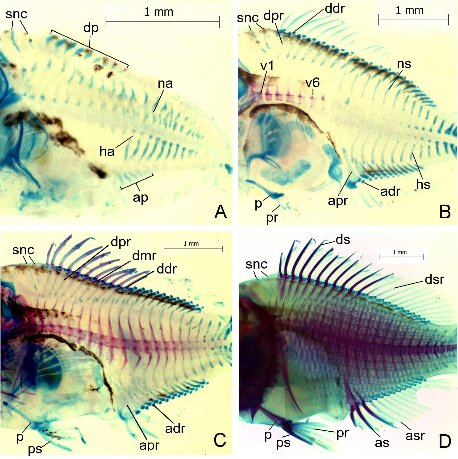
Vertebral column: All 22 haemal arches and their associated spines are present in the smallest specimen ( Fig. 3A, D View FIGURE 3 ). They are preformed in cartilage ( Fig. 3A View FIGURE 3 ) and ossify in an anteroposterior direction. In the flexion larva, the six anteriormost vertebrae are ossified ( Fig. 3B View FIGURE 3 ). The neural arches and their spines are preformed in cartilage, and the first fully formed neural arch appears on vertebra 11 ( Fig. 3B View FIGURE 3 ). The centra and their associated arches and spines are fully ossified in the largest specimen ( Fig. 3D View FIGURE 3 ).
Two cartilaginous supraneurals are present, and ossify perichondrally in the larger stages ( Fig. 3A, D View FIGURE 3 ). Both are obliquely oriented with the distal tip pointing dorsoanterad ( Fig. 3A, D View FIGURE 3 ). The proximal ends are associated with the second interneural space.
Dorsal and anal fins: The full complement of dorsal and anal fin pterygiophores, spines and rays are present in the largest specimen (D: XI + 11; A: III + 11; Fig. 3C View FIGURE 3 ). The proximal-middle radials as well as the distal radials appear to develop in a caudad direction ( Fig. 3A, D View FIGURE 3 ). The first pterygiophore in the dorsal fin is the largest and bears two spines, a serially associated and a smaller supernumerary spine anterior to it ( Fig. 3B, D View FIGURE 3 ). The cartilaginous precursor of the last pterygiophore has a small extension ( Fig. 3C, D View FIGURE 3 ). The first pterygiophore in the anal fin is the largest and bears two supernumerary spines and a serially associated spine ( Fig. 3C, D View FIGURE 3 ). All pterygiophores consist of a distal and a proximal-middle radial. We are uncertain whether middle radials are present in the pterygiophores that are associated with the soft rays (10 to 21). Similar to the last pterygiophore in the dorsal fin, the last pterygiophore in the anal fin has a cartilaginous extension ( Fig. 3C, D View FIGURE 3 ).
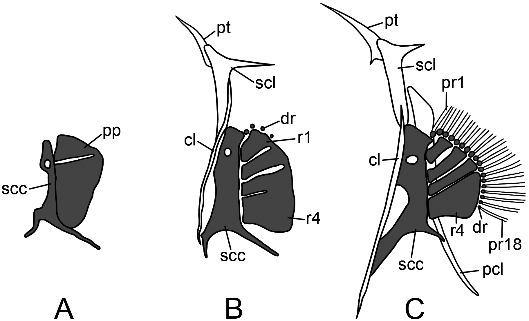
Pectoral fin and girdle: We were not able to trace any dermal elements in the smallest larva ( Fig. 4A View FIGURE 4 ). The endoskeletal scapulocoracoid and the pectoral plate are present ( Figs. 3A View FIGURE 3 , 4A View FIGURE 4 ). The scapulocoracoid has a foramen dorsally. The pectoral plate has an indentation dorsally indicating the separation of the most dorsal radial ( Fig. 3A View FIGURE 3 , 4A View FIGURE 4 ). The separation of the radials and the development of the distal radials follow a ventral direction ( Fig. 4B View FIGURE 4 ). The first distal radial is associated with the scapulocoracoid ( Fig. 4B, C View FIGURE 4 ). The posttemporal develops as a small pointed element ( Fig. 4B View FIGURE 4 ) that just develops its ventral arm in the early postflexion larva ( Fig. 4C View FIGURE 4 ). The supracleithrum has a posterior oriented spine at its dorsal part, and the cleithrum is a long needle-shaped ossification that runs in an anteroventral direction ( Fig. 4B, C View FIGURE 4 ). The postcleithrum is a large element that articulates with the supracleithrum and becomes needle-shaped dorsoventrally ( Fig. 4C View FIGURE 4 ). In the largest specimens the full complement of 18 fin rays is present ( Fig. 4C View FIGURE 4 ).
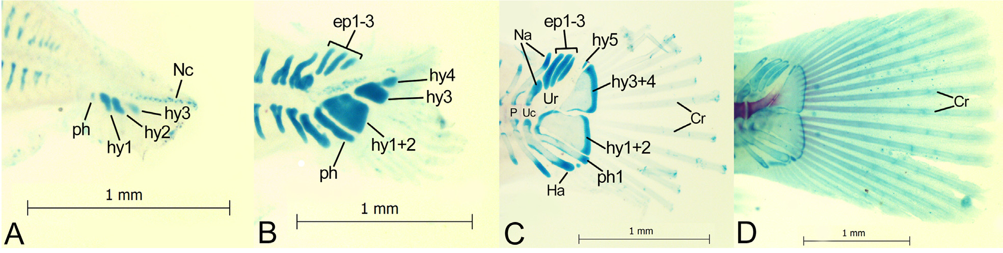
Caudal fin: The elements of the caudal skeleton consist of a parhypural, five hypurals, and three epurals ( Fig. 5 View FIGURE 5 ). In the smallest larva, only hypurals 1-3 are present as small, independent cartilaginous structures, as well as the parahypural ( Fig. 5A View FIGURE 5 ). In the flexion larva, hypural 4 is already visible ( Fig 5B View FIGURE 5 ). However, in older stages hypurals 1 and 2 and hypurals 3 and 4 fuse to a single plate, respectively ( Fig. 5C, D View FIGURE 5 ). The uroneurals are not present or did not pick up the alizarin stain in all our specimens. The first elements that are present in the smallest specimen are the parhypural and hypurals 1, 2 and 3. In the flexion-stage specimen, hypurals 1+2 and 3+4 are partially fused ( Fig. 5B View FIGURE 5 ). A fifth, smaller hypural appears in the postflexion larva ( Fig. 5C, D View FIGURE 5 ). The three epurals are present dorsally to the notochord in the flexion larva ( Fig. 5B View FIGURE 5 ). Preural centrum 2 and ural centrum have started to ossify in the younger postflexion stage ( Fig. 5C View FIGURE 5 ). The ural centrum has a small cartilaginous neural arch ( Fig. 5 View FIGURE 5 B-D). The fin rays have started to form during the flexion of the notochord ( Fig. 5B View FIGURE 5 ), and the full complement of 17 principal caudal fin rays are present (9+8; Fig. 5D View FIGURE 5 ) in the larger postflexion stage.
Pelvic fins: The pelvic element is formed by the flexion stage ( Fig. 3B View FIGURE 3 ) and shows signs of perichondral ossification. The anterior process is in contact with the ventral part of the cleithrum ( Fig. 3C, D View FIGURE 3 ). The posterior process is short (not shown in figures), and the strong spine is the first element of the fin to form ( Fig. 3B View FIGURE 3 ). The full complement of fin elements (I+5) is present in the larger postflexion stage ( Fig. 3D View FIGURE 3 ).
Squamation: In the two largest (5.31 and 6.10 mm SL) collected individuals, the first signs of squamation were observed. Scales are cycloid, each ornamented with two concentric lines and one central spine ( Fig. 6), forming rows along the midline of the body.
No known copyright restrictions apply. See Agosti, D., Egloff, W., 2009. Taxonomic information exchange and copyright: the Plazi approach. BMC Research Notes 2009, 2:53 for further explanation.