
Sinhomidia bicolor ( Yosii, 1965 ) Zhang, Deharveng, Greenslade & Chen, 2009
|
publication ID |
https://doi.org/ 10.11646/zootaxa.4358.3.10 |
|
publication LSID |
lsid:zoobank.org:pub:F55E459E-F215-4C88-BBC9-A78CB8039EA1 |
|
DOI |
https://doi.org/10.5281/zenodo.6029365 |
|
persistent identifier |
https://treatment.plazi.org/id/03B61377-FFAB-FF94-EDAE-FEDEFD23B588 |
|
treatment provided by |
Plazi |
|
scientific name |
Sinhomidia bicolor ( Yosii, 1965 ) Zhang, Deharveng, Greenslade & Chen, 2009 |
| status |
|
Sinhomidia bicolor ( Yosii, 1965) Zhang, Deharveng, Greenslade & Chen, 2009 View in CoL
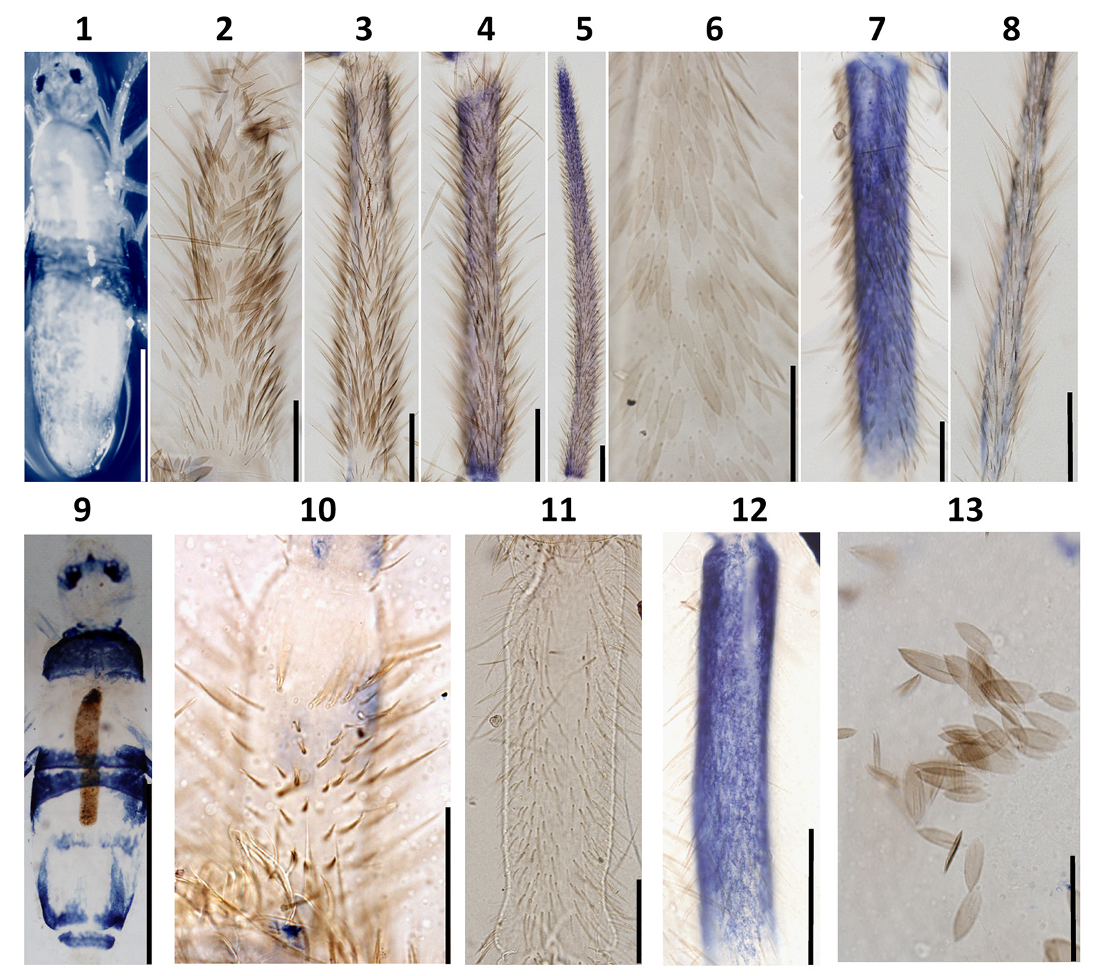
Figs 1–8 View FIGURES 1–13 , Figs 14–18 View FIGURES 14–18 , Table 1
Locality. CHINA, Baiyun Mount, Guangdong Province, 171m asl, coordinates 23°10'N, 113°17'E. On litter, leg. Jia, Yan & Jin, September 20, 2016. One male: collection code number CGZ161010504 J. Two females: collection code number CGZ161005507J, CGZ161010503 J. Deposition School of Life Sciences, Liaocheng University. GoogleMaps
Description of male. Body length up to 3.3 mm. Ground colour pale yellow in alcohol. Eye patches dark purple. Ant I and II lightly pigmented, Ant III and IV deeply pigmented ( Figs 4, 5 View FIGURES 1–13 ). Deep purple pigment present dorso-laterally on mesothorax, whole of Abd II and III; Abd IV with scattered pigment on anterior and posteriorlaterally and on posterior side of manubrium ( Fig. 1 View FIGURES 1–13 ). Hind femur deep purple ( Fig. 7 View FIGURES 1–13 ). Differences in the distribution of colour of females from the same locality to those in Zhang et al. (2009).
Head. Ant five times as long as cephalic diagonal. Ant I–II with scales ( Figs 2, 3 View FIGURES 1–13 ).
Thorax. Trochanteral organ with about 38–45 smooth spiny chaetae ( Fig. 14 View FIGURES 14–18 ). Coxal macrochaetal formula: 3, 3/4+2, 2+2+2 ( Fig. 15 View FIGURES 14–18 ). Leg covered with scales to femur ( Fig. 7 View FIGURES 1–13 ), tibiotarsi without scales ( Fig. 8 View FIGURES 1–13 ).
Abdomen. Dorsal face of manubrium covered with scales ( Fig. 6 View FIGURES 1–13 ). Basal two thirds of dens with 40–43 spines arranged in one or two rows along inner edge, scales absent. Dental basal chaetae bs1 and bs2 multilaterally ciliate, bs1 much thicker and longer than bs2. A pair of testes on both sides of thorax and abdomen, with medial unpaired duct connecting to genital tract and extending anteriorly to Abd I. Genital tract gradually narrowing from anterior to posterior, following a 360° twist to connect with genital plate ( Fig. 17 View FIGURES 14–18 ). Genital plate with 15 circinate chaetae around the genital pore.
Chaetotaxy. Dorsal chaetotaxy of head with the absence of An2e compared to females ( Fig. 18 View FIGURES 14–18 ). Dorsal Th III with about 20 mac. Lateral sensilla probably present in its normal location but not all seen S formula:?2/122, mS formula:?0/101
Body scales. Scales narrow, pointed and fusiform with coarse striations, present on Ant. I–II, head, thorax, basal segments of legs but not on tibiotarsus, abdomen and on the posterior side of the manubrium.
Remarks. Males were not included in previous descriptions of this species so are described here. Our male specimen agrees with the description in female specimens except An chaetal number of head and number of Th III chaetae. The colour differs from the female as there is no colour on Abd IV, only scattered pigment. The females in our sample are similar to that described in Zhang et al. (2009). Only one male has been found, which has some sexual dimorphism compared to females, so could be only an individual variation. This shortened description above emphasises the distinction of male from female.

No known copyright restrictions apply. See Agosti, D., Egloff, W., 2009. Taxonomic information exchange and copyright: the Plazi approach. BMC Research Notes 2009, 2:53 for further explanation.
|
Kingdom |
|
|
Phylum |
|
|
Class |
|
|
Order |
|
|
Family |
|
|
Genus |