
Ferrequitherium sweeti, Scott, 2019
|
publication ID |
https://doi.org/ 10.1093/zoolinnean/zly040 |
|
publication LSID |
lsid:zoobank.org:pub:D50F5678-824D-477B-9DC9-F39DD40002B9 |
|
DOI |
https://doi.org/10.5281/zenodo.5730329 |
|
persistent identifier |
https://treatment.plazi.org/id/03B6879E-FFB1-D907-FC2F-AD4FDD0CF886 |
|
treatment provided by |
Carolina |
|
scientific name |
Ferrequitherium sweeti |
| status |
gen. et sp. nov. |
FERREQUITHERIUM SWEETI SP. NOV.
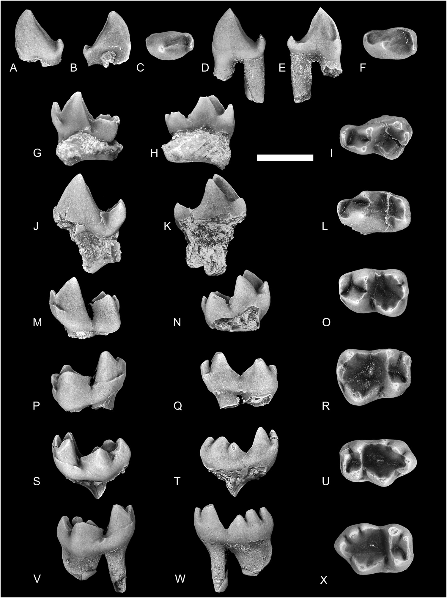
( FIGS 2–7 View Figure 2 View Figure 3 View Figure 4 View Figure 5 View Figure 6 View Figure 7 ; TABLES 1–2; SUPPORTING INFORMATION, APPENDIX S2)
Z o o b a n k L S I D. — u r n: l s i d: z o o b a n k. org:act: 9CEF0352-D12F-46DD-A325-1231C3EFA4C7
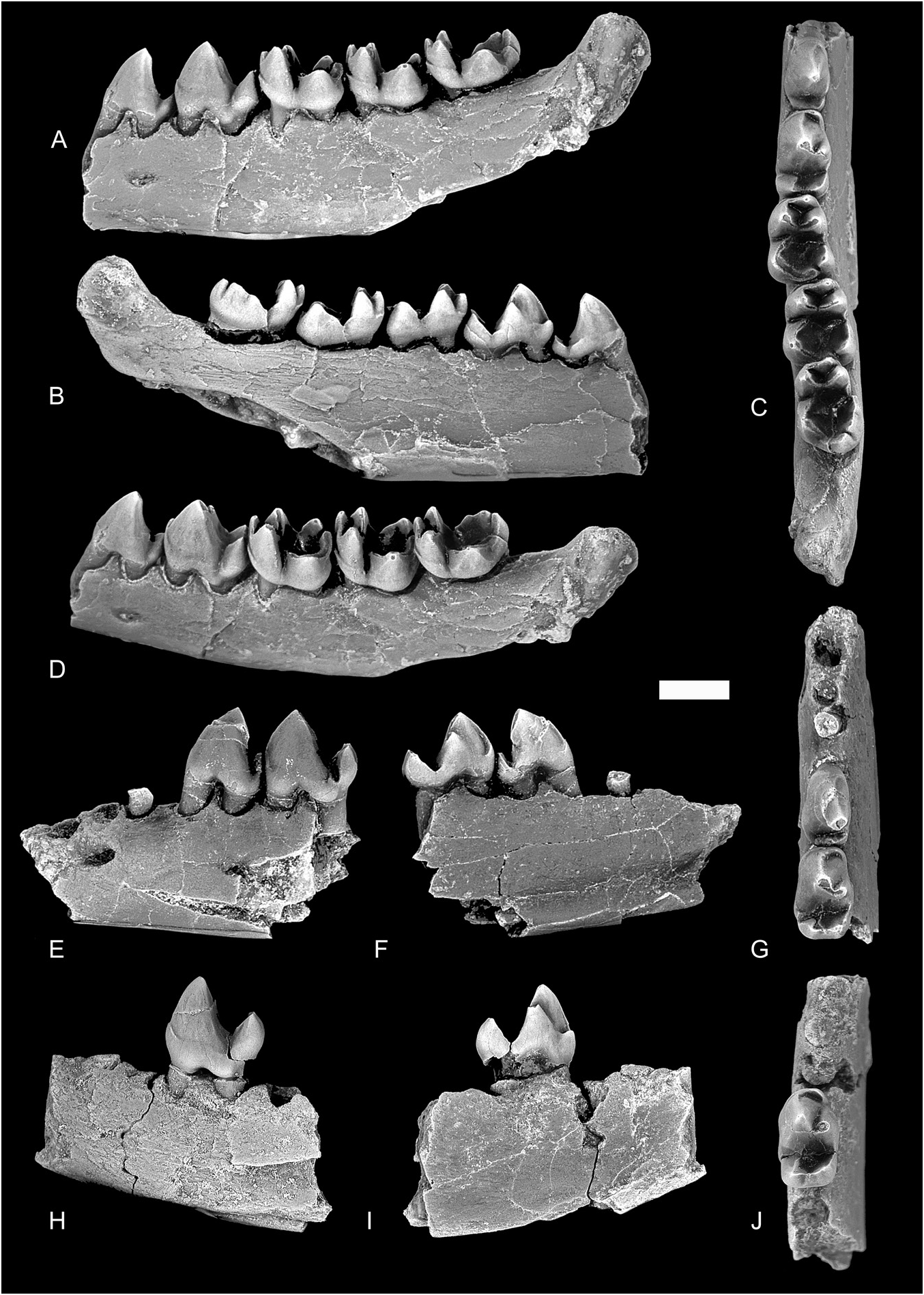
Holotype: TMP 2013.048 View Materials .0075, incomplete left dentary with p3–4, m1–3 ( Fig. 3A–D View Figure 3 ).
Typelocalityandhorizon: Trainspottinglocality,Paskapoo Formation of south-west Alberta, Late Palaeocene (earliest Tiffanian, Ti1, Plesiadapis praecursor / P. anceps Lineage Zone of Lofgren et al., 2004).
Hypodigm: See Supporting Information, Appendix S2.
Etymology: Named in honour of the late Dr Arthur R. Sweet (Geological Survey of Canada) for his many contributions to Late Cretaceous and Palaeogene palynology.
Occurrence: Earliest Tiffanian (Ti1, Late Palaeocene) of south-western Alberta, Canada.
Diagnosis: Differs from Horolodectes in having lower premolars with more nearly vertical and less trenchant protoconid and better-developed lower premolar talonid, in p4 having metaconid and basined talonid with more distinct entoconid and hypoconid. Differs further from Horolodectes in P4 having T-shaped occlusal outline, with weaker ectocingulum and postparacrista, and in P4 and lacking neomorphic crest extending labially from paracone apex. Differs further from Horolodectes in upper molars having less swollen conules, weaker notches in the protocristae at their junction with the conules, weaker notch in the ectocingulum labial to the deepest part of the centrocrista, and lacking neomorphic crest extending labially from paracone apex.
Description
Specimens of F.sweeti include isolated teeth and incomplete jaws with teeth. Associating the upper and lower teeth was justified primarily on the close resemblance to homologous teeth in H. sunae (see below), a strong occlusal fit between opposing upper and lower molars, and frequency of occurrence (for example, F. sweeti is the best represented eutherian in the Trainspotting mammalian fauna).
Maxilla and upper dentition
The maxilla is poorly preserved on TMP 2017.025.0317, and preserves little in the way of informative features. Although parts of the facial process remain, there is no evidence for the position of the infraorbital foramen, or for the position of the maxillary process of the zygomatic arch. The upper incisors, canine, and pre-ultimate premolars of F. sweeti are unknown.
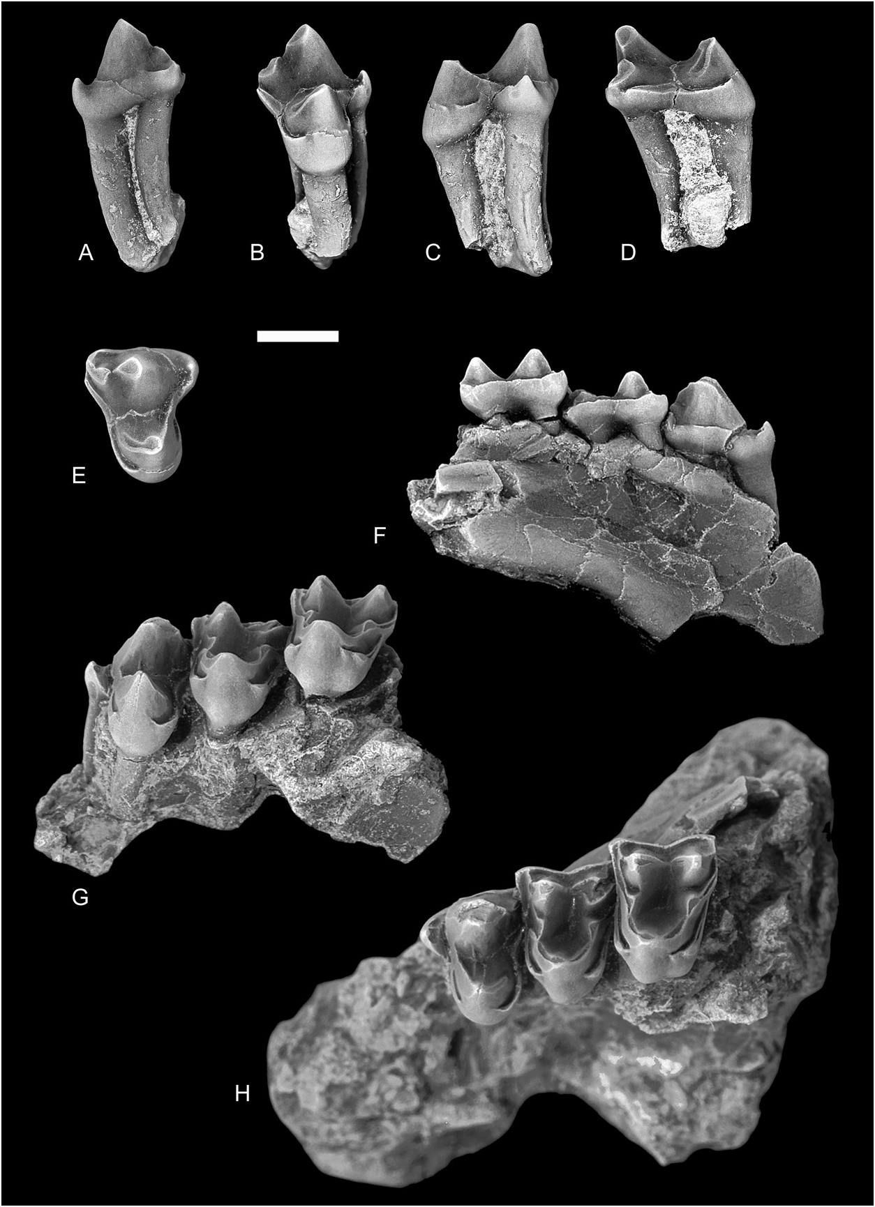
P4 ( Fig. 2 View Figure 2 ): The P4 of F. sweeti is known from 11 specimens, one of which (TMP 2017.025.0317) is an incomplete maxilla containing P4 and the first two molars ( Fig. 2F–H View Figure 2 ). The crown is T-shaped in occlusal outline, and is dominated by an inflated paracone and smaller protocone. The paracone is subcircular in cross-section and leans somewhat posteriorly, with long, gently sloping anterior and shorter, more nearly vertical posterior surfaces. The stylar shelf is undeveloped, and the ectocingulum is complete labially. A faint preparacrista extends along the anterior surface of the paracone to the parastylar corner, where it unites with the ectocingulum and precingulum. The parastylar region is drawn out in a distinct lobe, and supports a low but prominent parastyle. The postparacrista runs posteriorly from the apex of the paracone, uniting with a short, low premetacrista and forming a sharp notch. The metacone is small but distinct, and is developed on the posterior shoulder of the paracone approximately halfway between the apex of the paracone and the metastylar corner of the crown. The postmetacrista bends sharply labially as it nears the posterior margin of the crown where it unites with the ectocingulum, but not the postcingulum, which terminates just lingual to the metastylar corner. A weak swelling in the region of the metastyle occurs on several specimens (e.g. TMP 2015.069.0169). The protocone is both smaller and lower than the paracone, oriented more nearly vertically, and is positioned slightly anterior to the midwidth of the crown. The preprotocrista extends anterolabially towards the base of the paracone, uniting with the prominent precingulum; the stout postprotocrista curves posteriorly and slightly labially from the protocone apex, but fades away before reaching the postcingulum. A third crest, weaker than either of the protocone cristae, extends directly labially from the protocone apex towards the base of the paracone. Neither paraconule nor metaconule is developed.
M1 ( Figs 2F–H View Figure 2 , 3A–E View Figure 3 ): The crown of M1 is subtriangular in occlusal outline, with the labial margin slightly longer than the lingual margin. The low, robust ectocingulum extends along the labial margin of the crown, and the stylar shelf is narrow. A shallow ectoflexus divides the labial margin of the crown into weak para- and metastylar lobes, with the metastylar lobe projecting further labially. The preparacrista is stout and extends anteriorly from the apex of the paracone, joining the preparaconule crista+paracingulum at the parastylar corner. The centrocrista is straight, with no deflection of either the postparacrista or premetacrista towards the ectocingulum. The low postmetacrista curves sharply labially from the apex of the metacone to the metastylar corner of the crown, where it joins the ectocingulum. The paracone is slightly taller than the metacone, and the two cusps are subequal in size; both cusps are weakly convex labially and strongly swollen lingually. The paraconule and metaconule are well developed, somewhat swollen, and positioned near the base of the paracone and metacone, respectively. The short preparaconule crista extends anteriorly and slightly labially before continuing as the paracingulum towards the parastylar corner of the crown. The postmetaconule crista curves posterolabially and continues as the metacingulum before ending abruptly near the base of the metacone, rather than joining the postmetacrista. The postparaconule crista meets a short, lingually directed crest arising from low on the lingual side of the paracone; a weak notch is formed at their union. The premetaconule crista originates very low on the metaconule, and extends labially a short distance before fading away. A massive protocone dominates the lingual side of the crown, its apex leaning slightly anteriorly; the cusp is positioned slightly anterior to the midwidth of the crown. The protocone cristae are well developed, and form slit-like notches at their junction with the paraconule and metaconule. The precingulum and postcingulum are both robust, and although both cingula extend labially, neither joins the conule cristae and instead fade out near the base of the paraconule and metaconule, respectively. The postcingulum is somewhat swollen in the area of where a hypocone might be expected, but no distinct cusp is developed.
M2 ( Figs 2F–H View Figure 2 , 3F–J View Figure 3 ): The M1 and M2 of F. sweeti are closely similar in size and morphology. The crown of M2 is wider than that of M1, and the occlusal outline is more similar to that of an isosceles, rather than equilateral triangle. The crown of M2 differs most notably from that of M 1 in having a more prominent and labially extending parastylar lobe, and a slightly deeper ectoflexus that divides the labial margin into more distinct para- and metastylar lobes. The M2 differs further from M 1 in having a proportionally larger paracone relative to metacone, a slightly shorter postmetacrista, and a lingual slope of the protocone that is both longer and shallower than that on M1.
M3 ( Fig. 3K–T View Figure 3 ): The M3 of F. sweeti is smaller than either M1 or M2, subtriangular in occlusal outline, and lacks any development of a metastylar lobe. As with M1 and M2, the stylar shelf on M3 is undeveloped, and the ectoflexus is very shallow, faintly dividing the labial margin into anterolabially projecting parastylar and smoothly rounded metastylar areas. The ectocingulum is robust anteriorly, but diminishes posteriorly, fading away as it approaches the metacone. The preparacrista is stout and extends anteriorly and labially, uniting with the paracingulum and ectocingulum. The paracone is large, subconical, and leans slightly anteriorly. The metacone is about half the size and height of the paracone, and is joined to the latter by a broadly notched centrocrista. Both conules are strongly developed and appressed to the bases of the paracone and metacone, and although the paraconule cristae are both well developed, only the postmetaconule crista is present at the metaconule. The protocone is about the same height as the paracone, but is much more massive, constituting nearly one-third the width of the crown. As on M1 and M2, the protocone cristae on M3 form slit-like notches at their union with the conules. The protocone cingula are robust but incomplete lingually. A hypocone is not developed.
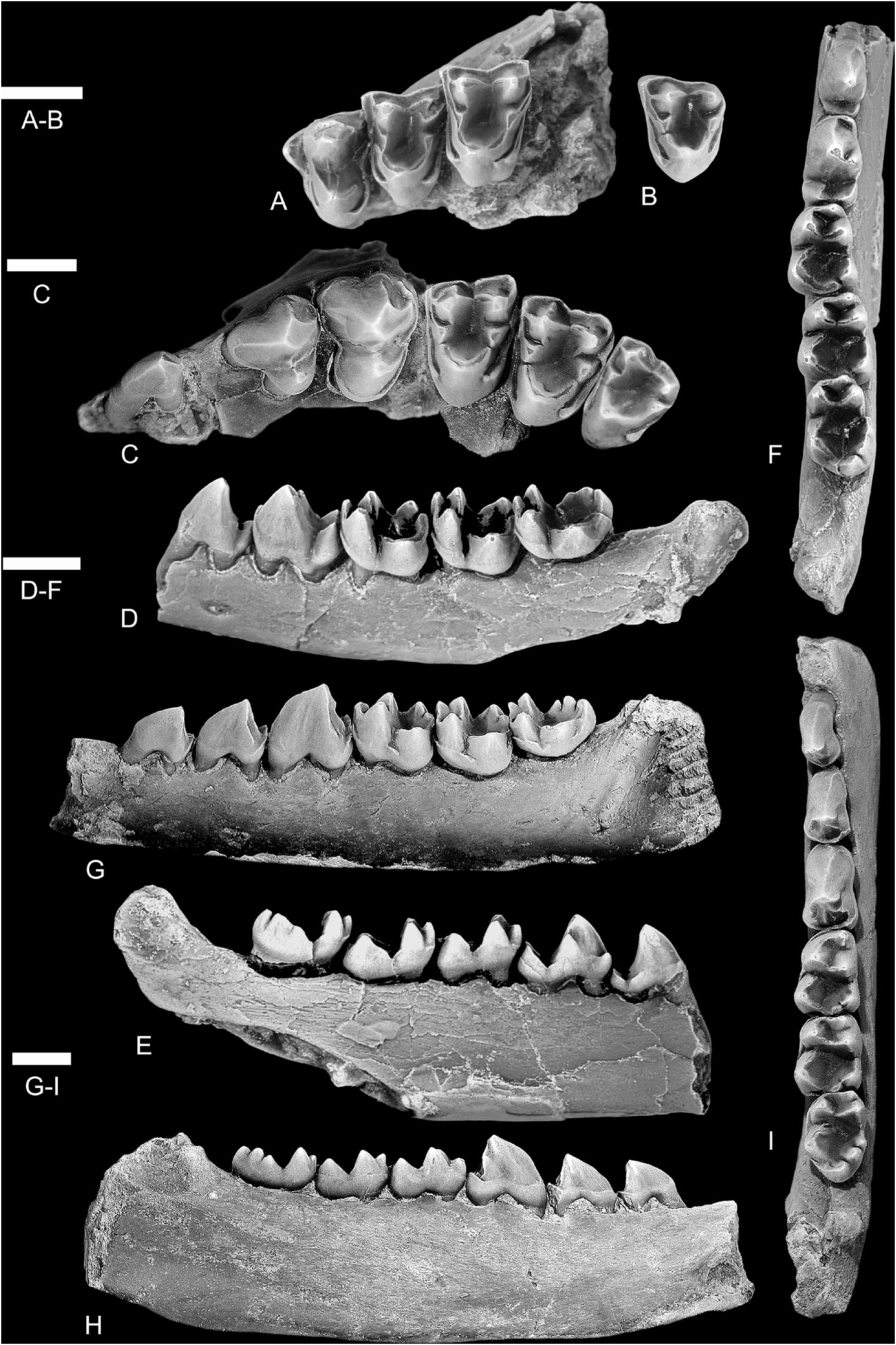
Dentary and lower dentition
The lower dentition of F. sweeti is known from incomplete jaws and isolated teeth that together document p2–4, m1–3. Nothing is known of the lower incisors or canine, and little can be said of the dentary other than the size and positions of the two mental foramina. The more anterior foramen opens ventral to the anterior root of p2, and its large, subcircular aperture faces anteriorly and laterally ( Fig. 4E View Figure 4 ). The more posterior foramen opens ventral to the posterior root of p3 and faces laterally ( Fig. 4A View Figure 4 ).
p1: The p1 remains undiscovered in F. sweeti , although its presence is confirmed by its alveolus in TMP 2015.069.0039 ( Fig. 4E–G View Figure 4 ). The alveolus for p1 is subovate, being somewhat bilaterally compressed, and is oriented posteromedially-anterolaterally, suggesting the tooth may have projected anteriorly and laterally. A short diastema separates the p1 alveolus from p2.
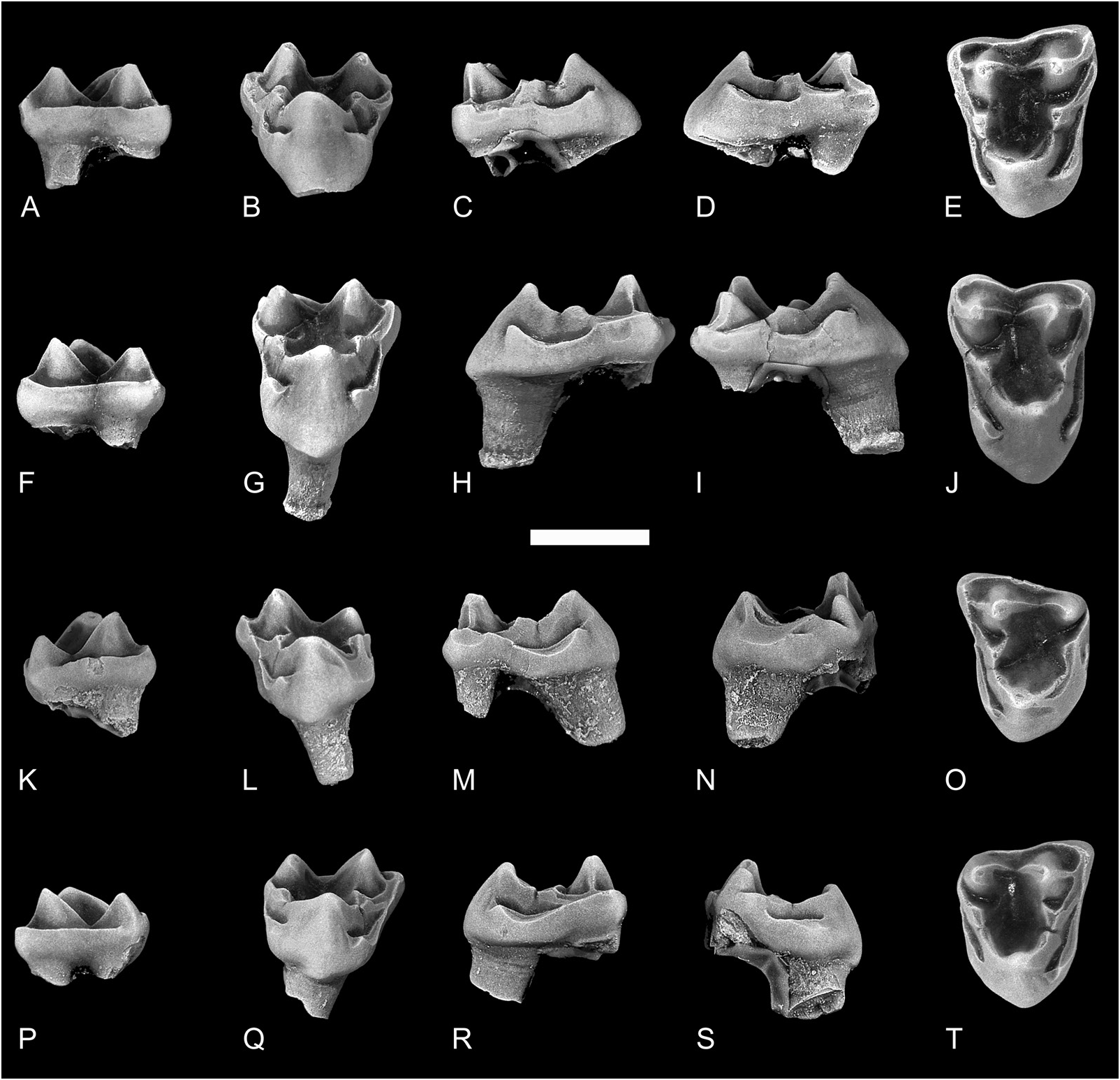
p2: p2 is known from a tentatively referred specimen, TMP 2015.069.1134 ( Fig. 6A–D View Figure 6 ), as well as from its two alveoli in TMP 2015.069.0039. The crown of p2 is dominated by a tall and slightly recurved protoconid anteriorly, and a low transversely oriented crest posteriorly. The protoconid is mildly inflated towards its base labially, but is only weakly convex lingually; the postvallid surface is virtually flat. A weak paracristid originating at the protoconid apex extends anterolingually towards the base of the crown, but neither paraconid nor metaconid is developed. The talonid consists of a low, transversely oriented crest: the crest is approximately half the width of the protoconid, slightly higher labially than lingually, and curves anteriorly at the lingual margin of the crown, enclosing a shallow basin. Weak exodaenodonty is developed at the posterior root. A diastema, nearly twice the length of the more anterior diastema, separates p2 and p3.
p3: The p3 of Ferrequitherium is known from three specimens. The crown is larger and taller than that of p2, and the protoconid is more inflated ( Figs 4 View Figure 4 , 6D–F View Figure 6 ). As with p2, the crown of p3 consists of a large, recurved protoconid and a low transverse crest. A weak paracristid arises from the protoconid apex and curves anterolingually, becoming more pronounced as it descends the protoconid; a tiny paraconid can be developed at the terminus of the paracristid. A metaconid is not developed. One or two poorly defined crests extend posteroventrally from the apex of the protoconid, but fade away before reaching the talonid. The talonid is low, approximately one-third the height of the protoconid, and consists primarily of a transverse crest; as with p2, the talonid crest on p3 is higher labially, and extends to the lingual margin of the crown where it turns abruptly anteriorly, enclosing a shallow basin. The crest becomes slightly thicker at the posterolingual margin of the crown, in the position of an entoconid, but distinct talonid cusps are otherwise not developed. Weak exodaenodonty can be developed at the posterior root.
dp4 ( Fig. 6G–I View Figure 6 ): Three dp4s are provisionally referred to Ferrequitherium . The crown of dp4 is molariform, with a tricuspid trigonid and wide, deeply basined talonid. The protoconid is the largest and tallest trigonid cusp, followed by a slightly smaller and lower metaconid, and a still lower but prominent paraconid. The protoconid and metaconid are inflated, subconical, and are connected by a deeply notched protocristid. A sharp paracristid extends anteriorly from the protoconid, forming a deep notch at its union with the paraconid. The paraconid is anteroposteriorly compressed and offset anteriorly from the protoconid and metaconid, with its apex aligned with
the deepest part of the protocristid notch. A short but prominent crest extends lingually from the paraconid apex, but fades away, leaving the trigonid open lingually. The talonid is significantly wider than the trigonid, and consists of a massive hypoconid and smaller, subequal
entoconid and hypoconulid. The cristid obliqua contacts the postvallid wall low and labial to the protocristid notch, and the hypoconid is connected to the hypoconulid by a well-developed hypocristid; both cristid obliqua and hypocristid are heavily worn. The hypoconulid is slightly closer to the entoconid than to the hypoconid, but a shallow notch separates the two cusps, and the postcristid is undeveloped. A weak precingulid occurs below the paracristid notch, but the postcingulid is undeveloped.
p4 ( Figs 4 View Figure 4 , 6J–L View Figure 6 ): The p4 of Ferrequitherium is known from ten specimens. The crown is slightly taller than that of p3, but is similarly subrectangular in occlusal outline, with a weak constriction developed at the hypoflexid. The trigonid consists primarily of a massive, inflated and recurved protoconid, and a smaller metaconid. A stout paracristid extends anteriorly from the protoconid apex, becoming more prominent and blade-like as it descends along the anterior surface of the protoconid. As the crest nears the base of the protoconid, it can curve gently lingually before fading away, or it can turn sharply lingually, forming a low platform; in either case, a paraconid is not developed. A weak precingulid is sometimes developed, but does not extend far onto the labial side of the protoconid. The metaconid is considerably smaller and shorter than the protoconid, varying from about one-third to one-half the size of the latter, and occurs directly lingual or slightly posterolingual to the apex of the protoconid. The metaconid and protoconid are connected by a weak but sharply notched protocristid. The talonid is about half the height of the trigonid, and consists primarily of a small hypoconid and larger, taller, but more poorly differentiated entoconid. The hypoconid occurs at the posterolabial corner of the talonid and is connected to the postvallid wall by an anterolingually directed cristid obliqua; the cristid obliqua can meet a short crest low on the postvallid wall near the midline. The entoconid is nearly twice the size of the hypoconid, with inflated lingual and posterior walls, but with a more poorly differentiated apex; it is connected to the hypoconid by a high, transverse crest. On one specimen, TMP 2014.047.0172, a small, weakly differentiated hypoconulid is present. A low entocristid connects the entoconid to the postvallid wall. Together, the talonid cusps and crests enclose a shallow basin that is skewed slightly labially. Exodaenodonty is developed at both roots, but is especially prominent posteriorly.
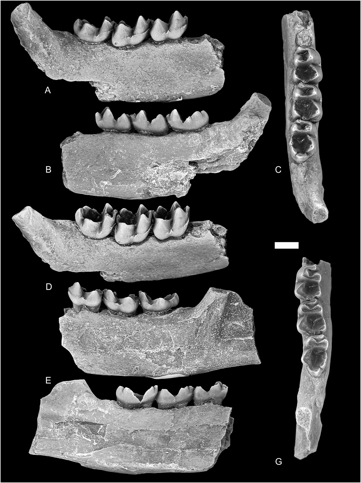
m1 ( Figs 4 View Figure 4 , 5 View Figure 5 , 6M–O View Figure 6 ): The crown of m1 is weakly hourglass-shaped in occlusal outline, with the talonid bulging labially and lingually past the level of the trigonid. The trigonid consists of an equidistant protoconid, paraconid and metaconid, with the three cusps forming a near-equilateral triangle in outline. The principal cusps are subconical and sharply pointed, have inflated interior walls and appear swollen towards their bases. The metaconid is slightly taller than the protoconid, although the cusps are subequal in size, and with the metaconid occurring slightly posterior to the level of the protoconid; the two cusps are connected by a deeply notched protocristid, of which the protoconid arm is shorter than the metaconid arm. The paraconid is about half the size and height of the protoconid and is positioned slightly labial of the lingual margin of the crown. The paracristid extends anteriorly from the protoconid apex, and then turns sharply lingually, continuing to the paraconid. The talonid is approximately two-thirds to three-quarters the height of the trigonid, and the slightly concave valley-facing sides of the hypoconid and entoconid form a deep, weakly V-shaped floor that becomes increasingly concave with wear. The hypoconid is the largest talonid cusp, occupying nearly half the width of the talonid; the cusp is connected to the postvallid wall by a cristid obliqua that contacts the trigonid low and labial to the level of the protocristid notch. The entoconid is slightly taller but smaller than the hypoconid, and connects to the trigonid by a prominent, moderately notched entocristid. Faint swellings in the position of a mesoconid and entoconulid can be present on the cristid obliqua and entocristid, respectively, but distinct cuspids are not developed. The hypoconulid is lower than the entoconid, is anteroposteriorly compressed with a flat basinfacing wall and is positioned close to the entoconid; on one specimen, TMP 2014.047.0179, a small cuspid is developed off of the labial side of the hypoconulid. The postcristid is short, steep and incised by a narrow notch, separating the entoconid and hypoconulid; the hypocristid curves posterolingually from the hypoconid, joining that cusp with the hypoconulid. A short but prominent precingulid is present, but does not continue labially as an ectocingulid; the postcingulid is weak or undeveloped.
m2 ( Figs 4 View Figure 4 , 5 View Figure 5 , 6P–R View Figure 6 ): The crown of m2 resembles that of m1, but is larger overall, with a more anteroposteriorly compressed trigonid and considerably wider talonid. The trigonid cusps are less robust than their counterparts on m1, but are nonetheless inflated, particularly the internal walls, and the postvallid wall is oriented transversely, rather than oblique to the anteroposterior axis of the crown. The metaconid is the largest trigonid cusp, and is connected to the shorter protoconid by a deeply notched protocristid; as on m1, the protocristid is asymmetric, with the metaconid arm being longer than the protoconid arm. A stout paracristid extends a short distance anteriorly from the apex of the protoconid, and then bends sharply lingually, continuing to the subconical paraconid, which is both taller and closer to the metaconid than on m1. The paracristid is both longer and more transverse than on m1, and the paraconid is more nearly vertical in its orientation and more fully incorporated into the paracristid. The talonid resembles that on m1, but is longer and wider, and the entoconid is taller. The hypoconid is the largest talonid cusp, followed by a smaller but equally tall entoconid and still smaller and lower hypoconulid; the hypoconulid is somewhat appressed to the entoconid, but is separated from the latter by a slit-like notch in the postcristid. On some specimens (e.g. TMP 2015.069.0943, not figured) a small cusp is developed off of the labial shoulder of the hypoconulid, effectively ‘doubling’ the hypoconulid. The cristid obliqua contacts the postvallid wall labial to the level of the protocristid notch, and the sharply notched entocristid continues dorsally along the postvallid wall as a metastylid crest. As on m1, a poorly defined mesoconid and entoconulid can be present on the cristid obliqua and entocristid, respectively. The precingulid is more prominent and labially extending than on m1, but an ectocingulid remains undeveloped.
m3 ( Figs 4 View Figure 4 , 5 View Figure 5 , 6S–X View Figure 6 ): The m3 of F. sweeti is longer than either m1 or m2, but is narrower and slightly lower crowned. The trigonid is anteroposteriorly compressed, even more so than on m2, and the paraconid is positioned closer to the metaconid. The trigonid cusps are less robust than those on m2, but their internal walls are likewise inflated, the metaconid larger and taller than the protoconid, and the protocristid is similarly notched and asymmetric in its proportions. The paracristid extends a short distance anteriorly from the protoconid apex before turning sharply lingually; the paraconid arm of the paracristid is longer and more transversely oriented than on m1 or m2, and the paraconid is more fully incorporated into the paracristid. The talonid is longer and slightly narrower than on m2, but is nonetheless wider than the trigonid, and the principal cusps are well developed and together enclose a weakly V-shaped floor that becomes more evenly concave with wear. The talonid is dominated by a massive hypoconid that occupies greater than half of its width; a robust cristid obliqua connects the hypoconid to the postvallid wall, and a weak swelling marks the position of an incipient mesoconid. The entoconid, slightly smaller than the hypoconid, is positioned posterolingually and close to the hypoconulid; both cusps are swollen and are separated by a notched postcristid. A prominent entoconulid is developed on the entocristid immediately anterior and lingual to the entoconid; the cuspid can be slightly smaller than either the entoconid or hypoconulid, or as large, but has similarly swollen walls, and is separated from the former by a shallow notch. The entoconulid, together with the entoconid and hypoconulid, defines the posterolingual margin of the talonid basin. The precingulid is conspicuous, and can continue along the labial surface of the protoconid as an ectocingulid to the hypoflexid. The postcingulid is weak or undeveloped.
No known copyright restrictions apply. See Agosti, D., Egloff, W., 2009. Taxonomic information exchange and copyright: the Plazi approach. BMC Research Notes 2009, 2:53 for further explanation.
|
Kingdom |
|
|
Phylum |
|
|
Class |
|
|
Order |
|
|
Family |
|
|
Genus |
Ferrequitherium sweeti
| Scott, Craig S. 2019 |
Plesiadapis praecursor
| Gingerich 1975 |