
Rhizomyces ramosus W.Rossi & Feijen, 2018
|
publication ID |
https://doi.org/ 10.5852/ejt.2018.474 |
|
DOI |
https://doi.org/10.5281/zenodo.3845935 |
|
persistent identifier |
https://treatment.plazi.org/id/03B687CF-5122-FF87-EDD6-F93F1D5F7C23 |
|
treatment provided by |
Valdenar |
|
scientific name |
Rhizomyces ramosus W.Rossi & Feijen |
| status |
sp. nov. |
Rhizomyces ramosus W.Rossi & Feijen sp. nov.
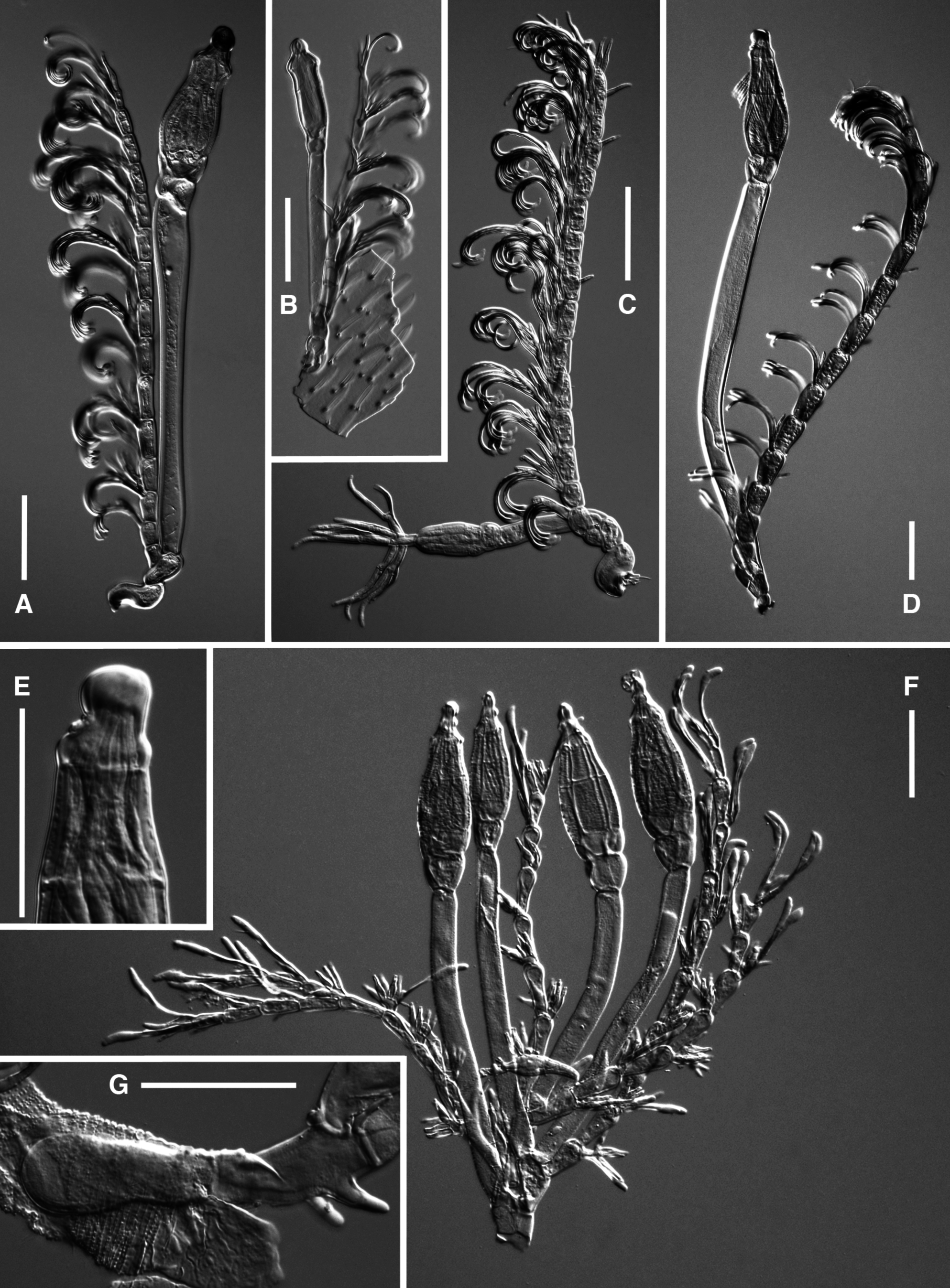
MycoBank No: MB827886 Fig. 1 View Fig F–G
Description
Basal cell hyaline, usually subcylindrical, distinctly longer than broad, producing more than one suprabasal cell in sequence. The basal cell extends under the integument of the host in a relatively small, ovoid or pear-shaped haustorium. Suprabasal cells irregularly trapezoidal, about as long as the basal, bearing distally the stalk cell of the perithecium paired either with the appendage or with another cell similar in shape and size, from which arise a second perithecium and the appendage.Appendages slender and flexuous, reaching the tip of the perithecium when erect, the main axis consisting of 8–11 clubshaped cells separated by conspicuous constrictions; the appendages can be either simple or bifurcate from the base, sometimes also bearing 1–3 short lateral ramifications made of 1–4 cells. Each cell of the axis bears close to the base of the following a relatively large cell irregularly shaped, which gives rise to antheridia and sterile branchlets. The antheridia are more numerous in the lower portion of the appendage (up to 5 per cell) and are bottle-shaped with darker necks. The sterile branchlets are gradually longer and more numerous in the upper portion (up to 3 and exceptionally 4 per cell): are slender and chestnut brown, with a hyaline distal portion variably enlarged and variably curved. Perithecial stalkcell (cell VI) very long and slender, of nearly constant diameter, pale amber yellow, with a roughened surface, abruptly distinguished distally from the relatively large and externally prominent basal cell region. Perithecium brownish yellow with the surface roughened by transverse ridges formed by wartlike elevations, broadly pear-shaped, its margins concave distally below a crown of small protuberances which subtend the paler and abruptly tapering tip; the apex is nipple-shaped, subtended on one side by two hemispherical prominences. Up to seven perithecia (six mature and one immature) were seen in a single thallus. Length from the external tegument of the host insect to perithecial apex up to 415 µm; longest appendages 365 µm; perithecia 85–102 × 30–40 µm; haustorium 20–62 × 15–25 µm.
Etymology
From the Latin ‘ ramosus ’, branched.
Types
UGANDA: Bushenyi distr., Kazinga Channel SE of Katunguru , 0.1292° S, 30.0525° E, 925 m a.s.l., on Diopsina nitida ( Adams, 1903) , 26 Mar. 2012, M. von Tschirnhaus leg. (holo-: FI 4109a; iso-: FI 4109b); same data as preceding except different host specimen (para-: FI 4093).
GoogleMapsRemarks
Rhizomyces ramosus sp. nov. can be distinguished at first glance from all the other species in the same genus by its ramified thallus, but also the shape and the roughened surface of the perithecia are very characteristic.
The presence of this fungus was already reported on Diopsina nitida by Feijen & Feijen (2013).
No known copyright restrictions apply. See Agosti, D., Egloff, W., 2009. Taxonomic information exchange and copyright: the Plazi approach. BMC Research Notes 2009, 2:53 for further explanation.
|
Kingdom |
|
|
Phylum |
|
|
Class |
|
|
Order |
|
|
Family |
|
|
Genus |