
Sclerothyone reichi, Martins & Tavares, 2019
|
publication ID |
https://doi.org/ 10.11646/zootaxa.4658.2.11 |
|
publication LSID |
lsid:zoobank.org:pub:68706766-E74E-4044-882E-2BF2EE22AC8A |
|
DOI |
https://doi.org/10.5281/zenodo.5680362 |
|
persistent identifier |
https://treatment.plazi.org/id/BBE7DFA8-B1DA-4B25-937A-930F80F0138E |
|
taxon LSID |
lsid:zoobank.org:act:BBE7DFA8-B1DA-4B25-937A-930F80F0138E |
|
treatment provided by |
Plazi |
|
scientific name |
Sclerothyone reichi |
| status |
sp. nov. |
Sclerothyone reichi View in CoL sp. nov.
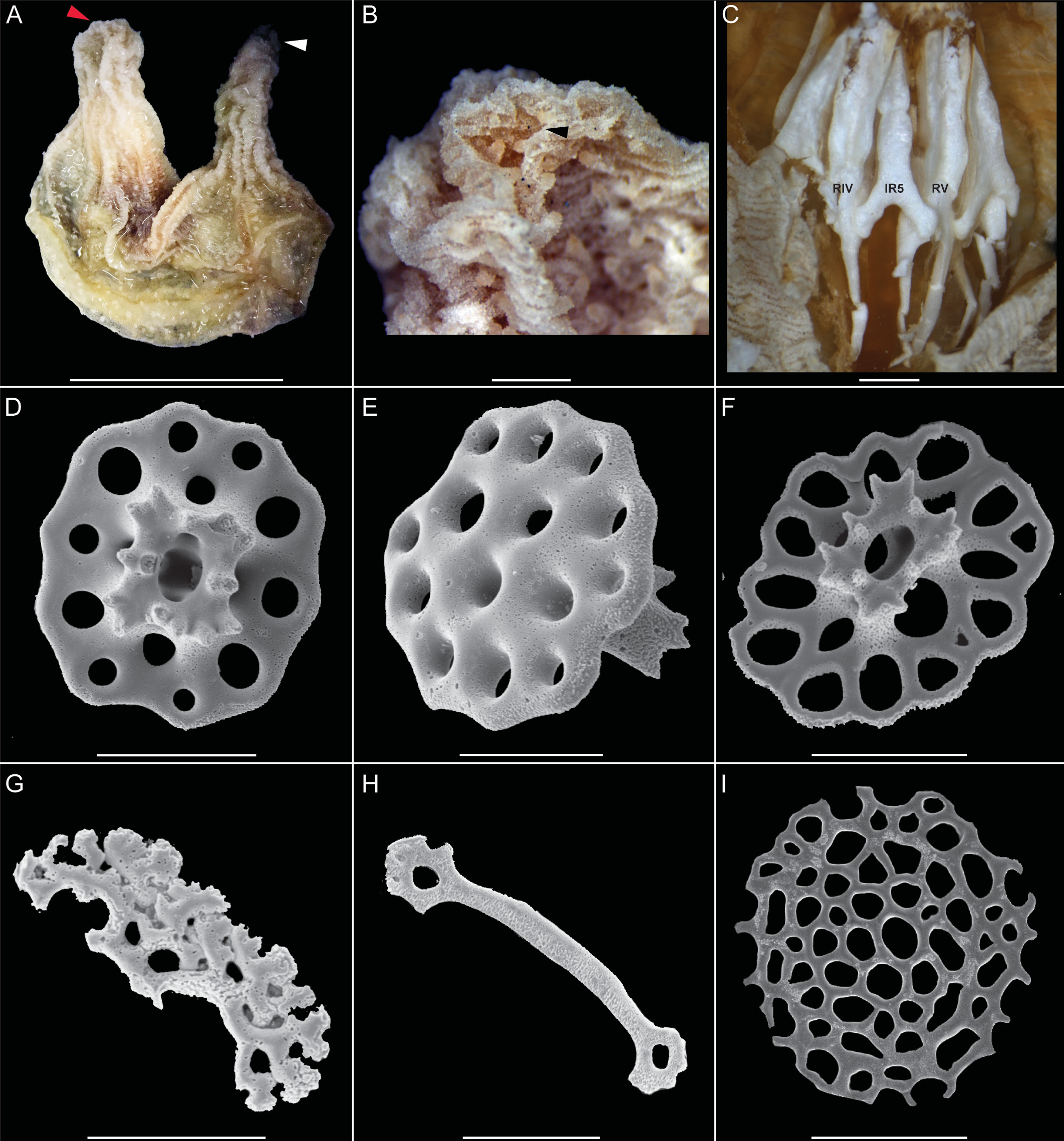
( Fig. 1 View FIGURE 1 , Table 1 View TABLE 1 )
Type material. REVIZEE, off coast of São Paulo, Brazil, 24°07’S, 44°42’W, 11.i. 1998, 101m, holotype 30 mm ( MZUSP 1644).
Etymology. This species is named in honor of Dr. Mike Reich in recognition for his contributions to the systematics of fossil Holothuroidea.
Diagnosis. Body U-shapped. Ten tentacles, ventral-most pair smaller. Tube feet scattered over body. Calcareous ring plates undivided; posterior processes subdivided and long, up to two times height of calcareous ring plate. Body wall with multilocular two pillared tables; tentacles with rods and introvert with two pillared tables and rosettes. Tube feet with only end plates.
Description. Body U-shaped, slightly upturned at both ends rough to touch ( Fig. 1a View FIGURE 1 ). Ten dendritic tentacles, ventral pair reduced. Anal papillae present ( Fig. 1b View FIGURE 1 ). Tube feet scattered throughout body. Radial and interradial plates of the calcareous ring of same length, entire, united at base only ( Fig. 1c View FIGURE 1 ). Radial plates notched anteriorly for the passage of the radial nerves and radial canal; posterior processes subdivided into about 4 pieces. Interradial plates arrow-shaped, with an anterior depression. Longitudinal muscles thin, split into two in the anterior region of body; retractor muscle short, flat, attached between the muscular processes. Internal organs degraded (i.e., Polian vesicle, stone canal and madreporitenot not observed). Color brown in ethanol.
Ossicles: Body wall with two-pillared tables with circular disc, multiperforated by several holes and undulating margin; spire short, ending in a crown of teeth (80–100 μm long, Fig. 1 View FIGURE 1 d–e). Introvert with two pillared tables with multiperforated, circular disc; undulating margin; spire short, ending in crown of teeth (40–60 μm long, ( Fig. 1f View FIGURE 1 ) and rosettes 30–50 μm long ( Fig. 1g View FIGURE 1 ). Tentacles with curved rods perforated at ends (60–80 μm long, Fig. 1h View FIGURE 1 ). Tube feet with only end plates circular (up to 100 μm long, Fig. 1i View FIGURE 1 )
Type locality. Brazil, off coast of São Paulo, 24°07’S, 44°42’W, 101 m. GoogleMaps
Distribution. Known only from the type locality.
Remarks. Sclerothyone reichi sp. nov. superficially resembles S. velligera and S. unicolumnus , but differs from both species by having (1) multilocular tables in the body wall ( Fig. 1d, e View FIGURE 1 ) and tentacles provided with rods only ( Fig. 1h View FIGURE 1 ) (vs. four-holed tables in the body wall and tentacles with rods and plates in S. velligera ; and (2) unfused pillars in the body wall tables ( Fig. 1d, e View FIGURE 1 ), and tables and rosettes in the introvert, ( Fig. 1f, g View FIGURE 1 ) (vs. fused pillars in the body wall tables and no ossicles in the introvert in S. unicolumnus ).
| MZUSP |
Museu de Zoologia da Universidade de Sao Paulo |
No known copyright restrictions apply. See Agosti, D., Egloff, W., 2009. Taxonomic information exchange and copyright: the Plazi approach. BMC Research Notes 2009, 2:53 for further explanation.
|
Kingdom |
|
|
Phylum |
|
|
Class |
|
|
Order |
|
|
Family |
|
|
SubFamily |
Sclerothyoninae |
|
Genus |