
Temnocephala catarinensis, Seixas & Amato & Amato & Damborenea, 2022
|
publication ID |
https://doi.org/10.11646/zootaxa.5209.1.8 |
|
publication LSID |
lsid:zoobank.org:pub:98ACEB95-636F-4836-80FA-496C503686FB |
|
DOI |
https://doi.org/10.5281/zenodo.7330396 |
|
persistent identifier |
https://treatment.plazi.org/id/03B6C56A-FFA5-1A0E-FF7E-F8BEFE22FE23 |
|
treatment provided by |
Plazi (2022-11-15 08:48:12, last updated 2024-11-29 03:55:10) |
|
scientific name |
Temnocephala catarinensis |
| status |
sp. nov. |
Temnocephala catarinensis sp. nov. Seixas, Amato & Damborenea
( Figs 1C View FIGURE 1 – 6 View FIGURE 6 )
Description. Based on 15 specimens collected from A. jarai ; 12 specimens mounted in toto (11 adults and 1 juvenile); 1 specimen mounted on stubs for SEM; 2 extracted cirri mounted in Faure: External characteristics. Body ( Figs 1C View FIGURE 1 , 3A View FIGURE 3 ), without tentacles 980–1817 (1408, 258) long, 820–1350 (1040, 192) wide; adhesive disk ventral, subterminal, partially covered by body 190–474 (317, 82) long, 260–572 (361, 88) wide; disc peduncle 150–300 (219, 61) diameter; ratio between total body length/adhesive disk length 4.4:1. Two dorsolateral, small epidermal ‘excretory’ syncytial plates (EPs), almost triangular; external margin in half circumference, sometimes reaching laterally the margin of body ( Fig. 2A View FIGURE 2 ); left plate 204 (n = 1) long, 200 (n = 1) wide; right plate 254 (n = 1) long, 154 (n = 1) wide; ratio between total body length, without tentacles/length of EPs 6.9: 1; ratio between total body width/width of the EPs 5:1. Nephridiopore (excretory pore) in the anterior half of the plate near the interior portion of the body ( Fig. 2A View FIGURE 2 ). Live specimens presented eyespots with red pigmentation. Digestive system. Pharynx 180–280 (223, 32) long, 250–510 (370, 75) wide, small, wider than long with a smaller anterior sphincter ( Fig. 3A View FIGURE 3 ); and a ‘bow-tie’-shaped intestine ( Figs 1C View FIGURE 1 , 3A View FIGURE 3 ). Glands. Rhabditogen glands small and numerous, with granular appearance 25–60 (43, n = 10, 9) in diameter, forming bunches (~ 70 cells) in lateral fields of body tending from the base of the lateral tentacles to below the posterior testes ( Fig. 1B View FIGURE 1 —black arrow head), ducts inconspicuous. Three pair of Haswell glands ( Fig. 3A View FIGURE 3 ) in front of the brain transverse band, largest cell diameter 15–75 (42, n = 10, 23). Disc glands 27.5–55 (38, n = 10, 9) in diameter, between adhesive disc and genital complex forming two lateral bunches extending from the posterior testes up to the middle of the back portion of the body ( Fig. 1B View FIGURE 1 —white arrow head), including one pair, of large, round and more central paranephrocytes ( Fig. 3A View FIGURE 3 ), 25–72.5 (60, n = 7, 17) in diameter. Reproductive system. Female. Vitellaria arborescent and slender extending in the limits of the intestine ( Figs 1C View FIGURE 1 , 3A View FIGURE 3 ); a small and ovoid ovary ( Figs 3D View FIGURE 3 , 5A, B View FIGURE 5 ), 60–135 (84, 25) long, 65–130 (92, 22) wide; a conspicuous vesicula intermedia ( Figs 3D View FIGURE 3 , 4A, B View FIGURE 4 ), 12.5–52.5 (34, n = 8, 12) long; a short vagina with a weak muscular wall ( Figs 3D View FIGURE 3 , 4A View FIGURE 4 ), 15–40 (26, n = 6, 10) long, 25–40 (30, n = 6, 5) maximum width; a single, well-developed and slightly asymmetrical vaginal sphincter ( Figs 3D View FIGURE 3 , 5A View FIGURE 5 ), 45–60 (52, n = 6, 5) in total diameter; diameter of anterior portion 22.5 (n = 1), diameter of posterior portion 27.5 (n = 1); vesicula resorbens large and usually full of sperm ( Figs 3D View FIGURE 3 , 4A, B View FIGURE 4 ) 72.5–155 (110, n = 9, 28) long, 112.5–190 (154, n = 9, 29) wide, wall thickness 2.5–10 (8, n = 7, 3); sessile and small eggs fixed on the external surface of the exoskeleton, especially in the chelipeds ( Fig. 1A View FIGURE 1 ), opercular plates not observed. Male. Four small and ovoid testes ( Figs 1C View FIGURE 1 , 3A View FIGURE 3 ), located two by two on each side of the body; right anterior testis 110–210 (145, n = 10, 30) long, 50–150 (95, n = 10, 29) wide; right posterior testis 115–330 (181, 60) long, 90–120 (155, n = 10, 44) wide; left anterior testis 100–180 (134, n = 10, 24) long, 60–100 (81, n = 10, 13) wide; left posterior testis 120–310 (166, 52) long, 80–240 (161, 46) wide; long and robust seminal vesicle ( Figs 3C View FIGURE 3 , 5C View FIGURE 5 ) 87.5–162.5 (127, n = 10, 29) long, 45–100 (65, n = 10, 17) wide; wall thickness 5–10 (7, n = 7, 2); large and wider prostatic bulb with a thin muscular wall ( Figs 3C View FIGURE 3 , 5C View FIGURE 5 ), 70–125 (99, 18) long, 52.5–75 (64, 8) wide; wall thickness 2.5–17.5 (5, n = 10, 5); small prostatic vesicle with abundant prostatic secretion ( Fig. 3C View FIGURE 3 ). Cirrus slightly curved ( Fig. 5 View FIGURE 5 ) 187.5–202.5 (195, n = 2, 11) long; shaft 157.5–172.5 (165, n = 2, 11) long, shaft base 80–85 (82.5, n = 2, 3) wide; introvert 30 (n = 2) long, 15–22.5 (19, n = 2, 5) wide at base; maximum introvert width at level of swelling, 20–25 (22.5, n = 2, 3). Introvert swelling with, approximately, 22 longitudinal rows of spines with ~ 10 spines each ( Fig. 5E View FIGURE 5 ). Introvert has two distinct portions, a proximal one with grooves and no spines and a distal one with small and sturdy spines 3.8–4.8 (4, n = 5, 0.4) ( Figs 5I View FIGURE 5 ). Ratio between total body length, without tentacles/ total length of cirrus 7: 1; ratio between total length of cirrus/maximum width of shaft’s base 2: 1; ratio between total length of cirrus/total length of introvert 6: 1.
Taxonomic summary.
Type host. Aegla jarai Bond-Buckup & Buckup, 1994 (Crustacea, Decapoda, Anomura ).
Type locality. Parque Nacional da Serra do Itajaí , Indaial, Santa Catarina, Brazil .
Site of infestation. Adults and juveniles in the branchial chamber and on the body surface, egg laying on the external and ventral surface of the exoskeleton: perioral area, pereiopods, chelipeds and in the first abdominal segments.
Additional host and localities. Samastacus spinifrons (Philippi, 1882) (Crustacea, Decapoda , Parastacidae ). Lago Lleu-Lleu; Río Donguil; Lago Rupanco; Río Duqueco; and Rio Contaco. All localities in Chile.
Helminth specimens deposited. ‘Coleção Helmintológica do Instituto Oswaldo Cruz (CHIOC)’— HOLOTYPE: CHIOC 39480 View Materials ; Paratype: CHIOC 39481 View Materials ; Additional material from S. spinifrons: CHIOC 39482, CHIOC 39483 View Materials and CHIOC 39484 View Materials . “ Colección de Invertebrados , División Zoologia Invertebrados, Museo de La Plata ( MLP)”— Paratypes: MLP-He 7721 (two specimens) ; Additional material from S. spinifrons : MLP-He 7722 and MLP-He 7723 (two specimens).
Host specimens deposited. Aegla jarai Bond-Buckup & Buckup, 1994 , “Coleção de Crustáceos do Departamento de Zoologia”, UFRGS 4833–4846.
Other helminth specimens examined. Temnocephala kingsleyae —“ Coleção de Invertebrados do Instituto Nacional de Pesquisas da Amazônia ( INPA)”, Manaus, Amazonas, Brazil: INPA 211 View Materials (seven specimens), and “ Colección de Invertebrados , División Zoologia Invertebrados, Museo de La Plata ( MLP)”, La Plata, Argentina: MLP-He 2226 (five specimens); Temnocephala mexicana —“ Colección Nacional de Helmintos , Laboratorio de Helmintologia , Instituto de Biología , Universidade Nacional Autónoma de México ( CNH-UNAM)”, Ciudad de México, México: 1311 and 1309 .
Diagnosis. Temnocephalid with an elongated-rounded body; eyespots with red pigmentation; almost triangular EPs with external margin in half circumference, sometimes reaching laterally the margin of body; a small pharynx, wider than long with a smaller anterior sphincter; a ‘bow-tie’-shaped intestine with thin wall and weak-marked septa; one pair of paranephrocytes, central and between the posterior testes; a small vagina with a weak muscular wall; a single, well-developed and slightly asymmetrical vaginal sphincter; a conspicuous vesicula intermedia; a sessile and small eggs fixed on the external surface of the exoskeleton, especially in the chelipeds with a thin subapical filament, opercular plates not observed; long and robust seminal vesicle that opens almost polarly into a large and wider prostatic bulb; four small and ovoid testes, located two by two on each side of the body, anterior testes smaller than the posterior ones; cirrus long and slightly curved (195μm on average), it appears less curved in lateral view; introvert with two distinct portions, a proximal one with grooves and no spines and a distal one with small and sturdy spines.

FIGURE 1. Aegla jarai A. Merus part of the cheliped showing unhatched (black head arrow) and hatched eggs (white head arrows) of Temnocephala catarinensis sp. nov. Scale bar: 1 mm. B. Posterior portion of the left side of the body showing posterior limit of the rhabditogenic glands (black head arrows) and the posterior limit of the disc glands (white head arrows) of Temnocephala catarinensis sp. nov., Scale bar: 250 μm. C. Ventral view of adult specimens of Temnocephala catarinensis sp. nov. from Aegla jarai. Scale bar: 250 μm.

FIGURE 2. Dorsal view of Temnocephala catarinensis sp. nov. observed with SEM, showing details of the epidermal ‘excretory’ syncytial plates (EPs). A. Specimen from Aegla jarai showing right EP and nephridiopore (n). B. Specimen from Samastacus spinifrons showing EPs and nephridiopores (n). C. Detail of the right EP from Samastacus spinifrons showing its limits and nephridiopore (n). Scale bars: 100 μm.

FIGURE 3. Line drawings of holotype of Temnocephala catarinensis sp. nov. from Aegla jarai. A. Adult specimen, ventral view, scale bar: 250 μm. B. Cirrus showing the proximal limit of the introvert (arrow), scale bar: 20μm. C. Partial line drawings of the male reproductive system. D. Partial line drawings of the female reproductive system. Scale bars: 50 μm. (adhesive disc (ad), anterior testes (at), anterior portion of the vaginal sphincter (avs), cirrus (c), deferent vessels (dd), excretory vesicle (ev), genital atrium (ga), genital pore (gp), Haswell glands (hg), intestine (i), mouth (m), ovary (ov), paranephrocytes (pa), prostatic bulb (pb), prostatic cells (pc), pharynx (ph), prostatic secretions (ps), posterior testis (pt), posterior portion of the vaginal sphincter (pvs), seminal vesicle (sv), tentacles (t), vagina (va), vitelline duct (vd), vitelline glands (vg), vesicula intermedia (vi), vesicula resorbens (vr)).
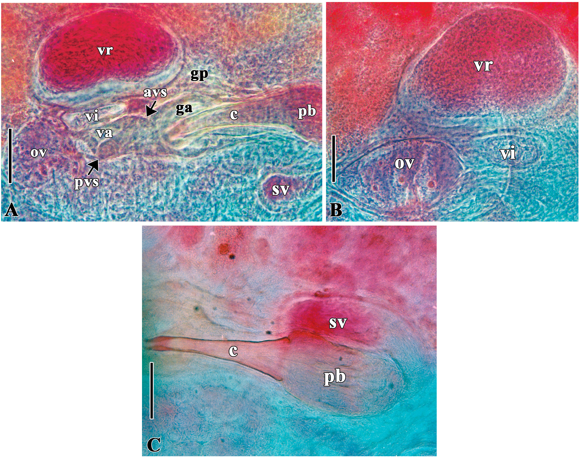
FIGURE 4. Partial reproductive system of an adult specimen of Temnocephala catarinensis sp. nov. from Aegla jarai. A. Partial female and male reproductive system B. Partial female reproductive system C. Partial male reproductive system. Scale bars: 50 μm. (anterior portion of the vaginal sphincter (avs), cirrus (c), genital atrium (ga), genital pore (gp), ovary (ov), prostatic bulb (pb), posterior portion of the vaginal sphincter (pvs), seminal vesicle (sv), vagina (va), vesicula intermedia (vi), vesicula resorbens (vr)).

FIGURE 5. Photomicrographs of cirri of Temnocephala catarinensis sp. nov. from Aegla jarai, seen with Nomarski’s DIC microscopy. A. Entire cirrus (lateral view), showing the evident curvature, scale bar: 25 μm. B. Entire cirrus (dorsal or ventral view), showing that the curvature is less evident when the cirrus is in this position, the proximal limit of the introvert (arrow) and the shaft rim (head arrow), typical of adult worms, scale bar: 20μm. C–K. Introvert seen in different focusing planes, showing the proximal limit of the introvert (arrow), scale bars: 10 μm.

FIGURE 6. Photomicrographs of cirri of Temnocephala catarinensis sp. nov. from Samastacus spinifrons, seen with Nomarski’s DIC microscopy. A. Entire cirrus (lateral view), showing the evident curvature. B. Entire cirrus (dorsal or ventral view), showing that the curvature is less evident when the cirrus is in this position and the proximal limit of the introvert (head arrow). Scale bars: 25 μm. C–H. Introvert seen in different focusing planes, showing the proximal limit of the introvert (arrow), scale bars: 10 μm.
No known copyright restrictions apply. See Agosti, D., Egloff, W., 2009. Taxonomic information exchange and copyright: the Plazi approach. BMC Research Notes 2009, 2:53 for further explanation.
|
Kingdom |
|
|
Phylum |
|
|
Order |
|
|
Family |
|
|
Genus |