
Hemilecanium cedrelus Hodgson, 2008
|
publication ID |
https://doi.org/ 10.3897/zookeys.3.45 |
|
publication LSID |
lsid:zoobank.org:pub:CBCD770C-79A0-4A53-9C35-DCD7A1A32A14 |
|
DOI |
https://doi.org/10.5281/zenodo.3792754 |
|
persistent identifier |
https://treatment.plazi.org/id/422B8F25-F836-43A3-B878-27DC51E0DE90 |
|
taxon LSID |
lsid:zoobank.org:act:422B8F25-F836-43A3-B878-27DC51E0DE90 |
|
treatment provided by |
Plazi |
|
scientific name |
Hemilecanium cedrelus Hodgson |
| status |
sp. nov. |
Hemilecanium cedrelus Hodgson , sp. n.
urn:lsid:zoobank.org:act:422B8F25-F836-43A3-B878-27DC51E0DE90
Material studied. Holotype female: Zambia, Luanshya , 26.VII.1955, on Cedrela toona (spelt tuna), G.G. Robinson ( BMNH): ad ♀ in good condition.
Paratype ♀. Data as for holotype ♀:14/14 ad ♀ (fair to good) ( BMNH, USNM) ; also 3/27 1st-instar nymphs (fair to good ( BMNH, USNM)) plus 1/1 3 rd- instar ♀ (good) + 1 2 nd- instar ♀ (with pharate 3 rd- instar ♀; fair, with inner hyphae ( BMNH)) .
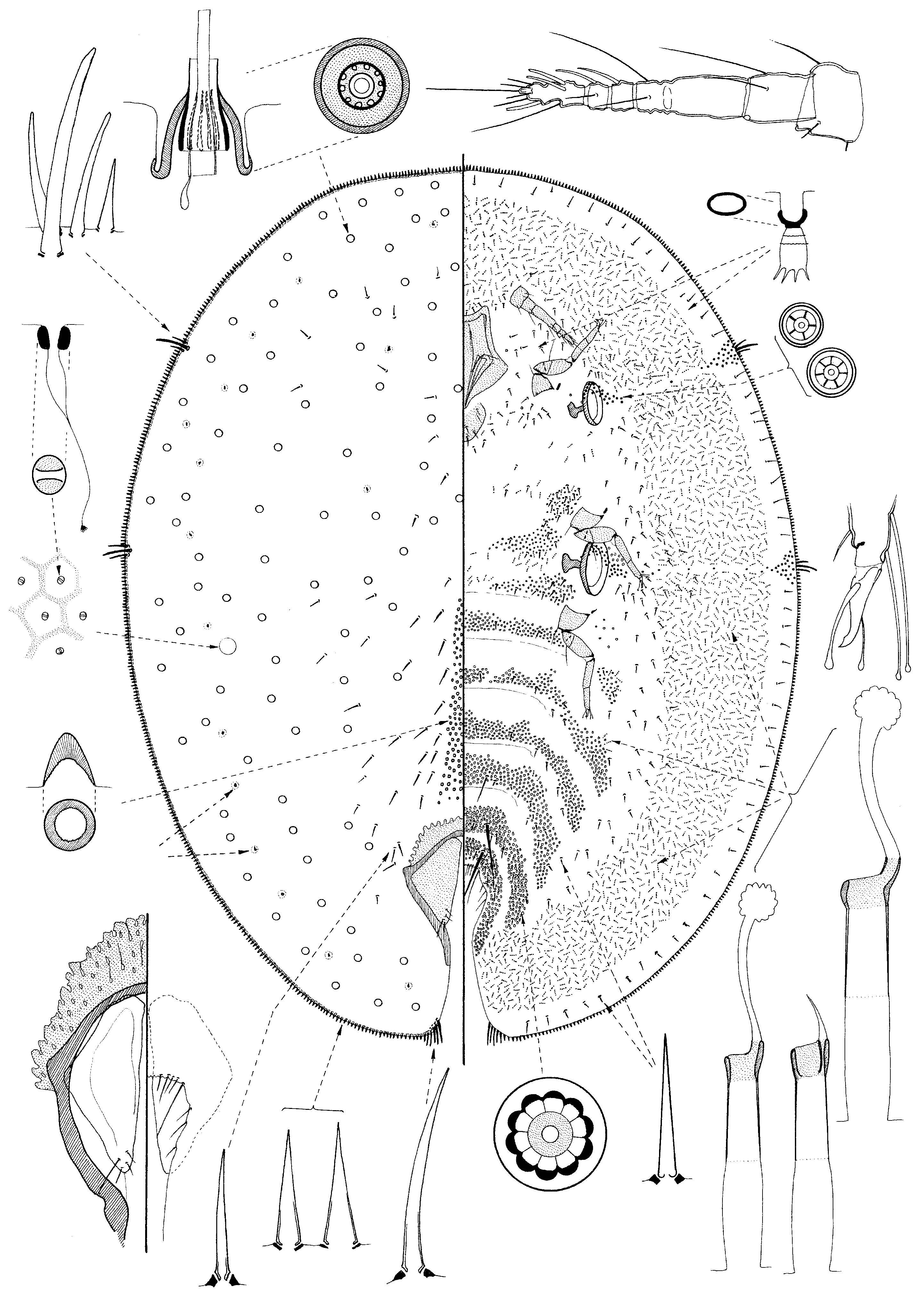
Adult female ( Fig. 2 View Fig ).
Described from 5 specimens in good condition, and with reference to the remaining 10 specimens.
Instar diagnosis. Dried material with many clear, round, brown-ochre-coloured spots present throughout dorsum indicating position of dorsal tubercles. Mounted material oval to almost round. Dorsum membranous apart from a narrow sclerotisation around anterior margin of anal cleft; derm with a reticulate pattern of areolations; also with about 150- 200 randomly distributed dorsal tubercles, distributed over entire dorsum, each without satellite tubular ducts; also “scars” (see discussion below) in position of dorsal tubercles of 3rd-instar. Conical preopercular pores and dorsal setae present. Anal plates roughly quadrate with 4 setae near apex. Margin with a single line of sharply spinose setae; with 3 stigmatic spines each clearly differentiated from marginal spinose setae, median stigmatic spines much longer than lateral spines. Venter with abundant 10-locular disc-pores on all abdominal and thoracic segments. Ventral microducts abundant throughout. Tubular ducts of probably 1 type, present in a wide submarginal band. Antennae 6 segmented. Limbs fully developed but relatively small; tibio-tarsal articulation without a sclerosis; claw digitules different; claw with a small denticle. Spiracles proportionately very large, width of peritreme much wider than width of coxae. Mouthparts relatively large.
Unmounted material. Dried material quite dark brown; younger specimens with a distinct shallow longitudinal dorsal ridge and a clear, shelf-like margin; older specimens becoming strongly convex with two strong shoulders medially; venter becoming highly concave, forming an inner egg chamber. Positions of dorsal tubercles indicated by clear round areas of more brown-ochre-coloured derm scattered over dorsum.
Mounted material. Oval to almost round, 2.8-6.0 mm long and 2.25-5.00 mm wide; anal cleft about 1/4th-1/5th body length. Basic structure as for diagnosis.
Dorsum. Derm mainly membranous but with a heavy sclerotised band around anterior margin of anal cleft, which expands anteriorly with age. Derm of available specimens with polygonal reticulations throughout, each reticulation with an inner areolation and a dorsal microductule; perhaps becoming sclerotised at maturity. Dorsal setae all rather spinose, quite sharply pointed, with parallel sides; frequent laterally and anteriorly to anal plates, but absent submarginally; those anterior to anal plates each 33-36 µm long, those more anteriorly and laterally smaller, down to 16-24 µm long. Preopercular pores present in an elongate triangular group anterior to anal plates, each pore conical and 8-11 µm wide; each group with 85-140 pores, extending anteriorly to about metathorax. Dorsal microductules oval, appearing bilocular but with single inner ductule arising medially, each about 2.5-3.0 µm widest, most ductules swollen proximally; abundant, present in each dorsal reticulation. Other dorsal pores absent. Dorsal tubercles large and convex, but sunken into derm; each 16-26 µm wide, with a heavily sclerotised outer cone plus 2 inner chimney-like tubes (one inside other); outermost tube with a ring of about 10-12 vertical ridges (see Discussion below adult female); innermost tube long, extending some way above tubercle; dorsum with a total of about 140-200 tubercles, randomly distributed throughout; also with about 16 submarginal “scars” and 2 pairs of submedial “scars” (in approximate positions of dorsal tubercles of 3 rd instar). Anal plates each about 275-330 µm long, width of single plate 115-170 µm; each plate triangular, with 4 apical setae, both inner margin setae and subapical seta 25-30 µm long, other seta appearing dorsal, about 60-72 µm long. Anogenital fold with a line of 6-8 setae along anterior margin, each fairly short but with a long seta at each corner, latter about 60-75 µm long; each lateral margin with 3 setose setae. Anal ring well developed, with many pores and probably 5 pairs of setae, each 270-360 µm long; anal tube about as long as anal plates. Eyespots not detected.
Margin. Marginal setae all sharply spinose, each 16-45 µm long, with a broad base, straight sides and narrow basal sockets; abundant, with 175-240 anteriorly between anterior stigmatic areas, 54-62 laterally between stigmatic areas and 125-195 on each side of abdomen; each anal lobe with a group of 5 or 6 longer, slightly curved setae, longest 105-115 µm long. Stigmatic clefts absent. Stigmatic spines 3, clearly differentiated from marginal spines, slightly curved and with a less pointed apex than marginal setae; median spine longest, 75-85 µm long, each lateral spine 28-65 µm long.
Venter. Derm membranous. Spiracular disc-pores each mainly with 5 loculi, in broad groups near margin and each peritreme but very few or even sometimes absent in between; with about 45-50 in each anterior band and 60-85 in each posterior band, latter with a small group of multilocular pores near each spiracle. Multilocular disc-pores each about 8-10 µm wide, mainly with 10 loculi, abundant across all abdominal segments and across meso- and metathorax; scarce on prothorax and head. Ventral microducts each about 3 µm wide, abundant throughout, except marginally. Ventral tubular ducts slightly variable but probably all of one type, each with an outer ductule 17-30 µm long, inner ductule 13-20 µm long, with or without a glandular end; abundant in a broad submarginal band and rather less frequently in bands across each thoracic segment; with 1 or 2 present medially on abdomen among multilocular disc-pores. Other pores types absent. Ventral setae mainly rather spinose, most about 20-26 µm long, present across each abdominal and thoracic segment but most abundant in a submedial band just laterad to spiracles and legs; with about 5 pairs of rather short inter-antennal setae, longest 40 µm long; abdominal segments V, VI and VII each with a pair of longer setae, longest on VI and VII, each about 150 µm long; submarginal setae frequent, each about 25 µm long.
Antennae each 6 segmented, total length 250-350 µm; scape with 3 setae, pedicel with 2 setae, other segments: III with 2 setae, IV with 1 fleshy seta, V with 1 fleshy seta + 1 flagellate seta and VI with 3 fleshy setae, about 5 stiff apical setae + 1 flagellate seta; length of apical seta 66-90 µm long. Clypeolabral shield 270-350 µm long, labium probably with 4 pairs of setae. Spiracles large, width of peritremes: anterior 125-165 µm, posterior 155-210 µm; muscle plate much shorter than width of peritreme. Legs well developed but small; lengths (µm) of metathoracic legs: coxae 120-130 (width of coxal base 80-100 µm); trochanter + femur 145-153; tibia + tarsus 185-210; claw 28-31; tibio-tarsal articulation not always clear, with no sclerosis; longest coxal seta about 70 µm; longest trochanter seta about 85 µm; other setae very sparse; tarsal digitules about equal to length of claw digitules; claw digitules longer than claw, with one slightly narrower than other; claw with a small denticle. Vulva probably present between abdominal segments VII and VIII.
Comment. The adult females of this species are superficially similar to those of Hemilecanium coriaceum Hall and H. uesatoi Kondo & Hardy , which also have dorsal tubercles randomly distributed throughout the dorsum,. However, H. cedrelus differs from H. coriaceum as follows (characters-states on H. coriaceum in brackets): (i) presence of 3 clearly differentiated stigmatic spines (absent or 1 barely differentiated); (ii) multilocular disc-pores abundant across all abdominal segments and also across meso- and metathorax (many fewer, restricted to abdomen); (iii) preopercular pores in an elongate group anterior to anal plates (in a broad group incorporating some dorsal tubercles anterior to anal plates); and (iv) large size of spiracular peritremes (small). Adult female H. cedrelus are also similar to the newly described H. uesatoi from the Ryukyu Archipelago, Japan, but the latter differs in having normal-sized spiracles; pocket-like sclerotisations restricted to the submargin, preopercular pores extending anteriorly onto head; 8-segmented antennae, and no denticle on the claw. In addition, the 1 st- instar nymphs are rather different (see under that stage below).
Initially it was assumed that the ring of sclerotised spots on each dorsal tubercle, which are clearly visible in dorsal views of each tubercle, were satellite tubular ducts similar to those on some Hemilecanium species. However, true satellite ducts have the structure of a small tubular duct, i.e. with a long outer ductule, a small cup-shaped invagination and sometimes an inner ductule, each duct opening onto the surface of the tubercle through an aperture some distance from the funnel-like central cone (see Etiennea halli , E. kellyi , E. petasus and E. villiersi in Hodgson, 1991 (now all in Hemilecanium )). This is quite different from what we see on H. cedrelus where, when seen from the side, these “sclerotised spots” appear to refer to vertical sclerotised ridges on the outer funnel-shaped tube; none of which have either an associated ductule or an outer aperture. It is therefore considered that these structures are not satellite tubular ducts.
Distribution. Hemilecanium cedrelus is currently only known from Zambia.
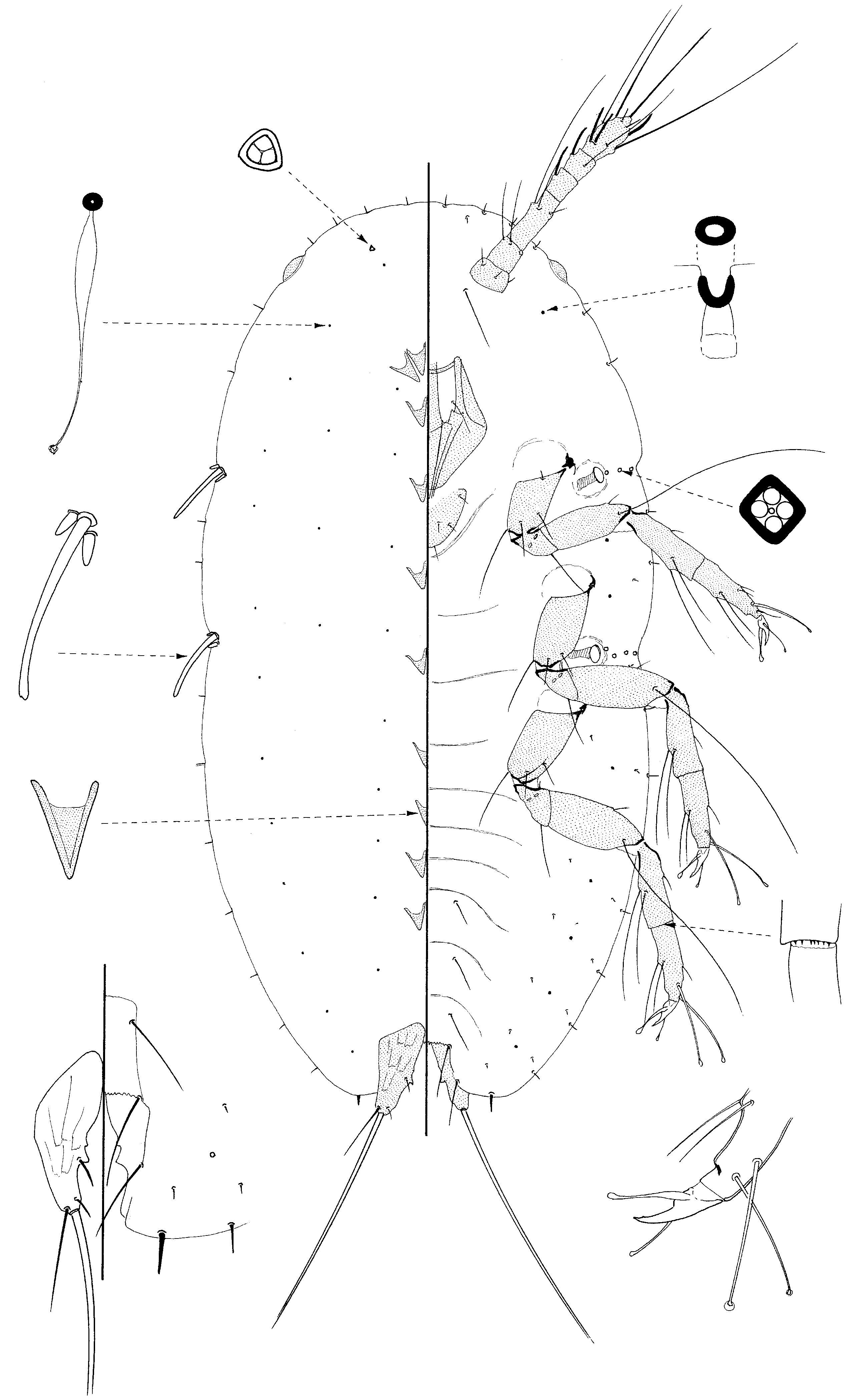
Third-instar female ( Fig. 3 View Fig )
Described from 1 specimen in good condition. (Note: the data in brackets for the number of dorsal tubercles are from the pharate 3rd-instar nymph – see discussion under 2nd-instar nymph below.)
Instar diagnosis. Oval and rather flat. Dorsum with a submarginal ring of large dorsal tubercles plus 2/3 submedially on thorax. Margin with a single line of sharply spinose setae; with 3 stigmatic spines clearly differentiated from marginal spinose setae. Venter with a small group of 5-locular disc-pores posterior to anal opening; also with a sparse submarginal band of tubular ducts. Antennae 5 or 6 segmented. Legs fully developed; claw digitules dissimilar; claw with a small denticle. Spiracles of normal proportions, width of peritreme smaller than width of coxae. Mouthparts relatively large.
Unmounted material. Dried material pale brown; oval, rather flat, with a few shallow, radial ridges. No sign of a wax test.
Mounted material. As in instar diagnosis. Body 1.33 mm long and 0.9 mm wide; anal cleft about 1/5th body length. Dorsum with a submarginal ring of about 30 large dorsal tubercles plus 2(3) submedially on thorax, plus “scars” left by the 12 dorsal tubercles of 2 nd- instar nymph.
Dorsum. Derm mainly membranous, without a sclerotised band around anterior margin of anal cleft and without a reticulate pattern of areolations. Dorsal setae very few, each short and finely spinose, each about 10 µm long; distribution uncertain but very sparse. Preopercular pores absent. Dorsal microductules small, each about 1.5 µm wide, with an inner ductule about 8 µm long, most ductules swollen proximally; abundant. Other dorsal pores absent. Dorsal tubercles of more or less two types: (i) large and convex, each about 20-24 µm wide, basically similar to those on adult female; with (on each side in a submarginal ring) 5 (5-7) on abdomen; 2 (3) between stigmatic clefts, 2 (3) between anterior cleft and eyespot and 4 anteriorly between eyespots; and (ii) what are here considered to be remains of dorsal tubercles of 2nd-instar female, structure very unclear (but probably similar to “scars” on adult female); with 3 on each side of abdomen, 1 between stigmatic clefts, between anterior cleft and eyespot and 2 anteriorly between eyespots. Anal plates each about 110 µm long, width of single plate about 55 µm; each plate triangular, with 4 setae: 2 inner margin setae, both short, and two other apical setae, positions uncertain, each possibly 16-23 µm long. Anogenital fold with a line of perhaps 8 setae along anterior margin, each about 28 µm long; each lateral margin with 2 setae. Anal ring well developed, with 4 pairs of setae, each about 100 µm long; anal tube about twice length of anal plates. Eyespots oval, 21-23 µm widest.
Margin. Marginal setae all sharply spinose, each 12-16 µm long, with a broad base, rather straight sides and narrow basal sockets; with a total of 26 anteriorly between eyespots, and (on each side) 15-19 between eyespots and anterior stigmatic areas, 20-22 laterally between stigmatic areas and 58-64 on each side of abdomen; each anal lobe with a group of 2 or 3 longer, slightly curved setae, longest 53-66 µm long. Stigmatic clefts shallow, each with 3 stigmatic spines, clearly differentiated from marginal spines, slightly curved and with a less pointed apex than marginal setae; median spine longest, 70-90 µm long, each lateral spine 20-40 µm long.
Venter. Derm membranous. Spiracular disc-pores, each with a broad sclerotised margin and perhaps mainly with 5 or 6 loculi, in broad bands between margin and each peritreme; with 20 or 21 in each anterior band and 24-26 in each posterior band. Preanal multilocular disc-pores each about 5 µm wide with 5 loculi, in a small group of 3 each side beneath anal plates. Ventral microducts each about 3 µm wide, abundant throughout apart from marginally, where absent. Ventral tubular ducts of one type, each with an outer ductule 15 µm long, inner ductule 10-12 µm long, with a glandular end; in a mainly narrow submarginal band but tending to be most concentrated on either side of spiracular disc-pore bands, with 3 anteriorly on head, 1-3 between eyespots and anterior stigmatic clefts, 11-12 on each side between stigmatic clefts and 11-18 on each side of abdomen. Other pore types absent. Ventral setae perhaps slightly larger medially than laterally, most about 10-12 µm long; with one longer pair and one shorter pair of inter-antennal setae, longest about 60 µm; with long setae medially on abdominal segments V–VII, longest about 85 µm; with 2 or 3 short setae associated with each coxa; other setae fairly frequent in a broad submarginal band, most abundant on abdomen, each about 6-7 µm long.
Antennae each either 5 segmented or slightly deformed (i.e. really 6 segmented), total length 200-215 µm; scape with 3 setae, pedicel with 2 setae + campaniform sensillum, setae on other segments uncertain. Clypeolabral shield 185 µm long; labium with 4 pairs of setae. Spiracles of normal size, width of peritremes: anterior 25 µm, posterior 30-32 µm. Legs well developed; lengths (µm) of metathoracic legs: coxae 80-85; trochanter + femur 100-110; tibia + tarsus 122-140; claw 23-26; tibio-tarsal articulation poorly defined; longest coxal seta about 58-60 µm; longest trochanteral seta about 55 µm; femur with 2 setae; tibia with 3 setae; tarsus with 3 setae; tarsal digitules perhaps extending further than claw digitules, each 28 µm long; claw digitules longer than claw, with one narrower than other, length 28-32 µm; claw with a small denticle.
Comment. This is the only immature stage of any Coccidae known to the author with preanal multilocular disc-pores.
Second- instar female ( Fig. 4 View Fig )
Described from a single specimen containing a pharate 3 rd- instar nymph.
Instar diagnosis. Oval. Similar to 3 rd- instar but venter without disc-pores posterior to anal opening and without tubular ducts; marginal setae and spiracular discpores fewer.
Unmounted material. Dried material pale brown; oval, rather flat, with a few shallow, radial ridges. No sign of a wax test.
Mounted material. As for instar diagnosis. Body 1.5 mm long and 1.08 mm wide; anal cleft about 1/6th body length. Submarginal ring of 12 dorsal tubercles.
Dorsum. Derm mainly membranous, without a sclerotised band around anterior margin of anal cleft and without a reticulate pattern of areolations. Dorsal setae possibly absent. Dorsal microductules small, each about 1.5 µm wide, with or without an inner ductule, most ductules swollen proximally; sparse. Other dorsal pores absent. Dorsal tubercles of 1 type, each 11-15 µm wide; structure basically similar to those on adult female but smaller, each with about 6 vertical ridges on outer inner tube; with 1 pair of tubercles anteriorly, and (on each side) 1 between eyespots and anterior stigmatic cleft, 1 laterally between stigmatic clefts and 3 or 4 on abdomen (plus dorsal tubercles of pharate 3rdinstar nymph (as indicated in brackets in the description above). Anal plates each about 90 µm long, width of single plate about 42 µm; each plate triangular, with 4 setae, all broken. Anogenital fold with 2 pairs of setae anteriorly, longest setae about 33 µm long; each lateral margin with 1 seta. Anal ring well developed, with 3 pairs of setae, each about 110 µm long; anal tube about twice length of anal plates. Eyespots oval, 15 µm widest.
Margin. Marginal setae all sharply spinose, each 8-20 µm long, with a broad base, slightly curved sides and narrow basal sockets; with 12 anteriorly between eyespots, and (on each side) 6 between eyespots and anterior stigmatic areas, 6 or 7 laterally between stigmatic areas and 19-22 on each side of abdomen; each anal lobe probably with a group of 2 or 3 longer setae but all broken. Stigmatic clefts shallow, each with 3 stigmatic spines, clearly differentiated from marginal spines, slightly curved and with a less pointed apex than marginal setae; median spine longest, 60 µm long (only one present), each lateral spine 15-18 µm long.
Venter. Derm membranous. Spiracular disc-pores each with a broad sclerotised margin and perhaps mainly with 5 loculi, in narrow bands between margin and each peritreme; with 7 in each anterior band and 10-12 in each posterior band. Preanal multilocular disc-pores absent. Ventral microducts each about 3 µm wide, sparse in a broad submarginal band and also occasional medially on head, thorax and abdomen. Ventral tubular ducts absent. Other pores types absent. Ventral setae few, with two pairs of interantennal setae, longest 33+ µm; with long setae medially on abdominal segments V–VII, longest about 80+ µm; each coxa with 1 minute associated seta; other setae in a submarginal line (1 laterally between stigmatic clefts), and an inner submarginal line on abdomen; each about 6-7 µm long.
Antennae both damaged but either 5 segmented or slightly deformed (i.e. really 6 segmented), total length perhaps 210 µm; scape with 3 setae, pedicel with 2 setae + campaniform sensillum; number on other segments uncertain. Clypeolabral shield 132 µm long; labium with 4 pairs of setae. Spiracles of normal size, all peritremes 17-19 µm wide. Legs well developed; lengths (µm) of metathoracic legs: coxae 78-80; trochanter + femur 116-120; tibia 80-85; tarsus 65-70; claw 20-22; tibio-tarsal articulation fairly clear; longest coxal seta about 60-66 µm; all long trochanter setae broken; femur 2 setae; tibia 3 setae; tarsus 3 setae; tarsal digitules perhaps extending further than claw digitules, each about 45 µm long; claw digitules longer than claw, with one clearly narrower than other, length 25-28 µm; claw with a small denticle.
Comment. Despite having a slightly larger body size than the 3 rd- instar nymph described above, this is clearly a female 2 nd- instar nymph. This is shown not only by the smaller limbs etc, smaller number of spiracular disc-pores and absence of preanal disc-pores, but also by the presence of the pharate 3 rd- instar and the distribution of the latter’s dorsal tubercles, which are clearly visible inside its cuticle.
Because this specimen had a pharate 3 rd- instar nymph within, it was possible to study the number and distribution of the dorsal tubercles, which were well developed and these data are given in brackets in the description of the 3 rd- instar nymph above. The number and distribution of the “scars” on the derm of the 3 rd- instar nymph agree with the number and positions of the dorsal tubercles on the 2 nd- instar nymph and therefore clearly refer to these (see also the Discussion beneath description of 1 st- instar nymph of H. cedrelus ).
First-instar ( Fig. 5 View Fig )
Instar diagnosis. Oval. Dorsum membranous but with a series of 1-4 large, triangular or cone-shaped protuberances medially on most segments. Dorsal setae absent. Margin with small spinose setae. Each stigmatic cleft with 3 stigmatic spines, median spine long. Venter with three pairs of long preanal setae. Ventral microducts in a sparse submarginal line. Legs well developed; each femur with an exceptionally long seta on anterior margin; long setae also present on tibia and tarsus; claw digitules different; claw with a small denticle.
Mounted material. As for instar diagnosis. Body 0.5-0.53 mm long and 0.26- 0.30 mm wide; anal cleft very short.
Dorsum. Derm mainly membranous, but with large triangular or cone-shaped protuberances medially, each margin of protuberance about 33-35 µm long and each 20-22 µm wide at base, distributed as follows: none on abdominal segments V-VII, pairs on abdominal segments III & IV, singles on abdominal segments I & II, each thoracic segment plus a pair posteriorly on head and 2 pairs together more anteriorly on head. Dorsal setae absent. Dorsal microductules small, each about 1.5 µm wide with a long inner ductule, most ductules swollen proximally; mainly in 2 pairs of longitudinal lines, one pair of lines medially (with 5 pores on abdomen, probably 3 on thorax and 1 on head) and other lines submarginal (with 7 pores on abdomen, 2 between stigmatic clefts and 4 anteriorly); also with single pores submedially in each thoracic segment. A pair of trilocular pores present on head some distance from anterior margin, each about 3.0 µm wide. Other dorsal pores absent. Anal plates each quite elongate, about 60-65 µm long, with a few shallow longitudinal ridges; each with a small spine on inner margin, 2 short setae along inner margin, each about 8 µm long; a very long apical seta, each 260-310 µm long, and a single seta on posterior margin, about 36 µm long. Anogenital fold with a single long seta at each corner, each 26-28 µm long, and a single similar seta on each lateral margin, 23-28 µm long. Anal ring with 2 rows of pores, each with 5-7 pores, plus 6 anal ring setae, each about 80-85 µm long; anal tube extending anterior to anal plates. Eyespots each 15-18 µm wide.
Margin. Marginal setae all finely spinose, most 6-8 µm long (that on each anal lobe 13-15 µm long), with well-developed socket, distributed as follows: with 8 anteriorly between eyespots, and (on each side) 2 between eyespots and anterior stigmatic cleft, 2 laterally between stigmatic clefts and 8 on abdomen. Stigmatic clefts shallow, each with 3 stigmatic spines clearly differentiated from marginal setae; median spinose seta very long, rather parallel sided and possibly with a slightly flattened apex, each 40- 45 µm long, with a broad basal socket; anterior lateral spine shortest, about 5 µm long, posterior spine about 6.5 µm long.
Venter. Derm membranous. Spiracular disc-pores each with very thick margins and perhaps with mainly 3 or 4 loculi (occasionally 5?), with 3 pores in each anterior pore band and 4 in each posterior band. Ventral microducts each about 1.5 µm wide, present in a submarginal line, with (on each side) 1 on head, 2 on thorax and probably 6 on abdomen. Ventral setae few; with 1 pair of interantennal setae, each about 40 µm long, and with pairs of long setae medially in abdominal segments V-VII, longest about 40-45 µm long; short setae in a submarginal line, with (on each side) 7 on abdomen, 1 on thorax and 1 anteriorly on head; also with an inner submarginal line of 7 setae on abdomen.
Antennae each 6 segmented, total length 150-175 µm; scape with 3 setae, pedicel with 2 quite long setae + campaniform sensillum segments: III with 3 setae, one very long, up to about 80-85 µm long, IV 1 fleshy seta, V 1 fleshy seta + 1 flagellate seta, and VI with 3 fleshy setae, about 4 stiff apical setae + 3 flagellate setae, longest at least 110 µm long; length of apical seta about 110 µm long. Clypeolabral shield 87-95 µm long; labium with 4 pairs of setae. Spiracles: all peritremes about 8 µm wide, in a shallow concavity. Legs well developed; lengths (µm) of metathoracic legs: coxae 60-66; trochanter + femur 83-85; tibia 55-60; tarsus 45-52; claw 20-23; longest coxal seta about 33-45 µm; longest trochanter seta about 40 µm; each femur with 3 setae, one exceptionally long seta on anterior margin, 140-150 µm long; tibia with 3 setae, 2 rather long, longest 75-80 µm long; tibia with a row of microspines along distal margin on each middle and hind leg; tarsus with 4 setae, 2 quite long, longest about 40 µm; tarsal digitules both capitate, offset, extending to about equal with claw digitules, each about 50 µm long; claw digitules longer than claw, with one distinctly narrower than other, each about 28 µm long; claw with a small denticle.
Comment. The 1 st- instar nymph of H. cedrelus is distinctive due to the presence of the triangular or cone-shaped protuberances medially on the dorsum of most segments, unknown on any other 1 st- instar nymphs as far as the author is aware, including those of H. uesatoi . However, Hodgson (1993), when describing the dorsum of Etiennea (Hemilecanium) petasus wrote “Derm entirely membranous, but thrown into small dermal nodules – in some specimens, these are rather pronounced and found throughout, in others they are few, but are always present around the margin and in pairs medially, probably one pair per segment” (my italics). In seems possible, therefore, that these “nodules” are just more pronounced on H. cedrelus . In addition, the presence of very long setae on each femoral segment of H. cedrelus is unusual, although similar setae are known on other species ( Hodgson, 1993) in the petasus group as defined by Kondo and Hardy (2008). They are also known on Protopulvinaria pyriformis (Cockerell) ( Ray & Williams, 1982) and Kilifia De Lotto ( Ray & Williams, 1982). In addition to the triangular or cone-shaped protuberances medially, which are absent on the 1 st- instar nymph of H. uesatoi , the 1 st- instar nymphs of H. cedrelus differ from those of H. uesatoi in having (character-states on H. uesatoi (from Kondo & Hardy, 2008) in brackets): (i) long femoral setae on all femora (restricted to the metafemur only); and (ii) claw digitules dissimilar (similar).
Etymology. The specific name cedrelus is taken from the generic name of the host plant, Cedrela toona (Meliaceae) .
A key to the adult females of Hemilecanium Newstead was included in Kondo and Hardy (2008, p. 195). This key can be modified to include H. cedrelus as follows:
6. Stigmatic spines not differentiated from marginal spines ..................................... 7
– Stigmatic spines clearly differentiated from marginal setae ................................ 9a
9a Dorsum with dorsal tubercles present throughout dorsum; antennae 6-segmented; spiracles very large, posterior peritreme generally more than 1.7 times wider than basal width of metacoxa ..................................................... H. cedrelus new species
– Dorsum with dorsal tubercles generally restricted to a submarginal band (except H. uesatoi ); antennae usually more than 6 segmented; spiracles smaller, width of peritremes of posterior spiracles usually less than basal width of metacoxa............. ..................................................................................9b (9b is original couplet 9)


| USNM |
Smithsonian Institution, National Museum of Natural History |
No known copyright restrictions apply. See Agosti, D., Egloff, W., 2009. Taxonomic information exchange and copyright: the Plazi approach. BMC Research Notes 2009, 2:53 for further explanation.