
Homidia leniseta, Pan & Yang, 2019
|
publication ID |
https://doi.org/ 10.11646/zootaxa.4671.3.3 |
|
publication LSID |
lsid:zoobank.org:pub:607D8157-8F1A-4452-9091-BAA07F227A66 |
|
persistent identifier |
https://treatment.plazi.org/id/03B76020-6827-4D05-9EFB-FF631998745E |
|
treatment provided by |
Plazi |
|
scientific name |
Homidia leniseta |
| status |
sp. nov. |
Homidia leniseta sp. nov.
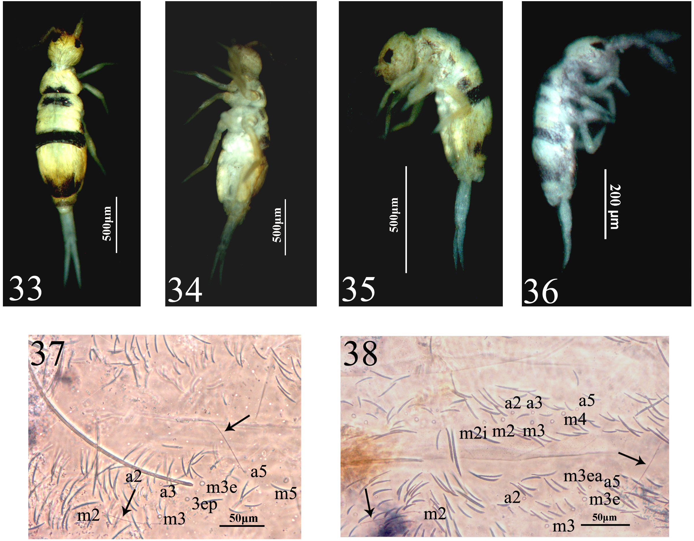
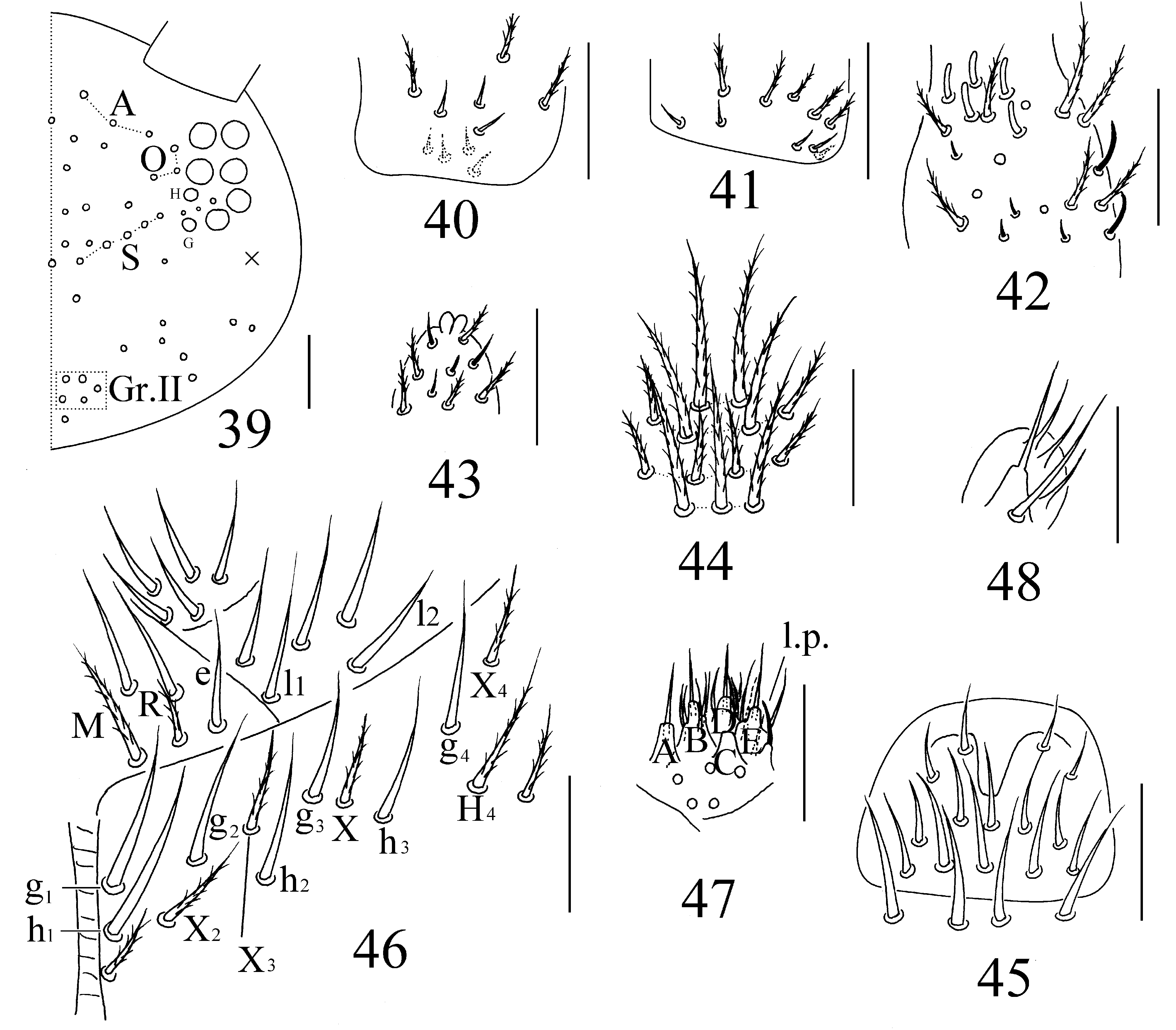
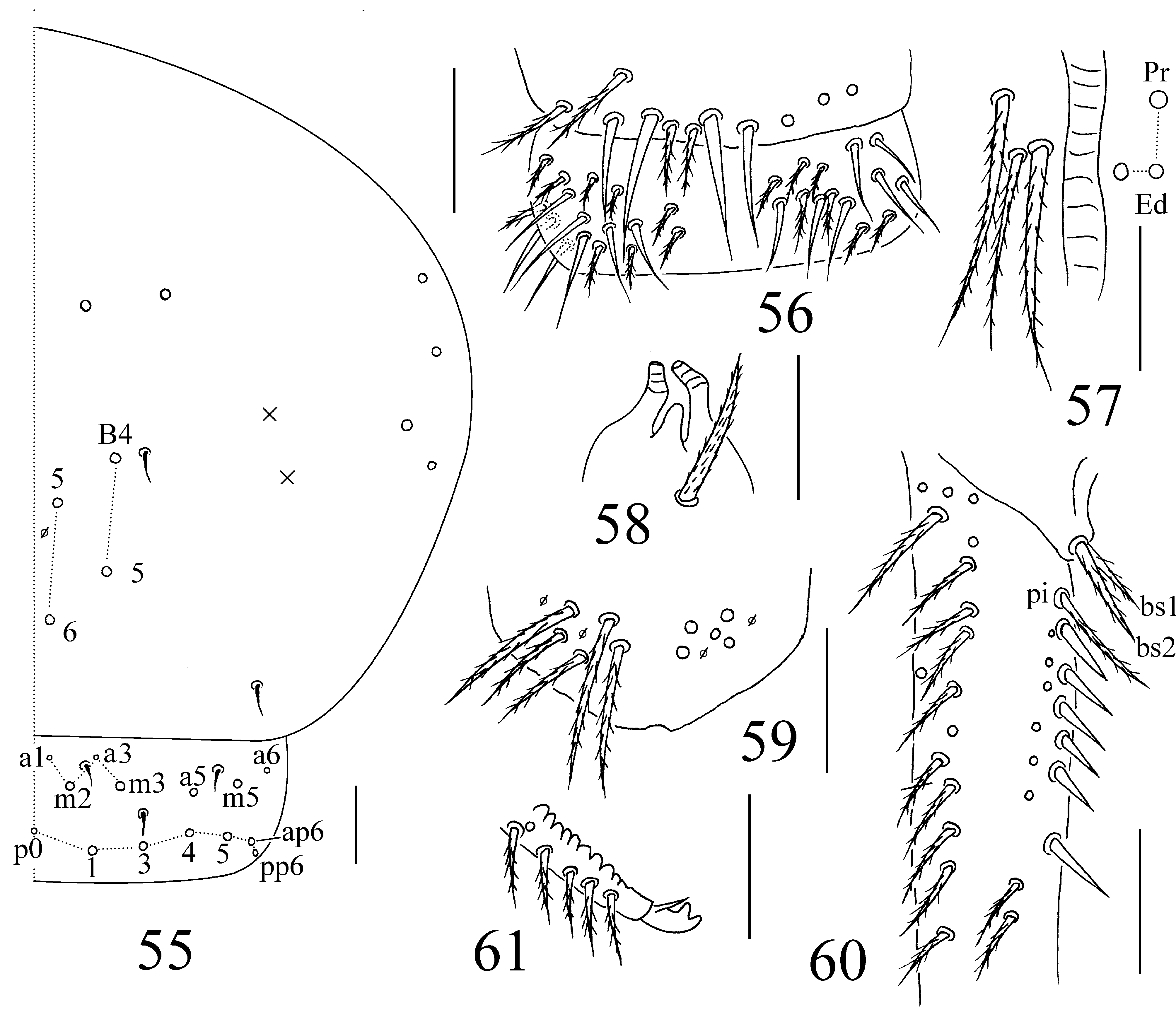
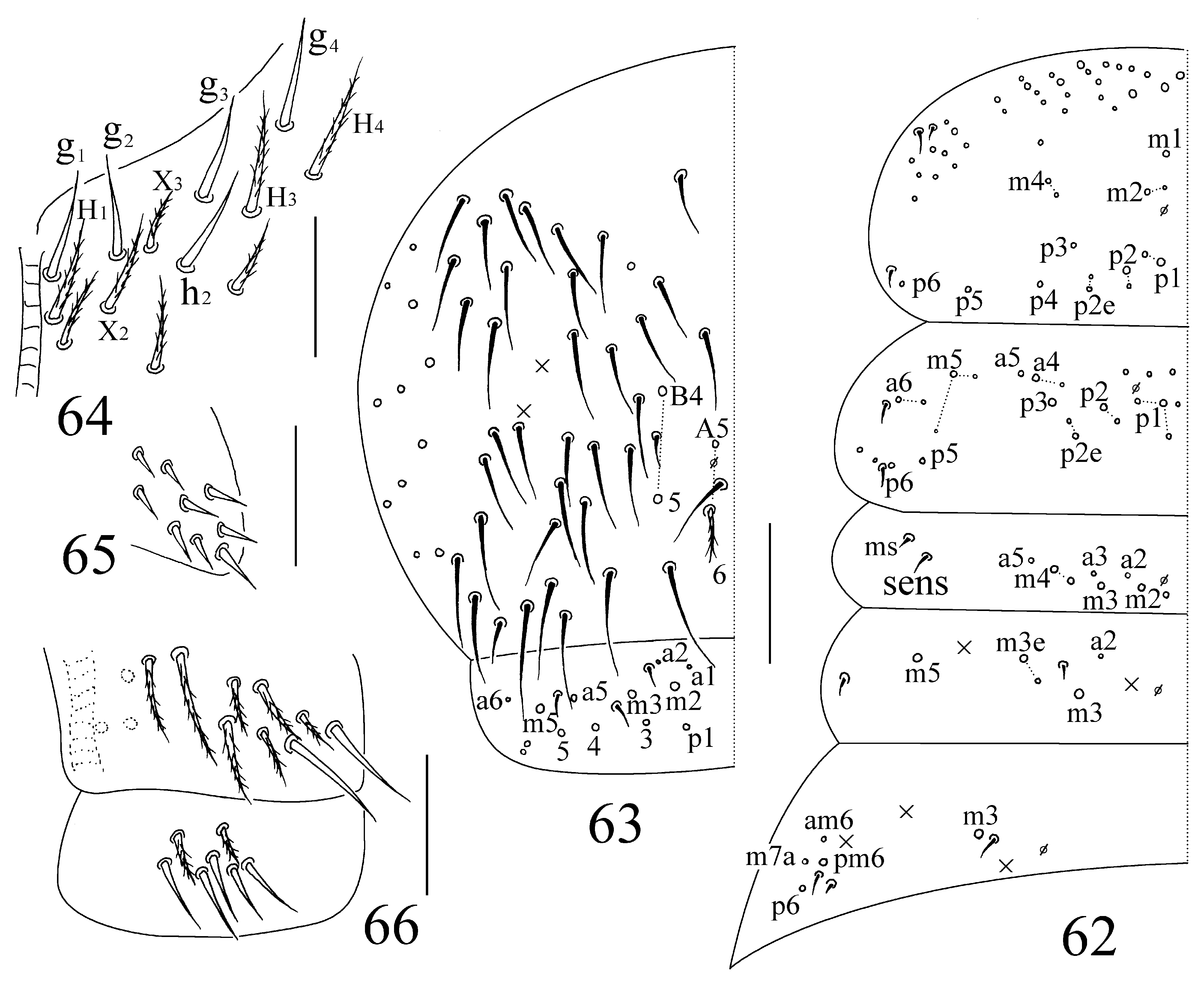
Figs 33–66 View FIGURES 33–38 View FIGURES 39–48 View FIGURES 49–54 View FIGURES 55–61 View FIGURES 62–66 , table 2
Type material. Holotype: female on slide, China, Guangdong Province, Guangzhou City, Tianhe District, Long- dong Reservoir , 23.235566N 113.399017E, alt. 127± 5 m, sample number 4661, 25.III.2018, collected by Z-X Pan and S-S Zhang. GoogleMaps
Paratypes. Five female adults and one subadult (maybe the second instar) on slide, two in ethanol. Same date as holotype. All types deposited in TZU .
Etymology. Named after the smooth chaetae l 2, g 1-4 and h 1-3 present on labium (latin: “lenis”, smooth, “seta”, chaetae).
Description. Adults. Size. Body length up to 2.18 mm. Colour pattern. Ground colour pale yellow. eye patches dark blue. Antennae pale, without dark pigment. Postero-central margins of Th. II–III and Abd. IV with M-shaped dark patches. Lateral side of Th. II–Abd. II and posterior margin of Abd. V slight pigmented. Abd. III mostly dark pigment, except lateral side with small unpigmented patch. Coxae, VT, furcula and ventral side of body without pigment ( Figs 33–35 View FIGURES 33–38 ).
Head. Eyes 8+8, G and H smaller than others; eye patches with three chaetae ( Fig. 39 View FIGURES 39–48 ). Antennal length 1.42– 1.71 times as long as head; antennal segment ratio as I: II: III: IV=1: 1.43–1.84: 1.21–1.34: 2.42–2.89. Basal part of Ant. I with three dorsal four ventral smooth mic ( Fig. 40 View FIGURES 39–48 ), Ant. II with five basal smooth mic ( Fig. 41 View FIGURES 39–48 ), five distal rod-like S-chaetae ( Fig. 42 View FIGURES 39–48 ). Ant. III organ with two rod-like and three short guard S-chaetae. Apical bulb of Ant. IV bilobed ( Fig. 43 View FIGURES 39–48 ). Dorsal cephalic chaetotaxy with three A, three O and five S mac, Gr. II with five mac ( Fig. 39 View FIGURES 39–48 ). Clypeus with 13 chaetae, arranged in four lines (3/4/4/2) ( Fig. 44 View FIGURES 39–48 ). Prelabral and labral chaetae as 4/5, 5, 4, all smooth ( Fig. 45 View FIGURES 39–48 ). Chaetotaxic formula of labial base as MRel 1 l 2, M and R ciliate and others smooth; postlabial chaetae not expanded with g 1–4, h 1–3 smooth (one examined individual with G 4 ciliate), H 4, X, X 2, X 3 and X 4 ciliate (one individual without X 3) ( Fig. 46 View FIGURES 39–48 ). Five papillae A–E on labial palp with 0, 5, 0, 4, 4 guard chaetae, respectively; the tip of lateral process (l.p.) slight exceeding apex of papilla E; hypostoma with two guard chaetae; five proximal chaetae ( Fig. 47 View FIGURES 39–48 ). Maxillary outer lobe with one apical, one subapical chaetae and three sublobal hairs on sublobal plate, subapical chaeta slightly larger than apical one ( Fig. 48 View FIGURES 39–48 ).
Thorax. Complete body sens as 2, 2/1, 2, 2, (at least 29), 3, ms as 1, 0/1, 0, 1, 0, 0. Th. II with three centro-medial mac (m1, m2 and m 2i), three centro-sublateral mac (m4, m 4i and m4p) and three S-chaetae (ms antero-internal to sens); posterior part with 23–27 mac; p4, p 4i, p5 and p6 as mac. Th. III with 35–37 mac and two sens; a5 series only one chaeta; p5 and p6 as mac, p4 as mic ( Fig. 49 View FIGURES 49–54 ). Coxal macrochaetal formula as 3 (pseudopores unclear)/4+1, 3 (two pseudopores)/ 4+2 (two pseudopores) ( Figs 50–52 View FIGURES 49–54 ). Trochanteral organ with 27–38 smooth chaetae ( Fig. 53 View FIGURES 49–54 ). Tenent hair clavate, slight shorter than inner edge of unguis in length; unguis with three inner, two lateral and one outer teeth; unguiculus lanceolate with one basal tiny tooth ( Fig. 54 View FIGURES 49–54 ).
Abdomen. Abd. IV length 3.96–5.72 times as long as Abd. III. Abd. I with 11 mac (a1–3, a 1i, m2–4, m 2i, m 4i, m4p and a5) and two S-chaetae (ms antero-external to sens). Abd. II with six central mac (a2, a3, m3, m3e, m3ea and m3ep) and one lateral mac (m5); trichobothria chaetae shorter than mac ( Figs 37–38 View FIGURES 33–38 ). Abd. III with one central mac (m3) and four lateral mac (am6, pm6, p6 and m7a) ( Fig. 49 View FIGURES 49–54 ). Abd. IV with at least 36 elongate (present anterior to “eyebrow” mac) and two normal length sens; “eyebrow line” with 2+2 mac; posterior central with four mac (A5–6, B4–5). Abd. V with three sens, middle one postero-external to m3; a1 and a3 as mic ( Fig. 55 View FIGURES 55–61 ). Posterior face of VT with four (2+2) apical smooth chaetae; lateral flap with 7–9 smooth and 7–14 ciliate chaetae on each side ( Fig. 56 View FIGURES 55–61 ); anterior face with many ciliate chaetae, 3+3 of them as mac, line connecting mac proximal (Pr) and external-distal (Ed) parallel to median furrow ( Fig. 57 View FIGURES 55–61 ). Tenaculum with 4+4 teeth and one large, multi-laterally basal ciliate chaeta ( Fig. 58 View FIGURES 55–61 ). Manubrial plaque with 2 pseudopores and 5–8 ciliate chaetae ( Fig. 59 View FIGURES 55–61 ). Dens with 10–15 spines, chaetae bs1 shorter than bs2, and pi slight longer than bs ( Fig. 60 View FIGURES 55–61 ). Mucro bidentate with subapical tooth larger than apical one; basal spine short, with tip reaching subapical tooth; distal smooth part of dens slightly shorter than mucro in length ( Fig. 61 View FIGURES 55–61 ).
Subadult. The examined specimen with well-developed trochanteral organ, ciliate chaetae on lateral flap of VT, and secondary chaetae on dorsal side of the body. So, this specimen is latter than the second instar larvae, but its exact instar is unclear.
Ground colour pale white. Colour pattern same with adults, significantly dark pigment on Th. II–III, Abd. III–V ( Fig. 36 View FIGURES 33–38 ).
Chaetotaxy shown in Figs 62–66 View FIGURES 62–66 . Abd. III with one (m3) central mac ( Fig. 62 View FIGURES 62–66 ). Chaetae g 1–4, h 2 on postlabial base smooth; H 1, H 3 and H 4 ciliate; X and X 4 absent ( Fig. 64 View FIGURES 62–66 ).
Ecology. Same as H. chroma sp. nov., they are found in same sample.
Remarks. This new species can easily distinguished from other known species of genus Homidia by its unique colour pattern, 2+2 mac in eyebrow and 4+4 mac on postero-central part of Abd. IV, short trichobothria and adult individuals with labial chaetae l 2, g 1–4 and h 1–3 smooth. It is mostly similar to Homidia sichuanensis Jia, Zhang & Jordana, 2010 in colour pattern (medial part of Th. II–III, whole Abd. III and posterior margin of Abd. IV with dark pigment), one medial mac on Abd. III. However, it can be distinguished from H. sichuanenis by the absence of longitudinal stripes behind each eye patch (along lateral borders of the head), smooth labial chaetae and 2+2 eyebrow-like mac. Also, this new species similar to Homidia quadriseta Pan, 2018 (in: Zhuo et al. 2018) in colour pattern and 2+2 eyebrow-like mac on anterior part of Abd. IV, but obviously differ from the latter in Th. II–III by central dark pigment and smooth labial chaetae present in new species. Detailed comparison between these similar species is listed in table 2.

No known copyright restrictions apply. See Agosti, D., Egloff, W., 2009. Taxonomic information exchange and copyright: the Plazi approach. BMC Research Notes 2009, 2:53 for further explanation.
|
Kingdom |
|
|
Phylum |
|
|
Class |
|
|
Order |
|
|
Family |
|
|
Genus |