
Cylomissus glabratus Broun, 1903
|
publication ID |
https://doi.org/ 10.1649/0010-065x-69.4.687 |
|
persistent identifier |
https://treatment.plazi.org/id/03B78799-FF8B-691E-FF11-F9ACFCD501D6 |
|
treatment provided by |
Diego |
|
scientific name |
Cylomissus glabratus Broun, 1903 |
| status |
|
Cylomissus glabratus Broun, 1903 View in CoL ( Figs. 9–14 View Fig View Fig View Fig View Fig View Fig View Fig )
Material Examined. N E W Z E A L A N D:
Marlborough: 5 L1, 4 L3 ( NMPC, includes molecu-
lar vouchers COL1774 and COL1854 ): Dead Horse Creek south of Canvastown , 41°19.59′S 173°39.57′E, wet stones with algae & moss along exposed stream ( RL1511 – RL1514 ), 30.xi.2010, lgt. Fikáček & Leschen. Gisborne: 1 L3 ( NMPC): Te Urewera National Park ( Fig. 9G View Fig ), 0.2 km NW of “Aniwaniwa” below Aniwaniwa Falls , in moss on the rock wall overflow by small stream ( NZ 08), 38°44.6′S 177°9.7′E, 600 m, 23–26.xi.2012, lgt. Becker, Fikáček & Hájek. Nelson: 2 L3 ( NMPC): Owen River , Sunrise Ridge Track, Halfway Creek , moss and rocks in/along the stream ( RL1519 ), 1.xii. 2010, lgt. Fikáček & Leschen. Taranaki: 1 L1, 3 L3 ( NMPC): unnamed stream 0.2 km S of Pukeiti Garden 9 km E Okato, 39°12.1′S 173°58.9′E, 370 m, in moss on stones in/along the stream ( Fig. 9J View Fig , white bar) in lowland Nothofagus forest ( NZ 27), 30.xi.2012, lgt. Becker, Fikáček & Hájek;3 L2,3 L3 ( NMPC): Mt. Egmont National Park, Te Popo stream at Curtis Falls Track ( Fig. 9F View Fig ) near “Mountain House”, 39°18.3′S 174°7.6′E, 830 m, in moss on stones in and along partly exposed mountain stream in montane forest and at wet rock covered by moss with water flowing over it ( NZ 21), 28–29.xi.2012, lgt. Becker, Fikáček & Hájek GoogleMaps .
Description. General Morphology. Third Instar. Body ( Fig. 9B–D View Fig ) moderately thick, nearly parallel-sided, lobate laterally; widest between abdominal segments 2–5. Color brown with slightly paler tubercles and yellowish sclerites. Head: Head
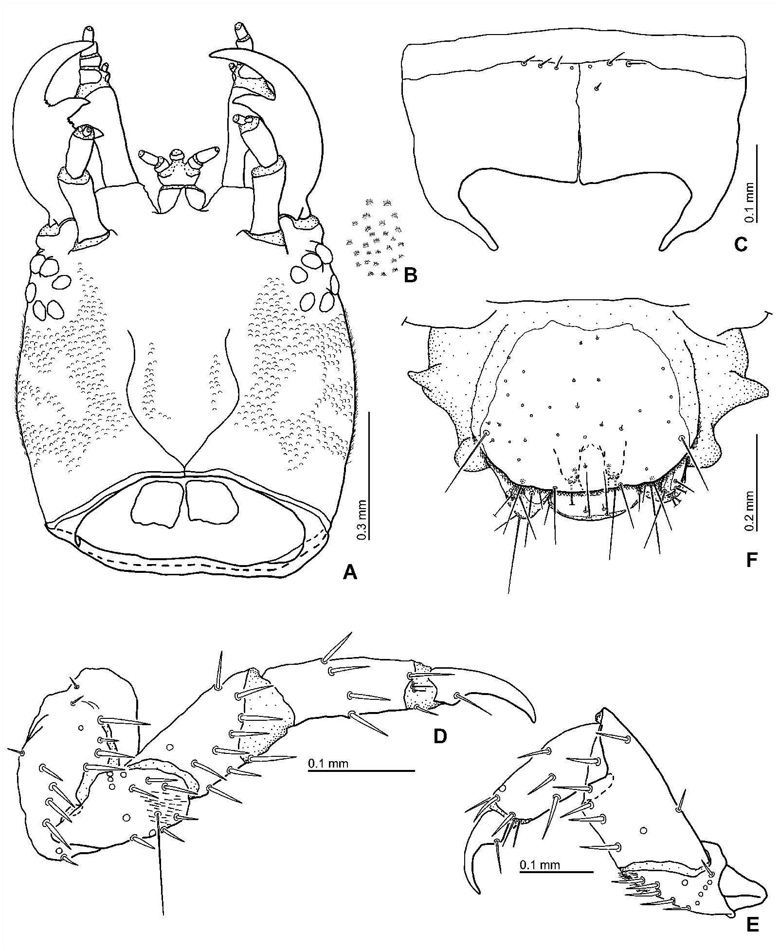
capsule ( Fig. 14A View Fig ) subtrapezoidal, widest medially. Cervical sclerite large, subpentagonal. Frontal lines indistinct, only basal half slightly visible; coronal line short. Basal part of frontale and dorsal and lateral surface of parietale bearing densely arranged small scale-like projections, thus look like bearing very finely granules; each scale-like projection bearing fine trichoid cuticular projections. Six stemmata on each anterolateral portion of head capsule. Posterior tentorial pits present close to junction of submental sulcus and gular sulcus ( Fig. 12B View Fig ). Clypeolabrum ( Fig. 12C View Fig ) slightly asymmetrical. Nasale with 1 large median projection, its apical portion more or less abraded. Lateral lobes of epistome large, strongly projecting anteriorly, both lobes projecting as far as nasale. Inner edge of left epistomal lobe strongly serrate; lateral part of epistomal lobe bearing fine, trichoid cuticular projections. Antenna ( Fig. 13A–B View Fig ) 3-segmented, rather slender; surface of antenna smooth. Antennomere 1 wider and slightly longer than antennomere 2; antennomere 3 shortest and narrowest. Antennal sensorium present, small. Mandibles ( Fig. 13C–D View Fig ) moderately wide, almost symmetrical, with 2 inner teeth each. Apical inner tooth larger than basal tooth. Incisor area of right mandible extremely finely serrate. Basal part of incisors area close to distal inner tooth and inner margin between inner teeth serrate. Inner face of mandible basally of basal inner tooth almost straight. Maxilla ( Fig. 13E–F View Fig ) 6-segmented, longer than antenna. Cardo large, irregularly shaped. Stipes longer and wider, distinctly longer than palpomere 1–4 combined. Maxillary palpus short, 4-segmented; surface of maxillary palpus bare. Palpomere 1 widest, incompletely cylindrically sclerotized dorsally. Inner process sclerotized. Palpomere 2 wider than palpomere 3, palpomere 4 narrowest. Labium ( Fig. 13G–H View Fig ) well-developed. Hypopharyngeal lobe absent. Submentum fused to head capsule, large, subpentagonal; submental sulcus visible. Mentum subquadrate, slightly longer than wide. Dorsal surface with densely arranged cuticular teeth. Prementum subquadrate, transverse, anterior part wider than posterior part. Anterior membranous area of prementum with densely arranged, short trichoid cuticular projections behind palpomere 1. Ligula well-developed, median part sclerotized as ring. Labial palpus long, longer than ligula; palpomere 1 slightly wider than palpomere 2 and shorter than ligula, bearing fine, trichoid cuticular projections; intersegmental membrane between palpomere 1 and 2 bearing rather short, trichoid cuticular projections dorsally and laterally. Thorax: With several pairs of sclerites; meso- and metanotum lobate laterally, 2 pairs of apically pointed lobes on each side. Prothorax slightly wider than head capsule. Proscutum formed by 1 large plate subdivided by fine sagittal line, anterior part weakly sclerotized; median part with transverse groove, lateral part with convex portions. Proscutal plate mostly covered with fine granules. Prosternal sclerite (e.g., Fig. 14C View Fig ) subpentagonal, subdivided by fine sagittal line, anterior part rather weakly sclerotized; bearing a few short setae anteromedially. Mesonotum with 3 pairs of sclerites. Anterior 2 sclerites small, narrow, transverse; 1 mesally, 1 laterally. Posterior sclerite large, transverse, subquadrate. Sclerites adjacent mesally, posterior part bearing granules. Metanotum with 3 pairs of sclerites. Anterior sclerite very small, narrow, transverse, hardly visible but present on anterolateral part; median sclerite large, subquadrate, transverse. Sclerite adjacent mesally; posteromesal part bearing granules; posterior sclerite rather small, semi-oval, transverse, on posterolateral part. Legs (e.g., Fig. 14D–E View Fig ) moderate in length, slightly visible in dorsal view, 5-segmented; all 3 pairs similar in shape. Abdomen: Fig. 9B–D View Fig . Ten-segmented, slightly tapering posteriorly, densely covered with granules, bearing tubercles of several sizes, lobate laterally, the lobes pointed apically; segments 1–7 similar in shape and size; dorsal sclerite present on segment 1 only. Segment 1 with 1 pair of small dorsal sclerites medially on anterior part. Segments 2–7 similar to segment 1, with dorsal sclerites seemingly absent, but with a pair of very small bare portions on anteromesally. Spiracular atrium ( Fig. 14F View Fig ) on segment 8 with large oval dorsal plate bearing small setae and covering granules; posterior margin of sclerite with 3 pairs of long setae; procercus incompletely sclerotized, with 2 long and 1 short seta. Spiracular atrium on segment 9 rather small, trilobed, partly sclerotized dorsally. Median lobe transverse; lateral lobe without acrocercus. Urogomphi rather short, 1-segmented; prostyli absent.
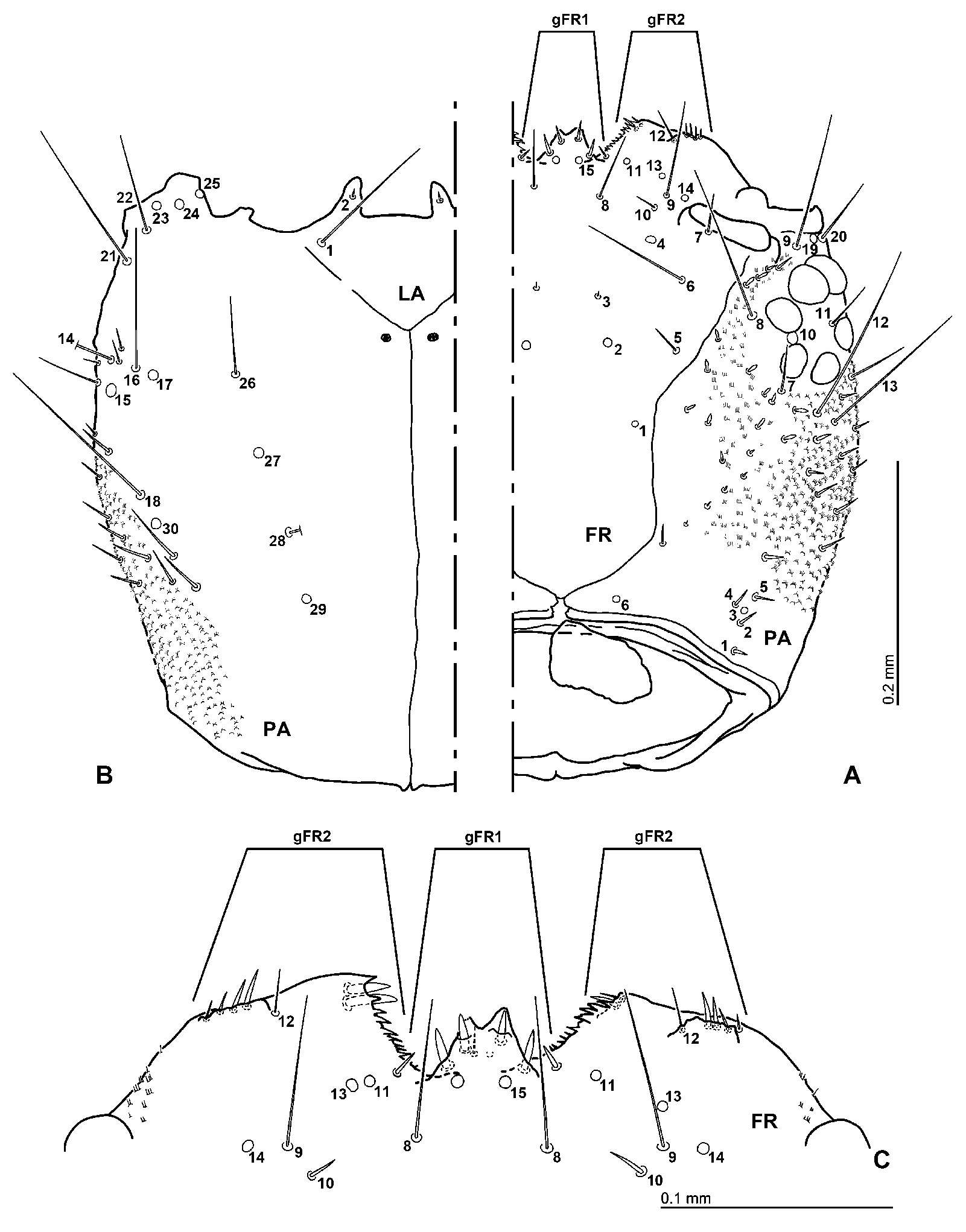
First Instar. Very similar to third instar ( Fig. 9A View Fig ). Head: Frontal lines clearly visible, lyriform, abruptly converged at base, coronal line very short ( Fig. 10 View Fig ). Nasale with one median tooth but emarginate medially at apex; epistomal lobe projecting further than nasale; left lobe projecting slightly farther than right lobe. Antenna ( Fig. 11A–B View Fig ) slightly proportionally stouter than in third instar. Mandibles ( Fig. 11C–D View Fig ) with inner face more weakly serrate than in third instar, or not serrated; basal portion projecting mesad just below basal inner tooth. Maxilla ( Fig. 11E–F View Fig ) with outer face of stipes bearing densely arranged, short, trichoid cuticular projections dorsally (this part of third instar bearing a series of stout setae). Intersegmental membrane between palpomeres 1, 2, and 3 with few fine trichoid cuticular projections dorsally. Thorax and abdomen: Lateral face weakly lobate, not as distinct as in third instar ( Fig. 9A View Fig ).
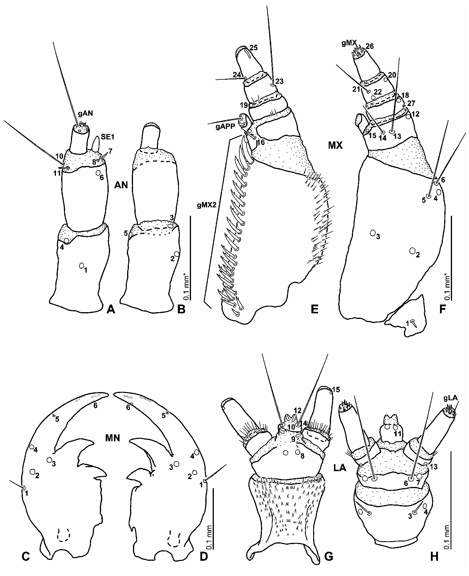
Chaetotaxy of Head of First Instar. Frontale: Fig. 10A, C View Fig . Central part with 3 pairs of sensilla (FR1–3) divergent posteriad; FR1 seta (broken in first instar examined, but present in third instar), FR2 pore-like situated between FR1 and FR3, close to FR3; FR3 very short seta, situated anteromesally to FR2. Rather short seta FR5 and very long seta FR6 lateral to FR2 and FR3, respectively. Slightly oval pore-like sensillum FR4 situated anteromesally to FR6, mesally to antennal socket. Rather short seta FR7 close to posterior margin of antennal socket. Short seta FR10, long seta FR9, and pore-like sensillum FR14 slightly anteromesal to antennal socket; FR10 anterior to FR4; FR9 between FR10 and FR14; FR14 laterally to FR9. Distribution of sensilla on epistomal lobes asymmetrical – left lobe: pore-like sensilla FR11 and FR13 closely aggregated on posteromesal portion, FR11 mesal to FR13, close to lateral-most seta of gFR1; FR12 short seta on median portion of anterior margin of epistomal lobe; – right lobe: pore-like sensilla FR11 and FR13 more distant than those on left, on basal portion; FR13 situated laterally to FR11, anteromesally to FR9; short seta FR12 situated medially on anterior margin of epistomal lobe. Rather long seta FR8 posteriorly to nasale. Pore-like sensillum FR15 on base of nasale. Nasale with 4 short, very stout setae on projection, 2 short stout setae lateral to projection, and (presumably) 2 short ventral setae (one of them broken in the examined specimen) (gFR1). Left epistomal lobe with 6 setae excluding FR12 (gFR2); 4 setae short to rather short, slender to stout setae on mesal portion of anterior margin, close and lateral to FR12; 2 setae short, very stout setae on ventral side of mesal corner. Right lobe probably with 6 setae excluding FR12; 4 short slender to stout setae on mesal portion of anterior margin close and lateral to FR12, 2 short setae on ventral side of mesal corner (the setae are very indistinct in examined specimen). Parietale: Fig. 10A–B View Fig . Dorsal to lateroventral surface with numerous, minute to rather long additional sensilla of variable shape: posteromesal portion with a irregular row of short setae, anteromesal part of parietale with sparsely arranged short scale-like setae, a few short scale-like to simple stout additional setae on posterolateral to antennal socket. Median portion of lateral surface of parietale with numerous short to rather long additional setae. Dorsal surface with a group of 5 sensilla (PA1–5) forming irregular longitudinal row in posterior part of parietale; PA1–2 and 4–5 short stout setae, PA3 pore-like. PA6 porelike, laterally to coronal line. PA8 very long seta, lateral to mesal-most anterior stemma, close to frontal line. PA10 pore-like, between mesal-most anterior and posterior stemmata, rather long seta PA11 on between lateral stemmata. PA7 and PA12–17 forming irregular transverse row on median part of lateral face of parietale. PA7 rather short seta, behind mesal-most posterior stemma; PA12 very long seta, behind median stemma of posterior series; very long seta PA13 close and lateral to PA13. PA14–17 on lateroventral surface; PA14 seta, PA15 pore-like posterior to PA14; PA16 very long seta mesally to PA14–15, between PA14 and PA17; PA17 pore-like. PA18 and PA30 on ca. posterior third of head capsule posterior to PA16–17; PA18 very long seta, PA30 pore-like sensillum. PA9 and PA19–22 situated on anterior corner of head capsule, PA9 very long seta, PA19 pore-like sensillum, PA20 long seta, PA21 very long seta, PA22 long seta. PA9 dorsal to remaining sensilla, PA19 between PA9 and PA20; PA21 ventral to PA20; PA22 anterior to PA21. PA23–25 on ventral mandibular articulation; PA23–24 laterally to PA25, PA24 between PA23 and PA25. PA26–29 on median part of ventral surface of parietale, forming longitudinal row divergent anteriad, sensilla almost equidistant from each other; PA26 long seta, PA28 seta, possibly very long (compared to third instar), PA27 and PA29 pore-like sensilla; from anterior to posterior, PA26, PA27, PA28, PA29. Antenna: Fig. 11A–B View Fig . Antennomere 1 with 5 porelike sensilla (AN1–5); AN1 situated dorsally on median part, AN2 on outer face laterally to AN1; AN3–5 subapical on intersegmentary membrane, AN3 on outer face, AN4–5 on inner face. Antennomere 2 with 1 pore-like sensillum (AN6) situated dorsally very close to distal margin of sclerite; setae AN7–8 and AN10–11 on intersegmental membrane between antennomeres 2 and 3; sensorium SE1 on the intersegmental membrane, AN9 absent; AN7–8 and SE1 on lateral face, AN7 small, AN8 minute, basally to SE1, SE1 slightly shorter than antennomere 3, stout, conical; AN10–11 on inner face, AN10 very long, AN11 short, both setae close to each other. Antennomere 3 with apical sensilla (gAN) in apical membranous area; 1 very long seta, 1 long seta, others minute (or possibly broken). Mandibles: Fig. 11C–D View Fig . With 3 pore-like sensilla (MN2–4) on median part, forming triangular group; MN3 mesal to MN2 and MN4, MN4 on lateral face, anterior to MN2–3; MN2 between MN1 and MN3; MN1 moderately seta on ca. midlength of lateral face. Minute seta MN5 subapical on lateral face, minute pore MN6 on subapical part of inner face. Maxilla: Fig. 11E–F View Fig . Cardo with 1 short stout ventral seta ( MX 1). Inner face of stipes with a row of rather short and stout setae increasing in size toward distal margin, each bearing subapical tooth, densely arranged on dorsal part (gMX2), MX 7 rather short and stout dorsal seta at base of gMX2, distinguishable from gMX2 setae by absence of subapical tooth. Ventral surface of outer face of stipes with 3 primary sensilla ( MX 4–6) closely aggregated on distal part of sclerite, MX 4 pore-like, MX 5–6 long setae, MX 4 lateral to MX 5, MX 6 distal to MX 4. Pore-like sensilla MX 2–3 situated ventrally, both ca. at midlength, MX 3 mesally to MX 2. Dorsal surface of palpomere 1 with 1 rather short, stout seta ( MX 16) situated on inner face. Three sensilla ( MX 12–14) ventrally on distal part of sclerite; MX 12 pore-like on lateral face, MX 13–14 on median part, MX 13 very long seta, between MX 12 and MX 14, MX 14 moderately long seta. Pore-like sensilla MX 15 and MX 17 situated on membrane behind inner appendage, MX 17 dorsally, MX 15 ventrally. Inner appendage with 1 very long seta and 1 short stout seta apically (gAPP). Palpomere 2 with 2 porelike sensilla ( MX 18 and MX 19) and 1 minute seta ( MX 27); MX 18 situated ventrally on lateral part of anterior margin of sclerite; MX 19 situated dorsally on inner face of intersegmental membrane between palpomeres 2 and 3; MX 27 situated basally on lateral face. Palpomere 3 with 2 long setae ( MX 21 and MX 23) and 2 pore-like sensilla ( MX 20 and MX 22); MX 21 and MX 22 closely aggregated, situated ventrally on inner face, MX 22 basal to MX 21; MX 20 on lateral part of ventral surface, close to anterior margin of sclerite; MX 23 dorsally on lateral face, in basal third of sclerite. Palpomere 4 with 1 moderately long seta ( MX 24) situated basally on inner face, and with digitiform sensillum ( MX 25) and pore-like sensillum ( MX 26) apically on outer face of sclerite; MX 25 dorsal, MX 26 ventral at distal margin of sclerite. Apical membranous area of palpomere 4 with several minute setae (gMX). Labium: Figs. 11G–H View Fig . Submentum with 2 pairs of setae (LA1–2) on anterolateral portion; LA1 very long on lateral margin, LA2 short on anterior margin. Ventral surface of mentum with 1 pair of rather short setae (LA3) and pore-like sensilla (LA4) situated on anterior margin of sclerite; LA4 lateral to LA3. Prementum and its anterior membranous area with 5 pairs of sensilla (LA5–9). LA5 not found in examined larva but probably just overlooked as it is present in third instar, LA6–7 ventral, closely situated on anterior margin of sclerite, LA6 very long seta, mesal to LA7, LA7 porelike. LA8–9 dorsal on median part, LA8 pore-like, on anterior margin of sclerite, LA9 pore-like, on median portion of membranous area. Ligula with 3 pairs of sensilla (LA10–12); LA10 very long seta, lateral to midline; LA11 pore-like, subapical on ventral surface; LA12 pore-like, digitiform, apical on dorsal surface. Palpomere 1 with 2 sensilla (LA13–14); LA13 minute seta, situated ventrally on basal part; LA14 pore-like, located dorsally on intersegmental membrane between palpomeres 1 and 2. Palpomere 2 with pore-like sensillum LA15 situated on distal margin on outer face of sclerite. Apical membranous area of palpomere 2 with several minute setae of variable shape (gLA).
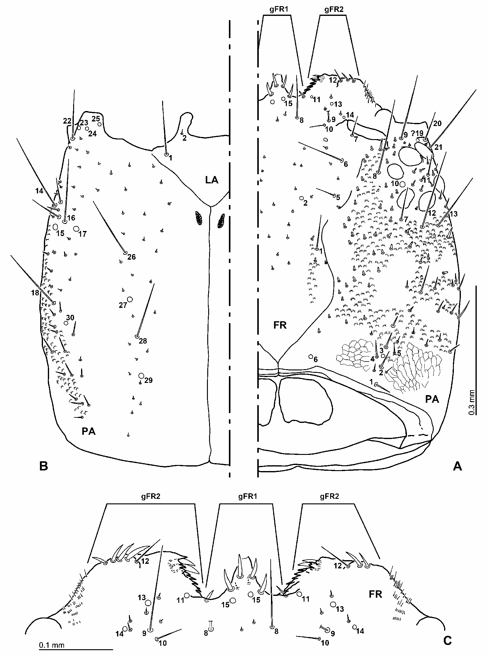
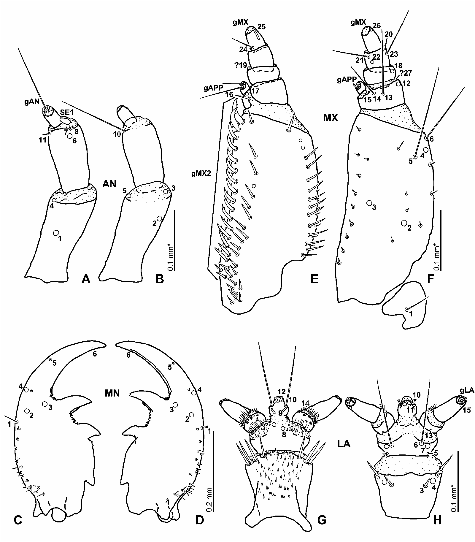
Chaetotaxy of Head of Third Instar. Head: Arrangement of primary sensilla very similar to that of first instar, but head capsule bearing larger number of additional/secondary sensilla of variable length. Ventral surface around sensilla PA26–29 without short additional setae present in first instar. Antenna: Fig. 13A–B View Fig . Similar to first instar, sensorium (SE1) proportionally shorter. Mandibles: Fig. 13C–D View Fig . Similar to first instar, outer basal face of each mandible with numerous additional short setae. Maxilla: Fig. 13E–F View Fig . Similar to first instar. Stipes with an irregular row of additional stout setae dorsally of gMX2, MX 7 indistinguishable from these setae; ventral surface with a row of short stout setae along inner face and with scattered additional minute setae on ventral surface and lateral face. Maxillary palpus without additional sensilla. Labium: Fig. 13G–H View Fig . Similar to first instar, LA15 distinct, present in basolateral portion of prementum. Distal margin of mentum with series of moderately long stout additional setae laterally and dorsolaterally.

| NMPC |
National Museum Prague |
No known copyright restrictions apply. See Agosti, D., Egloff, W., 2009. Taxonomic information exchange and copyright: the Plazi approach. BMC Research Notes 2009, 2:53 for further explanation.
|
Kingdom |
|
|
Phylum |
|
|
Class |
|
|
Order |
|
|
Family |
|
|
Genus |